What are cardiovascular diseases?
Cardiovascular diseases (CVD) are the leading cause of mortality in many economically developed countries, including Russia, accounting for 55% of total mortality.
According to statistics from recent years, in the structure of mortality from CVD, 85.5% are due to ischemic heart disease (46.8%) and cerebral stroke (38.7%). In the Russian Federation in 2000, the mortality rate from diseases of the circulatory system was 800.9 per 100,000 thousand population. The main cause of cardiovascular disease is atherosclerosis. And although it is impossible to eliminate many risk factors for the development of atherosclerosis, we can change, for example, our lifestyle and diet.
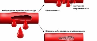
Natural age-related “wear and tear” of blood vessels creates conditions for cholesterol deposition inside and on the walls of arteries. This leads to a gradual narrowing of the lumen of the blood vessels and reduces the ability of the heart to pump blood through them to the body. The moment the vessel is completely blocked, a heart attack (myocardial infarction) or stroke occurs if the blockage occurs in a vessel in the brain. It is important to note that high blood cholesterol does not always have an unfavorable prognosis. The fact is that cholesterol is associated with proteins - proteins, together they make up lipoproteins. So, cholesterol can be in high (HDL-CHOL.) and low (LDL-CHOL.) density lipoproteins. Cholesterol in HDL-CHOL. prevents the progression of atherosclerosis - “beneficial lipoproteins”, and cholesterol in LDL-CHOL. promotes the progression of atherosclerosis.
What diseases can electrocardiography (ECG) tell about?
An electrocardiogram (ECG) is considered the main diagnostic method for identifying various diseases of the cardiovascular system. Our heart works in the body under the control of its own pacemaker, which produces electrical impulses and directs them to the conduction system, and they are recorded on the ECG. It turns out that using an electrocardiogram, we can record a peculiar language of our myocardium. Based on the deviations of the main teeth: P, Q, R, S and T, it is possible to determine which disease is the basis of cardiovascular pathology.
Hypertrophy of the heart
Hypertrophy of the heart chambers occurs as a result of hemodynamic disturbances in the bloodstream, which provoke overload of the ventricles or atria. On the ECG you can see seven main signs of cardiac muscle hypertrophy:
- An increase in the time of internal deviation, since in the hypertrophied myocardium the excitation spreads longer in the area from the endocardium to the epicardium.
- An increase in the amplitude of the R wave, while the excitation vector is larger in magnitude.
- Ischemia of the subendocardial layers of the heart, due to the fact that they lack blood flowing through the coronary arteries.
- Conduction disturbance.
- Deviation of the electrical axis of the heart towards the hypertrophied section, as its mass increases due to the growth of cardiomyocytes.
- Changes in the electrical position of the heart.
- Displacement of the transition zone (V3), manifested by a change in the ratio of the R and S waves in the third chest lead.
Angina pectoris
The disease is characterized by attacks of anginal pain lasting from a few seconds to twenty minutes. This disease is one of the formischemic heart diseases. In the classic form of angina pectoris, electrocardiographic signs are manifested by changes in the final part of the ventricular QRS complex:
- Depression of the S-T segment.
- Various changes in the T wave, such as decreased amplitude, biphasicity, isoelectricity, or negativity.
- The focal nature of these changes: they are recorded in one or two leads, since the observed hypoxia is local in nature, developing in the basin of a separate branch of the coronary artery.
During the periods between attacks, the ECG often shows no pathological changes at all. In addition, the above-described deviations are possible in many other heart diseases and pathological conditions. That is why in some cases the diagnosis of angina pectoris can be difficult.
Arrhythmia
Pathology of the cardiovascular system associated with a violation of the formation of an excitation impulse or its spread throughout the myocardium. In most cases, it manifests itself as an interruption in the rhythm of heart contractions, with periods of acceleration and gradual deceleration. Typically, your heart rate increases as you inhale and decreases as you exhale. The features of the ECG are as follows:
- The frequency of changes in the R - R intervals is more than 0.1 seconds.
- Unlike other rhythm disturbances, there is a gradual change in the duration of the R – R interval, usually due to the T – P segment.
- Small fluctuations of P – Q and Q – T are characteristic.
The most reliable electrocardiographic sign of sinus arrhythmia is considered to be a gradual periodic shortening of the R - R section against the background of an increased rhythm and, conversely, a lengthening of the R - R intervals when the rhythm slows down.
Tachycardia
An increase in heart rate is called tachycardia. In this case, the heart rate accelerates to 100-150 beats per minute. A similar disorder can develop due to increased automatism of the sinus node. The pathology is also inherent in healthy individuals during physical exertion or emotional stress. The cause is often ischemia, dystrophic changes, various infections and toxic effects. Main ECG signs:
- There is a decrease in the R–R interval as the T–P interval shortens.
- With severe tachycardia, the P–Q segment shortens.
- The degree of increase in heart rate is directly proportional to the decrease in Q – T.
- An upward displacement of the RS – T segment downward from the isoelectric line.
- The amplitude and direction of the teeth correspond to the norm.
Bradycardia
A deviation that is manifested by a reduced heart rate (less than 60 per minute). It occurs with reduced automatism of the sinus node; it can occur even in healthy people, for example in athletes, when exposed to various factors. A common cause is considered to be an increase in the tone of the vagus nerve. The electrocardiographic picture, in principle, differs little from the norm, only the rhythm is slowed down. The following changes are noted on the ECG:
- The R interval increases due to the T – P shift.
- Q – T increases according to the decrease in rhythm frequency.
- The amplitude and vector of the teeth change slightly.
Heart aneurysm
A cardiac aneurysm is an enlargement of the myocardial cavity due to pathological changes in the muscle layers or abnormalities in the development of the organ at the stage of embryogenesis. The main signs of a cardiac aneurysm include protrusions in its area due to thinning of the wall, which can rupture. This is what can lead to irreparable consequences, which ECG research helps prevent. There are two leading signs that allow you to diagnose an aneurysm:
- The QS wave is present in leads where high R is usually recorded.
- A “frozen” ECG curve: instead of Q, a dome-shaped RS-T segment appears, shifted upward from the isoline, and sometimes a negative coronary T-wave appears.
Extrasystole
Extrasystole is the most common heart rhythm disorder. The pathology develops due to the appearance of an active heterotopic focus capable of generating an electrical impulse that interrupts the work of the sinus node. Manifested by extraordinary excitation and subsequent contraction of parts of the heart or its entirety:
- On an ECG, extrasystoles differ in shape, relation to the isoline, location of the P wave or width, direction of the QRST complex teeth.
- The presence of an increased pause (compensatory) after the extrasystole.
- Atrial extrasystole is characterized by: interval R(c)-R(e) <interval R(c)-R(c), there is a wave P(e), different from the wave P(c), an incomplete compensatory pause.
- The signs of ventricular extrasystole are as follows: interval R(s)—R(e) <interval R(s)—R(s), the P(e) wave is absent, the QRS complex is deformed, a complete compensatory pause.
Pulmonary embolism
Pulmonary embolism is accompanied by the development of pulmonary heart syndrome, since the resulting hypertension of the pulmonary circulation leads to acute overload of the right ventricle. In this case, the myocardium is in a state of hypoxia, and its hypertrophy develops. All of the above determines several ECG options for pulmonary embolism:
- SI-QIII-TIII syndrome: deep S waves in standard I and Q waves in leads III, while T waves in lead III become negative.
- acute hypertrophy of the right parts of the heart muscle, which is manifested by a high pointed P wave in standard lead II.
- acute supraventricular tachyarrhythmias.
In some cases, with acutely developing cor pulmonale, metabolic changes in the area of the right ventricle provoke the occurrence of complete or partial blockade of the right bundle branch.
Pericarditis
The dynamic picture of the ECG during pericarditis depends on its etiology. However, there are also common characteristics, for example: inflammation of the pericardial tissue changes its electrical status, which leads to the emergence of so-called “inflammatory currents” coming from the heart. It is these “currents” that are recorded by the electrode located above the myocardial area:
- This is graphically displayed on the ECG by the rise of the S-T segment in all leads (concordant rise of the S-T segment).
- There is no displacement of the RS segment – Tbelow the isoline level.
- Pathological Q-waves do not appear dynamically.
The above differences form and disappear in the case of acute pericarditis much more slowly than in the case of myocardial infarction. The appearance of effusion in the pericardial cavity is accompanied by a noticeable decrease in the voltage of all electrocardiographic waves, especially in the leads from the limbs of the subject.
Myocarditis
In all cases of myocarditis, parenchymal inflammation occurs and progresses in the wall of the ventricles, which is focal or diffuse in nature, affecting a certain area of the heart muscle:
- The total T vector tilts in the direction opposite to the affected area, and a low or negative T wave is displayed on the ECG.
- The S–T vector is directed towards the lesion.
- The RST segment is displaced up and down from the isoelectric line.
- Negative T can become symmetrical in acute myocarditis, it becomes pointed, as in coronary insufficiency. The localization of all noted disorders depends on the location of the inflammatory reaction.
These electrocardiographic shifts are best determined in the chest leads. A concomitant pathology is rhythm and conduction disturbances.
Myocardial dystrophy
Myocardial dystrophy is part of the complex of pathological clinical symptoms of menopausal hormonal changes in older people. The pathology is described by pain in the heart area, different from angina pectoris, and a disturbance in heart rhythm. There are cases when these symptoms occur before the onset of menopause. The most common electrocardiographic signs are the following:
- A negative but not deep “coronal” T wave or it may be biphasic with a negative second phase.
- Dynamic changes in electrocardiographic data.
- A slight shift of the RS – T segment downward from the position of the isoelectric line.
- More pronounced changes in the middle chest and right leads.
The main problem of ECG diagnostics is that many deviations and changes in the electrocardiographic picture are similar for a number of diseases. For example, coronary insufficiency, some forms of myocarditis and myocardial dystrophy are characterized by similar electrocardiograms. As practice shows, clinical symptoms need to coincide with the dynamics of ECG abnormalities. In this regard, it is important to note the development of the promising direction presented in the Kardi.ru project. The technique of monitoring the state of your heart allows you to register micro changes in the work of the heart muscle even before the appearance of serious disorders, which is displayed graphically by the CardiRu device and facilitates timely decision-making on appropriate therapeutic measures.
Risk factors for cardiovascular disease
Some risk factors for cardiovascular disease are potentially reversible or can be changed. They include:
- Smoking cigarettes
- Elevated LDL (low density) cholesterol levels
- High triglyceride levels in the blood
- Low HDL (high density) cholesterol
- Overweight
- Large waist circumference (“apple shape”)
- High blood pressure
- Sedentary lifestyle
- Diabetes
Diabetes
Diabetes mellitus leads in mortality from diseases of the circulatory system among other endocrine diseases.
With diabetes mellitus, various lesions of the cardiovascular system develop:
- changes in the blood vessels of the heart - diabetic macroangiopathy;
- arterial hypertension (due to diabetic kidney damage, metabolic syndrome - depending on the type of diabetes);
- diabetic cardiomyopathy with rhythm disturbances and circulatory failure;
- IHD, sometimes with painless angina;
- increased levels of cholesterol and triglycerides in the blood;
- increased risk of ventricular fibrillation, myocardial infarction, and cerebrovascular accidents.
Treatment:
- compensation of carbohydrate metabolism (insulin therapy, hypoglycemic drugs);
- antihypertensive drugs, antiplatelet agents;
- lipid-lowering drugs (statins).
Diet and cardiovascular disease
Making small changes to your diet is one of the easiest and most effective ways to reduce your risk of heart disease. You can do it like this:
- Reducing fat in your diet, especially saturated fat and trans fat
- More fruits and vegetables, whole grains and soluble fiber
- Drinking alcohol in moderation
- Reducing salt to maintain lower blood pressure
Cholesterol and cardiovascular disease
Because there is a proven direct link between high blood cholesterol and your risk of heart disease, reducing the amount of fat in your diet, especially saturated fat, can help lower your blood cholesterol. According to the official recommendations of the European Society of Atherosclerosis, “normal” levels of fatty fractions in the blood are: 1. Total cholesterol - less than 5.2 mmol/l. 2. Low-density lipoprotein cholesterol (LDL) - less than 3-3.5 mmol/l. 3. High-density lipoprotein cholesterol (HDL) - more than 1.0 mmol/l. 4. Triglycerides - less than 2.0 mmol/l. People with average energy expenditure should consume no more than 70g of fat per day and less than 20g of saturated fat.
Proper nutrition and lifestyle
Trans fats are fats containing trans isomers of unsaturated fatty acids. They are naturally present in small quantities in the meat and milk of ruminants. They are produced from vegetable oils using a special treatment method - hydrogenation: hydrogen is passed through liquid vegetable oil heated to high temperatures. The result is a solid fat mass that is resistant to oxidation. It is used to make margarines, confectionery and cooking fats, which are used to improve the taste and extend the shelf life of food products. Because these fats are cheap and easy to use, they are found in almost all industrially produced foods. Trans fats can also be formed during deep frying. Hydrogenated vegetable fats have recently been frequently introduced into many food products and are completely unjustifiably advertised as dietary fats. These fats behave in the human body like real fats of animal origin - they increase cholesterol levels and accelerate the aging process of the arteries. This type of fat is used to prepare products such as corn flakes, potato chips, and they are used in semi-finished products - pizzas, battered products, muffins, cookies, and candies.
When reducing the fat content in food, it is important not to exclude from the diet “heart-healthy” fats (mono- and polyunsaturated fats), mainly found in vegetable fats and seafood (fish oil).
How to change the ratio of fats in your diet:
- Use butter and spreads (butter substitutes) sparingly
- Choose lean meat
- Use grilling, steaming or baking meats. Avoid frying.
- Replace saturated fats such as butter with unsaturated oils such as sunflower, canola or olive oil
- Limit consumption of trans fats (such as margarine) for cooking
- Eat fatty fish (eg sardines, mackerel, fresh tuna, salmon)
Essential fatty acids (Omega-3, Omega-6). Previously known as vitamin F. Vitamin F is important for the cardiovascular system: it prevents the development of atherosclerosis, improves blood circulation, and has cardioprotective and antiarrhythmic effects. Polyunsaturated fatty acids reduce inflammatory processes in the body and improve tissue nutrition. Essential fatty acids are found in fatty fish. For heart health, you need to eat two servings of fish per week, one of which should be fatty.
Stanols and sterols
Plant sterols and stanols are the so-called phytoesterols - insignificant in content, but very important elements of the cell wall of plants. They are found in small amounts in some vegetable oils, nuts, grains, beans, fruits and vegetables. Plant sterols and stanols help lower blood cholesterol levels. Phytoesterols are added to foods - margarines, mayonnaise, yoghurts, orange juice, breakfast cereals, and much more. These fortified foods may be beneficial for people with high blood cholesterol. Clinical trials have shown that with regular use, they can reduce high blood cholesterol levels.
Fruits and vegetables
Fruits and vegetables are rich in many important nutrients, including vitamins C, E, and carotenoids (antioxidants). They may help protect the heart by limiting the harmful effects of cholesterol on body tissue. This confirms the need for daily inclusion of vegetables and fruits in the diet.
Whole grains and fiber
Research has shown that diets rich in whole grains can reduce the risk of heart disease by 30 percent. It is important to include whole grains at every meal, choosing wholemeal bread and using appropriate types of pasta and rice. Soluble dietary fiber found in oats and legumes can help lower LDL (low-density lipoprotein) cholesterol levels. These foods should be included as part of an overall healthy, balanced diet at least two to three times a week. Soy protein. Recommending a diet that includes at least 25 grams of soy per day has been associated with a reduction in cholesterol and cardiovascular disease. Soy isoflavones, in particular, reduce the risk of cardiovascular disease because they inhibit the growth of cells that form when arteries become clogged with atherosclerosis. Soy protein is also an excellent meat substitute and is available in a convenient and tasty form in many ready-made meals. Another good source of protein is soy milk and yogurt.
Rest and relaxation
While exercise can help lower blood pressure and strengthen the heart, rest and relaxation can reduce anxiety and improve your stress response. We all have to deal with situations such as divorce, bereavement, job loss or financial problems from time to time. There are also everyday events (traffic on the way to work, an argument with your partner, or a disagreement with someone at work) that can lead to stress—these everyday annoyances have an even greater impact because they are constant. Ways to cope with stress - Keep a journal - take note of stressful situations and how you responded to them. This will help you change how you react to stress in the future - Stay positive: Be in control of your feelings. Listen to your emotions. Replacing negative thoughts with positive ones will help you cope with stressful situations more calmly. — Learn to relax, pay attention to your posture, master yoga techniques, massage, meditation, etc. — Restore and regulate your sleep. Each person requires a different number of hours of sleep to feel rested. Try to get enough sleep every night.
Smoking
One of the most important steps you can take to reduce your risk of heart disease is to quit smoking. Smokers under 50 are five times more likely than non-smokers to die from coronary heart disease. Quitting smoking will reduce the risk of not only cardiovascular disease, but also lung diseases such as cancer and chronic obstructive pulmonary disease (COPD). Your doctor may prescribe nicotine replacement therapy or, for example, acupuncture to help you.
Thyrotoxicosis
Thyroid hormones (triiodothyronine, thyroxine) stimulate metabolism; their excess leads to an increase in the need for oxygen and energy. The heart muscle is especially sensitive to this and begins to suffer first: myocardial dystrophy develops, as a result - specific cardiomyopathy (“thyrotoxic heart”). Thyroid hormones also have a “stimulating” effect on the heart: they increase heart rate and cardiac output.
The following symptoms may develop:
- feeling of palpitations, rapid pulse even at rest, severe fatigue;
- increased blood pressure, especially systolic (“upper” pressure), usually stable, without crises;
- there is often a significant spread between the values of systolic and diastolic pressure;
- when listening to heart sounds, the doctor can identify characteristic noises, and ultrasound examination of the heart often reveals prolapse (sagging, protrusion) of the mitral valve;
- most often there is a so-called sinus tachycardia;
- if the patient initially had a cardiac pathology, against the background of thyrotoxicosis it worsens: attacks of angina pectoris, atrial fibrillation, resistant to drug therapy (in the elderly this is sometimes the only symptom of thyrotoxicosis).
Over time, the walls of the heart thicken, its cavities expand, the heart muscle is partially replaced by connective tissue, fibrosis forms, and the contractility of the heart is impaired, which can lead to heart failure and atrial fibrillation.
Treatment:
- medicinal – thyreostatics, beta blockers, cardiac glycosides, diuretics, antiplatelet agents, etc.;
- radioiodine therapy;
- Surgical intervention is resorted to in severe cases, with low effectiveness of conservative treatment - the so-called subtotal resection of the thyroid gland is performed - i.e. removing most of it. Hypothyroidism
With decreased thyroid function, heart rate, cardiac output, and blood pressure, on the contrary, decrease. The lipid composition of the blood changes, there is an increased cholesterol content, widespread atherosclerosis, especially of the heart vessels. A specific cardiomyopathy is formed - “hypothyroid” or “myxedematous heart”.
The following symptoms may appear:
- rare weak pulse;
- pain in the heart area;
- systolic pressure is usually low, while diastolic pressure may remain normal;
- sometimes it is possible to develop concomitant hypertension;
- general swelling;
- weakness, lethargy, sleep apnea;
- X-ray examination reveals an increase in the size of the heart.
The most common type of arrhythmia in hypothyroidism is sinus bradycardia.
The contractile function of the heart suffers, and there is a risk of developing heart failure. Mucous fluid accumulates in the muscle wall and in the heart sac, mucinous edema develops, which also impedes the blood supply to the heart, all this can lead to the appearance of foci of ischemia and an increased risk of heart attack.
Treatment:
- The main treatment is thyroid hormone replacement therapy;
- With concomitant arterial hypertension - antihypertensive drugs, diuretics;
- There are also surgical innovative methods - for example, transplantation of donor thyroid cells.