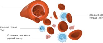
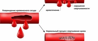
The two main tasks of hemostasis are to maintain blood in a fluid state and to prevent its loss in the event of injury. The operation of the system looks like a chain of complex biochemical reactions in which blood cells, plasma and vessel walls participate.
Bleeding disorders can be inherited. In some cases, they are acquired through medications, malfunctions of the immune system, malignant processes and liver diseases, in which the production and exchange of coagulation factors occurs.
Classification of disorders
There are three “branches” in the hemostasis system: coagulation - responsible for the formation of blood clots, anticoagulation - maintains the rheological properties of blood normal, and fibrinolytic - ensuring the dissolution of blood clots.
Disorders of the blood coagulation system occur in one of two directions:
- In the direction of hypocoagulation - decreased coagulation properties (thrombocytopenia, von Willebrand and Wergolf diseases, hemophilia).
- Or its increase - hypercoagulation (thrombophilia, DIC and APS syndrome).
According to the mechanism of development, disorders of the vascular-platelet unit and failures in coagulation hemostasis are distinguished.
How to suspect deterioration of coagulation hemostasis
The first signs indicating poor blood clotting
Prolonged bleeding occurs with minor skin injuries or after injections. Normally, cuts or injections should not bleed for more than 3-5 minutes, but if there is pathology, this time can increase significantly. Sometimes such people have hemorrhages under the skin.
Another symptom that indicates this condition is prolonged nosebleeds that are difficult to stop. Women with hemocoagulation disorders may experience menorrhagia and metrorrhagia. Sometimes traces of blood may even be present in urine and feces.
If these symptoms appear, it is recommended to donate blood for a coagulogram. Research conducted by our specialists will help identify disorders of coagulation hemostasis. All analyzes are carried out using modern equipment and reagents.
Hypocoagulation: symptoms, why the pathology is dangerous
Nosebleeds for no reason, bleeding of mucous membranes when brushing your teeth, spontaneous appearance of bruises and bleeding from your finger that you cannot stop for longer than usual - all these are still harmless signs of reduced coagulability.
The real danger awaits carriers of the pathology during surgical operations, increased blood pressure, after childbirth, and even routine tooth extraction at the dentist.
Diseases associated with blood hypocoagulation
| Name | Pathogenesis – mechanism of occurrence and development | Why is pathology dangerous? | How is it diagnosed? |
| Thrombocytopenia | A decrease in the number of platelets in plasma (less than 150 × 10 * 9 g/l), associated with a violation of their production or rapid decay. The pathology can be congenital, more often acquired due to bone marrow damage, deficiency of vitamins B9 and B12, severe infections, and splenomegaly. | Risk of massive blood loss, internal, uterine bleeding, miscarriage, hemorrhagic stroke. Prohibition on physical activity, restriction of vital activity. | Clinical blood test Hemostasiogram |
| Thrombocytopenic purpura, ITP (Wergolf's disease) | An autoimmune disease that occurs in a chronic form and is more common in young women. With ITP, the amount of IgG on the surface of platelets increases 10 times. The appearance is provoked by viruses, bacteria, and taking certain medications. | Ophthalmic hemorrhages, decreased visual acuity. Skin hemorrhages - petechial rash, purpura. Uterine bleeding, spontaneous abortion, stroke. | Titer for antibodies to platelets General blood test Coagulogram |
| Hemophilia | Inherited with the maternal X chromosome in an autosomal recessive manner. There are 3 forms of the disease, the most common is classical hemophilia A. | Hemorrhages in muscles, internal organs, and joints, and associated disability. | Thromboelastography, APTT Tests for antihemophilic globulin and D-dimer Prothrombin test Coagulogram |
| von Willebrand disease | Associated with a mutation in the hemostasis gene located on chromosome 12. There are 4 forms of pathology - from mild, with asymptomatic carriage, to severe, with gastric, uterine and other types of bleeding. | Genetic risk of bleeding from any organs, premature birth, hemorrhagic stroke - bleeding in the brain with increased blood pressure. | Study of the activity and properties of von Willebrand factor Activity of ADAMTS 13, 8th coagulation factor |
To treat hypocoagulable conditions, corticosteroids and immunosuppressants are used, and the missing antihemophilic globulin (coagulation factor VIII) or platelet concentrate is administered.
Blood clotting
Many people begin to fear blood clots after, when donating blood from a vein, the nurse reports that it is too viscous. Those who have too thin blood that successfully fills the test tube hope that they are protected from thrombosis. But viscosity and coagulability are two completely different concepts, and one does not always determine the other.
How viscous you are
Increased blood viscosity, due to which it becomes less fluid, most often occurs due to the predominance of its formed elements over liquid ones. This happens due to too strict adherence to the recommendations “do not eat 12 hours before taking the test” and because of the decision to add one more restriction to this – not to drink. Just to be sure. The end result is worse - and the blood flows poorly into the test tube, and some indicators (for example, hemoglobin, hematocrit, total number of red blood cells, white blood cells and platelets) turn out to be artificially high. Therefore, it is important to remember: before taking blood tests, you should not limit yourself to fluids.
Another common cause of increased viscosity is increased levels of red blood cells and hemoglobin, characteristic of smokers. After all, the more smoke and less air a person inhales, the greater the concentration of oxygen carriers required. Their compensatory increase is formed. Therefore, visually, the blood of smokers often appears more viscous.
According to hematologists, true diseases (thrombocytosis, erythrocytosis, etc.) associated with increased blood viscosity account for a small number of all cases of “viscous blood”. And this is clearly visible from a routine general blood test - the doctor will immediately notice that the number of red blood cells or platelets is too high.
Normally, the content of red blood cells is 3.7-5.1, platelets - 180-320.
Viscosity and coagulability - what's the difference?
The most important indicator is blood clotting. Unfortunately, it can be difficult to obtain accurate information about coagulation, even despite the level of development of medicine. On the one hand, obvious diseases with clotting disorders, such as hemophilia, have long been known. On the other hand, there is a lot of hidden pathology, which may not show itself for a long time, but once manifested, it can quickly lead to serious consequences.
Only in recent decades have researchers learned to identify these problems using high-tech genetic tests. Considering that, according to statistics, more than 1-3% of the world's population has a congenital pathology of the blood coagulation system, it is likely that in the future these tests will be carried out in the maternity hospital for every newborn. And absolutely for those who need to be prescribed certain medications that can increase the risk of blood clots.
Protection or danger?
For example, this is very important to do before prescribing hormonal contraceptives. Their ability to enhance blood clotting and occasionally lead to thrombosis (as it turns out, this almost always occurs in women who have a hidden clotting disorder) has been known for several decades. Due to the widespread use of this method of protection against unwanted pregnancy these days, the incidence of thrombosis in young, apparently healthy women has increased. At the same time, when prescribing COCs, gynecologists rarely focus their patients’ attention on this danger. They can be understood - the complication is infrequent in general, and I don’t want to once again intimidate patients, as if pushing them into an unwanted pregnancy. Meanwhile, the most advanced Western colleagues have already included a full blood clotting test in the mandatory examination algorithm before prescribing hormonal contraceptives.
What will the analysis show?
What tests need to be done to check blood clotting? The most common and familiar analysis to many, a coagulogram, may not answer all questions, especially in the prevention of thrombosis.
However, a classic coagulogram is the first stage of a screening examination of the coagulation system. If it reveals abnormalities, the next step will be more detailed studies of hemostasis - thromboelastography or thromboelastometry. A separate story is the determination of D-dimer , Leiden mutation and other genetic clotting disorders - tests that reveal a tendency to form blood clots in the future. What is necessary and in what cases?
The most common standard coagulogram today includes five components: PTI (prothrombin index); INR (International Normalized Ratio. Reflects the ratio of the patient’s blood clotting time to the blood clotting time of a healthy patient; APTT (activated partial thromboplastin time. Estimates the time it takes for a blood clot to form after special reagents are added to the plasma), FIBROGEN and PLATELET LEVEL.
At the same time, APTT is informative only in people undergoing treatment with heparin, and INR is important only for people constantly taking blood-thinning drugs from the neodicoumarin group (warfarin).
It turns out that two out of five indicators are not so important for screening. The total number of platelets is also not always indicative, because with most coagulopathies it is not their number that changes, but primarily the functional activity.
Therefore, the most informative method, which allows one to evaluate several stages of blood coagulation at once, is thromboelastography. This is a kind of detailed observation of the formation of a blood clot, and its subsequent dissolution (lysis) with the construction of graphs of each of the stages. Thromboelastometry is another version of this study, which is considered even more informative. Unfortunately, the instruments for conducting these studies are expensive and require special training of personnel, so not every laboratory can offer thromboelastography services.
Another important indicator is D-DIMER (this is a breakdown product of fibrin, a small fragment of protein present in the blood after the destruction of a blood clot).
It is actively used to determine the risk of blood clots. Those with even slightly elevated D-dimer are at much greater risk of developing blood clots than others. It is necessary to monitor D-dimer in case of venous diseases (thrombophlebitis), after surgical interventions and upon discharge from the hospital if you have been bedridden for a long time. Monitoring D-dimer levels is useful during pregnancy and when taking hormonal contraceptives (the risk of blood clots in the presence of the Leiden mutation while taking birth control pills increases almost 9 times). And now during COVID-19 and several weeks after recovery.
Today, in addition to D-dimer testing, genetic tests for congenital bleeding disorders are emerging. The most common of these is the Leiden mutation, which occurs in 2–6% of Europeans. The presence of a defective gene increases the likelihood of venous blood clots 6–8 times, and the risk of heart attack and stroke increases significantly. But other mutations, of which there are more than ten today, are no less dangerous. At the same time, timely prevention of thrombosis (mainly the constant use of anticoagulants, the exclusion of certain foods and medications, wearing compression stockings when flying and working on your feet, etc.) reduces the risk of dangerous complications tenfold.
Flickering problem
If everything is in order with the coagulation system, a possible cause of increased coagulation may be arrhythmia, namely atrial fibrillation. According to statistics, 2% of the planet's population suffers from paroxysmal (occurring in attacks) or permanent form of atrial fibrillation. This problem usually appears after thirty. The basis for the disorder is the appearance of pathological turbulence of the electrical impulse in the atria, which gives the myocardium extraordinary electrical stimuli. In medical language, this is called the “re-entry” or re-entry mechanism. As a result, the atria become a churn that churns blood into clots like milk into butter. The situation worsens even more in the case where the person initially had the same clotting disorders. Blood clots can form within 48 hours after the onset of an attack and “fly away” into the arteries of the brain, causing an ischemic stroke, into the intestines, leading to mesenteric thrombosis, into the arteries of the extremities, provoking their acute ischemia.
Von Willebrand factor and COVID -19.
Severe COVID-19 may be associated with elevated levels of one of the blood coagulation factors, von Willebrand factor. Anna Aksenova, a senior researcher at the Laboratory of Amyloid Biology at St. Petersburg State University . Her scientific article was published in the journal Ecological Genetics. It has already been proven that the SARS-Cov-2 virus can have a direct damaging effect on the inner wall of blood vessels. In response to damage, the body strives to “patch” the hole as quickly as possible, and the leading role in this is played by the von Willebrand factor, which is involved in the activation of platelets and, in fact, triggers the process of local thrombus formation. In the course of research, it turned out that some people are characterized by an increased concentration of this factor in their cells, so, as a rule, it is higher in people with blood group II. An individual characteristic of the body is also possible. As a result, in response to massive microdamage to blood vessels, massive microthrombosis occurs, which causes the appearance of larger and more dangerous blood clots.
Genetic mutations of the coagulation system identified during tests:
MUTATION OF COAGULATION FACTOR V OF BLOOD CLOTTING (LEIDEN FACTOR)
PLASMINOGEN ACTIVATOR INHIBITOR 1
MUTATION OF COAGULATION FACTOR II (MUTATION OF PROTHROMBIN)
MUTATION OF METHYLENETETRAHYDROPOLATE REDUCTASE (MTHFR C677T)
MUTATION OF COAGULATION FACTOR VII OF BLOOD CLOTTING (F7 ARG353GLN)
POLYMORPHISM OF THE METHIONINE SYNTHASE REDUCTASE GENE (MTRR A66G)
FIBRINOGEN MUTATION, BETA (FGB G-455A)
MUTATION OF THE PROMOTER OF THE COAGULATION FACTOR GENE FVII (-312 INS 10BP)
INSERTATION/DELETION OF ALU ELEMENT IN THE ANGIOTENSIN CONVERTING ENZYME GENE (ALU INS/DEL)
PLATELET GLYCOPROTEIN 1B ALPHA SUBUNIT MUTATION
PLATELET ADP RECEPTOR MUTATION (P2RY12 H1/H2)
MUTATION A1298C OF METHYLENETETRAFOLATE REDUCTASE GENE
D-dimer is significantly elevated in most patients with moderate to severe COVID-19. Therefore, all patients receive therapeutic doses of anticoagulants.
The Leiden mutation is the most common hidden bleeding disorder, occurring in 2-6% of Europeans.
Published: January 19, 2021
Thrombophilias and pregnancy
When pregnancy occurs, the total activity of all blood coagulation factors increases, the functional state of platelets changes and the level of antithrombin III decreases.
In a healthy woman, such transformations ensure the normal formation of the fetoplacental complex and prevent blood loss during childbirth. And in an expectant mother with thrombophilia, a pathological tendency to thrombosis and hypercoagulation, changes in hemostasis can lead to termination of pregnancy in the early stages.
Recent studies by hemostasiologists indicate the enormous role of increased blood clotting in reproductive losses and obstetric complications:
- 80% of patients with spontaneous abortion have hemostasis disorders such as hypercoagulation.
- Increased coagulability is recorded in 100% of cases of non-developing pregnancy.
In case of disorders of the blood coagulation system, pregnancy loss occurs as a result of placental infarction (develops in the form of symptoms of chorionic detachment) or pathology of the vascular wall.