Almost all of the images presented here were taken using a scanning electron microscope (SEM). The electron beam emitted by such a device interacts with the atoms of the desired object, resulting in 3D images of the highest resolution. Magnification of 250,000 times allows you to see details measuring 1-5 nanometers (that is, billionths of a meter).
The first SEM image was obtained in 1935 by Max Knoll, and already in 1965 the Cambridge Instrument Company offered its Stereoscan. Now such devices are widely used in research centers.
Looking at the pictures below, you will take a journey through your body, starting from your head and ending with your intestines and pelvic organs. You'll see what normal cells look like and what happens to them when they are attacked by cancer, and you'll also get a visual understanding of how, say, the first meeting of an egg and sperm occurs.
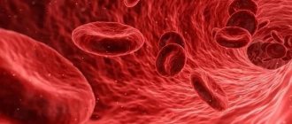
Red blood cells
This is what you might call the heart of your blood, the red blood cells (RBCs). These cute biconcave cells have the responsible task of carrying oxygen throughout the body. Typically, in one cubic millimeter of blood there are 4-5 million such cells in women and 5-6 million in men. People living at high altitudes, where there is a lack of oxygen, have even more red cells.
Types of human tissues and their functions
Tissues are an association of groups of cells according to their purpose (function). Organs are formed from tissues. Some organs are made up of the same type of cells (for example, heart muscle and skeletal muscle). Some cells have a high degree of proliferation (that is, multiplication by division), under the influence of hormones, for example.
Others, when mature, lose the ability to divide or mutate - nerve cells, blood cells.
Types of organic fabric:
- Epithelium - performs the integumentary function; the outer covering is formed from it - skin, mucous membranes, soft tissues. Forms the outer capsule of some organs.
- Connective – cartilaginous, fatty, bone.
- Muscular – it forms all types of muscles, performs a motor and contractile function.
- Nervous – consists of nerve cells (neurons). Provides communication between organs and tissues and the brain through electrical impulses.
Purkinje cells
Of the 100 billion neurons in your brain, Purkinje cells are some of the largest. Among other things, they are responsible in the cerebellar cortex for motor coordination. They are adversely affected by alcohol or lithium poisoning, as well as autoimmune diseases, genetic disorders (including autism), as well as neurodegenerative diseases (Alzheimer's, Parkinson's, multiple sclerosis, etc.).
Human cell: properties
Before considering the functions of the cell and its properties, let us pay attention to the composition of the human cell.
Composition of a human cell
Knowledge of its composition will help you understand the properties of a cell:
- Cells contain compounds of oxygen (O), sulfur (S), phosphorus (P), carbon (C), potassium (K), chlorine (Cl), hydrogen (H), iron (Fe), sodium (Na), nitrogen (N), calcium (Ca), magnesium (Mg).
- The main component is water. Nutrients are dissolved and transported in it, and all reactions take place. Water removes harmful metabolic products from cells. It regulates body temperature and makes up up to 85% of cellular composition.
- Carbohydrates supply energy for all intracellular processes.
- Fats are needed for the formation of membranes, and with a lack of carbohydrates they become an energy resource.
- All cell organelles, as well as part of the membrane, are built from proteins.
- The nucleic acid DNA stores and transmits genetic information, and RNA is involved in the synthesis of proteins.
- ATP serves as a source of energy.
Properties
Human cells are endowed with the following properties:
- They are capable of self-reproduction by division.
- May change during existence.
- Cells constantly maintain metabolism with the external environment and other cells of the body.
- Capable of using energy accumulated in chemicals (carbohydrates, fats, ATP).
- Cells respond to external and internal stimuli.
- Adapt to environmental conditions.
Human cell division process: Freepick
Villi of the small intestine
The villi of the small intestine increase its area, which promotes better absorption of food. These are irregularly cylindrical outgrowths up to 1.2 millimeters high. The basis of the villi is loose connective tissue. In the center, like a rod, runs a wide lymphatic capillary, or lacteal sinus, and on the sides of it there are blood vessels and capillaries. Fats pass through the milky sinus into the lymph and then into the blood, and proteins and carbohydrates enter the bloodstream through the blood capillaries of the villi. Upon careful examination, you can notice food debris in the grooves.
The structure of a human cell
The human cell is an elementary living system, the basic structural and functional unit of the body, which can self-renew, self-regulate and self-reproduce.
The human body contains tens of trillions of cells that together form tissues and organs. There are different types of human cells: the brain, heart and liver, for example, consist of specific cells.
But still, the general structure of the cells is very similar, and we will dwell on it in more detail. What does a cell consist of? The structure of a human cell is represented by components.
Cytoplasmic membrane
We begin to consider the structure of the cell from the membrane, since it is its basis. The following is known about this cell component:
- This is a kind of constructor, which, firstly, encloses the entire cell, and secondly, contains the nucleus and all membrane organelles (small organs of the cell).
- The membranes form a double lipid (fat) layer. On their outer side there are special protein molecules - receptors that interact with other cells and substances.
- All membranes have selective permeability, that is, they can let some substances in, but not others.
The membrane performs a protective function, regulates the exchange of substances between the cell and the environment, and also maintains its shape.
Human cell cytoplasm
This is the liquid environment of the cell in which all the organelles and various inclusions are located. Its main component is water. This is the environment for all chemical processes to take place. The cytoplasm also unites the entire cell into a single whole and serves as a field for the interconnection of all components.
Organoids
Each of these smallest parts is endowed with an important function and performs it smoothly.
Human Cell Organelles: Freepick
The main organelle is the nucleus. It consists of:
- nuclear membrane;
- nucleolus;
- karyoplasma;
- chromosomes.
With the help of a membrane, the nucleus is separated from the cytoplasm. Inside it is filled with nuclear juice (karyoplasm). The nucleolus is necessary for the process of protein synthesis. The most intimate part of the nucleus is the chromosomes, DNA with a record of all hereditary information.
It is worth noting that the number of chromosomes is different for each species and has nothing to do with the complexity of its organization. Thus, a human cell contains 46 chromosomes, a chimpanzee cell - 48, and a dog cell - 78.
The cell nucleus stores hereditary information about the cell, transmits it to daughter cells during division, and implements hereditary information through the synthesis of proteins that are characteristic of a given cell.
In addition to the nucleus, the body cell contains:
- Endoplasmic reticulum (ER). This system of channels penetrates the cytoplasm and is needed for the exchange of proteins and fats.
- The Golgi apparatus, which is located around the nucleus in the form of flat tanks. This organelle transfers, sorts and stores proteins, lipids and polysaccharides, and also forms lysosomes.
- Lysosomes are small sacs filled with digestive enzymes that perform the functions of protecting and digesting proteins, fats and carbohydrates.
- Mitochondria are involved in the synthesis of ATP, the substance from which the body obtains energy.
- Ribosomes are necessary for protein synthesis.
- The cell center is a dense cytoplasm with centrioles (a complex of microtubules) that is involved in cell division.
In certain groups of cells there are organelles for special purposes. These include:
- flagella in male reproductive cells, thanks to which they move;
- myofibrils in muscle cells, which are responsible for muscle contraction processes;
- neurofibrils in nerve cells that transmit nerve impulses;
- photoreceptors in eye cells.
Cells can also permanently or temporarily contain a number of inclusions:
- pigments that color cells (the brown pigment melanin is produced in the skin in the sun to protect it, and we see this process as the formation of a tan);
- trophic inclusions in which energy is stored;
- secretory inclusions are found in cells that secrete hormones;
- excretory inclusions. This group includes sweat in the sweat glands.
All this fits into 3-4 micrometers (µm) - this is the average size of a human cell!
Human embryo and sperm
It looks like a war of the worlds, but in fact, you have an egg in front of you 5 days after fertilization. Some sperm are still retained on its surface. The image was taken using a confocal microscope. The egg and sperm nuclei are purple, while the sperm flagella are green. The blue areas are nexuses, intercellular gap junctions that communicate between cells.
How is the cell membrane structured?
The cytoplasmic membrane is the membrane that separates each cell from the extracellular space and you, of course, are wondering what the plasma membrane of a cell consists of? It consists of two layers of phospholipids and glycolipids.
These molecules have a polar end (which attaches to other molecules and atoms) and are a hydrophilic end. This part comes into contact with the intercellular environment and the cell plasma. The ends of non-polar lipids (hydrophobic) form the double inner layer of the cell membrane.
Also built into the shell are large protein molecules that act as opening pores, electrolyte channels and connecting mechanisms. Such a protein molecule passes through the bi-lipid layer. There are also glycocalyx threads on the surface. The shell may have folds, roughness and bulges, and processes like flagella.
Basic organelles and organelles of an animal cell
Organelles and organelles are the “organs” responsible for the functioning of a microorganism.
Core
The nucleus is the source of deoxyribonucleic acid (DNA) genetic material. DNA is the source of the creation of proteins that control the state of the body. In the nucleus, strands of DNA wrap tightly around highly specialized proteins (histones) to form chromosomes.
The nucleus selects genes to control the activity and functioning of the tissue unit. Depending on the type of cell, it contains a different set of genes. DNA is found in the nucleoid region of the nucleus where ribosomes are formed. The nucleus is surrounded by a nuclear membrane (karyolemma), a double lipid bilayer that separates it from the other components.
The nucleus regulates cell growth and division. During mitosis, chromosomes are formed in the nucleus, which are duplicated during the process of reproduction, forming two daughter units. Organelles called centrosomes help organize DNA during division. The core is usually represented in the singular.
Ribosomes
Ribosomes are the site of protein synthesis. They are found in all tissue units, in plants and animals. In the nucleus, the DNA sequence that codes for a specific protein is copied into a free messenger RNA (mRNA) strand.
The mRNA strand travels to the ribosome via messenger RNA (tRNA), and its sequence is used to determine the arrangement of amino acids in the chain that makes up the protein. In animal tissue, ribosomes are located freely in the cytoplasm or attached to the membranes of the endoplasmic reticulum.
Endoplasmic reticulum
The endoplasmic reticulum (ER) is a network of membranous sacs (cisternae) extending from the outer nuclear membrane. It modifies and transports proteins created by ribosomes.
Distinctive features of plant and animal cells in the table
Plant and animal cells have both similarities and differences, which are briefly described in the table:
| Sign | Vegetable | Animal |
| Getting food | Autotrophic. Photosynthesizes nutrients | Heterotrophic. Does not produce organic matter. |
| Power storage | In vacuole | In the cytoplasm |
| Storage carbohydrate | starch | glycogen |
| Reproductive system | Formation of a septum in the maternal unit | Formation of constriction in the maternal unit |
| Cell center and centrioles | In lower plants | All types |
| Cell wall | Dense, retains its shape | Flexible, allows change |
The main components are similar for both plant and animal particles.
What does an animal cell look like under a microscope?
Under a standard optical microscope, the main components are visible. Due to the fact that they are connected into a constantly changing organism that is in motion, it can be difficult to identify individual organelles.
The following parts are not in doubt:
- core,
- cytoplasm,
- cell membrane.
A higher resolution microscope, a carefully prepared specimen, and some practice will help you study the cell in more detail.