Modern medicine allows you to get rid of hemorrhoids using minimally invasive methods. Coagulation of nodes is an effect on them without the need for painful surgery. The positive aspects of this method include the ability to get rid of nodes without the use of potent drugs and carry out a long period of rehabilitation. Before prescribing coagulation, the doctor conducts a complete diagnosis of the patient, determines the stage of the disease, the number and size of nodes.
What is the essence of coagulation of hemorrhoids?
Infrared coagulation of hemorrhoids involves the local thermal effect of waves on problem areas generated by an infrared ray. Using this method, the blood supply to the hemorrhoidal tumor is disrupted, which ultimately leads to its destruction and falling off.
Infrared coagulation is recommended for the treatment of stages 1 and 2 hemorrhoids
Radio wave coagulation of hemorrhoids involves the use of high-frequency waves, non-contact incision, and vaporization of blood vessels. Thanks to heating, a cutting effect is produced, which subsequently leads to the destruction of the node.
At stages 3-4 of hemorrhoids, radio wave coagulation is usually recommended
Both methods are quite effective. The specialist chooses the technique that he considers most optimal for a particular individual case.
Minimally invasive procedures are painless and have minimal risk of postoperative complications. The patient returns to his usual rhythm of life almost immediately after the procedure.
Statistics show that patients usually tolerate both manipulations well. For both procedures, specialized medical equipment is used.
To carry out infrared coagulation of hemorrhoids:
- electrical system,
- applicator holder
- applicator equipped with a hard LED based on quartz glass.
To carry out radio wave coagulation:
- apparatus emitting high-frequency waves
- ultrasonic device to control manipulation.
Indications for coagulation for hemorrhoids
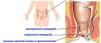
Infrared coagulation of hemorrhoidal tumors is recommended for patients with internal or combined forms of hemorrhoids.
The main indications in this case include:
- hemorrhoids at stage 1 or 2 of development,
- presence of hemorrhoids with bleeding,
- combined form of hemorrhoids,
- inability to achieve the desired therapeutic effect through ligation of hemorrhoids.
Indications for radio wave coagulation:
- hemorrhoids at stages 3 or 4 of development,
- the presence of internal hemorrhoidal neoplasms,
- the presence of external hemorrhoids.
Both types of coagulation are modern methods of treating hemorrhoids of various forms. The choice of method should be entrusted to a qualified specialist. He will take into account the peculiarities of the course of the disease, the shape and location of hemorrhoids, the type of complications, and evaluate the general symptoms. This will allow him to decide on the best method for his patient.
Principle of the method
An infrared coagulator is a device that allows tissue cauterization by contact method. It consists of a block and an applicator, which directly performs all manipulations on the rectum. The block is equipped with a timer that allows you to set coagulation parameters.
Using the applicator, the surgeon cauterizes the vascular pedicle of the modified cavernous bodies, causing their necrosis. Hemorrhoids treated in this way become empty and die, separating from the mucosa after a few days.
Advantages:
- minimal trauma to underlying tissues
- ability to set the time of exposure to infrared radiation with an accuracy of fractions of a second
- the ability to use minimal thermal energy for infrared irradiation
Flaws:
- limiting the number of nodes that can be coagulated in one manipulation (usually one intervention allows you to get rid of 1-3 nodes)
- high price
- limited indications for use.
Possible complications:
- bleeding – postoperative or during removal of a dead hemorrhoid
- hemorrhoidal thrombosis
- severe pain in the anus
- necrosis of mucous membranes
- addition of a secondary infection
Contraindications to coagulation of hemorrhoids
Infrared coagulation is not performed if the patient has an advanced form of the disease. In case of a purulent-inflammatory process, the procedure is most often not performed. With stage 3-4 hemorrhoids, radio wave coagulation is usually needed. Other contraindications include:
- damage to the rectal canal mucosa,
- presence of rectal fistulas,
- purulent inflammation,
- inflammation of a pronounced nature in the tissues of the pelvis,
- thrombosis.
Radio wave coagulation is not performed with the following contraindications:
- diabetes,
- old age of the patient,
- exacerbation of infectious diseases,
- epilepsy.
Make an appointment
Treatment of hemorrhoids
Appointment with a proctologist
Online booking of an appointment with a proctologist.
Phones:
+7 (812) 30-888-03
+7
Clinic address: St. Petersburg, Vyborg district, st. Asafieva, 9, k2, lit. A (metro station Ozerki, metro station Prospekt Prosveshcheniya)
Below we will discuss the main methods of treating hemorrhoids without surgery:
- Alloying hemorrhoids with latex rings
- Infrared coagulation of hemorrhoids
- Cryotherapy
- Sclerotherapy
Key principles of preparation for surgery
The patient receives full information about the preparation during a consultation with a proctologist. Before coagulation, the patient must undergo all necessary types of examinations:
- ECG,
- fluorography of the chest organs,
- general blood analysis,
- general urine analysis,
- anoscopy,
- palpation examination of the anus,
- Wasserman reaction,
- blood sugar test.
Additionally, colonoscopy or sigmoidoscopy may be prescribed.
The patient should also adhere to the recommendations: exclude fried, spicy and fatty foods from the diet 1 day before coagulation, as well as foods that promote gas formation. Before the procedure, you need to take a laxative. In some cases, an enema is used to cleanse the intestines.
What to expect from treatment with an infrared coagulator?
Bleeding from the anus occurs 7-10 days after the procedure, when the hemorrhoids disappear. Bleeding is usually minor and stops on its own.
- The patient is advised to use mild pain relievers and sit in a shallow bath of warm water (sitz bath) for 15 minutes at a time to reduce discomfort.
- To reduce the risk of bleeding, you should avoid taking aspirin and other nonsteroidal anti-inflammatory drugs (NSAIDs) for 4 to 5 days both before and after infrared coagulator treatment.
- It is recommended to take stool softeners containing fiber to ensure smooth bowel movements. If the patient strains during bowel movements, the likelihood of relapse increases.
How is coagulation of hemorrhoids performed?
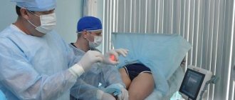
Infrared and radio wave coagulation is performed under local anesthesia to eliminate possible discomfort during the procedure.
Infrared coagulation of hemorrhoidal cones requires a special knee-elbow position for the patient. Sometimes the procedure is performed in a different position, when the patient sits in a proctology chair, spreads his legs and tucks them in.
The doctor uses a dilator for the tissues of the perianal area
A special device, called an “anoscope,” is inserted into the rectal canal. The next stage of the procedure is to bring a quartz LED to the stem of the hemorrhoid.
Coagulation lasts about 2 seconds. If the neoplasm is large, in addition to the stem, its upper part is also treated
If the patient has several nodes, they are removed simultaneously or on different days. Repeated coagulation is recommended in this case 14 days after 1 session.
Radio wave coagulation also involves a special positioning of the patient in a proctology chair or on a couch. The device used allows for non-contact cutting, coagulation and vaporization of blood vessels using a high-frequency wave.
Evaporation (or heating) of soft tissues leads to their dissection
It is important to note that the electrode does not contact tissue and cannot heat up. The surrounding layers are not destroyed during the procedure.
Rectal fistulas
According to statistics, approximately 95% of patients with rectal fistulas associate the onset of the disease with a history of purulent inflammation (acute paraproctitis). It is not advisable to postpone radical surgical treatment for a long time, since exacerbation of paraproctitis can recur and the inflammatory process can lead to deformation of the surrounding tissues, including the development of anal muscle insufficiency. For most rectal fistulas, excision of the fistula is performed (with or without suturing the wound). Surgical treatment of simple fistulas leads to a permanent cure in approximately 95% of cases and is not accompanied by any serious complications. Complex fistulas (so-called deep trans- and extrasphincteric) can also be cured without functional impairment.
Epithelial coccygeal tract
In proctological practice, every 10 patients (in men, on average, three times more often) are found to have fistulas in the coccyx area. This is a congenital anomaly - the epithelial coccygeal duct. Patients are often unaware of their illness, or their complaints are limited to a small constant discharge above the anus, wetness of the skin between the buttocks, and itching. In other cases, often after an injury, suppuration suddenly occurs in the coccyx area, after opening which a non-healing fistula remains. Prolonged course of the epithelial coccygeal tract without proper treatment or self-medication leads to the formation of secondary fistulas; the entire sacrococcygeal region is affected by the inflammatory process. The prognosis for radical treatment of the epithelial coccygeal tract at any stage of the disease is favorable, complete recovery occurs.
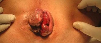
Perianal genital warts
Pointed perianal condylomas are gray-pink papillary formations between which unaffected skin is visible, sometimes in the form of papillae that merge and form whole conglomerates that can cover the anus. Sometimes there are giant condylomas up to 20 cm or more in diameter. It has now been established that condylomas are caused by human papillomavirus (HPV) types 6 and 11. The disease is usually sexually transmitted. They must be distinguished from flat condylomas, characteristic of syphilis and lesions in patients with AIDS. Surgical treatment is most often undertaken in the presence of large nodes and damage to the anal canal. Excision is performed with a scalpel, or an electric knife, or a laser. Sometimes, in the presence of large condylomas, surgical removal is not performed at once, since the resulting extensive wounds can lead to deformation in the area of the anus.
Patient consultations in our department are free and are held daily on weekdays. You can pre-agreed the time of your visit by phone. Read more on the website: https://www.surgery-future.ru/proktologiya
Possible complications after surgery
After coagulation of hemorrhoids, some patients experience pain for the first time. Their appearance may be due to the location of the hemorrhoidal tumor below the dentate line, where pain receptors are located. The proctologist will advise the patient to use rectal suppositories and drugs with an analgesic effect.
It is better not to self-medicate, so if pain occurs after coagulation, you should immediately contact a specialist and solve the problem together.
Pathological complications after coagulation also include anorectal thrombosis, rectal bleeding, and necrosis of hemorrhoids.
Rectal bleeding is possible several days after the procedure. This is usually associated with the hemorrhoid falling off. As for node necrosis and anorectal thrombosis, they usually manifest themselves with extensive coagulation.
To eliminate the risk of possible complications, it is recommended to strictly adhere to all doctor’s recommendations after coagulation.
Causes of hemorrhoids
There are many factors that can lead to hemorrhoids. Among them are:
- sedentary lifestyle;
- heavy physical activity associated with lifting weights;
- pregnancy and childbirth (postpartum hemorrhoids);
- excessive consumption of hot and spicy foods;
- prolonged constipation;
- overheating or hypothermia of the pelvis;
- hemodynamic and muscular dystrophy;
- diseases of the pelvic organs;
- congenital predisposition.
Thus, at risk are people who, due to the nature of their professional activities, lead a sedentary lifestyle, or who often lift heavy objects and experience serious physical exertion. Overweight people and those who do not watch their diet are also susceptible to hemorrhoids. A special category of patients diagnosed with hemorrhoids are pregnant women and women who have recently given birth, in whom the formation of hemorrhoids and bleeding is associated with complications during pregnancy. In general, this disease is typical for middle-aged and older people (45 years and above). But there are cases when younger patients also seek treatment for hemorrhoids.
Efficiency
Infrared coagulation of hemorrhoidal disease is highly effective, as it significantly reduces tumors, helps stop bleeding and improves overall well-being.
The patient who has undergone this procedure can return to normal activities within a day. Rehabilitation lasts on average about one week.
However, it should be noted that some patients may experience recurrence of the disease within five years after coagulation. Constant monitoring by a proctologist is required. For this, the patient needs to undergo regular examinations.
It is also important to note that infrared coagulation of hemorrhoids is not performed at stages 3 and 4 of the disease, since in this case it is ineffective.
Radio wave coagulation is often better tolerated and the risk of relapse is lower, but this technique is usually used for complicated and late stages of hemorrhoids.
After infrared photocoagulation
In most cases, the initial stages of hemorrhoids require a one-stage operation. Repeated infrared photocoagulation is carried out if the patient has 4 or more nodular formations. At the end of the procedure, the patient may experience minor pain and discomfort that lasts up to 6 hours. To relieve pain, it is enough to take non-narcotic analgesics (ibuprofen and paracetamol).
The first few days after infrared photocoagulation, the patient is recommended to adhere to a gentle diet. During this period, it is prohibited to drink carbonated and alcoholic drinks. The daily diet should be divided into 6-7 meals. This significantly reduces the mechanical pressure of food masses on the walls of the large intestine. It is advisable to avoid heavy physical activity and sports for two weeks.
After infrared photocoagulation of hemorrhoids, full restoration of functionality occurs within 2-3 days. During this period, the patient must strictly observe the rules of personal hygiene, especially after bowel movements.
Disadvantages of coagulation
The disadvantages of infrared and radio wave coagulation include the risk of possible complications and relapses. After the procedure, patients may have complaints:
- painful syndrome,
- problems with bowel movements,
- risk of thrombosis and rectal bleeding.
In addition, it should be noted that coagulation techniques can eliminate the consequences of the disease, but they do not directly affect the very cause of its development.
Cost of treatment
The price of infrared and radio wave coagulation of hemorrhoids depends on their number, shape, complexity of the case and volume of intervention. The approximate cost of one procedure is from three to ten thousand rubles.
When choosing between infrared and radio wave coagulation of hemorrhoids, it is better not to waste time and entrust the solution to this issue to a specialist.
Depending on the stage of the disease ( I , II , III , IV ), some features of its manifestation, the age and health status of the patient, the doctor will determine what will be the most effective and safe .
Modern technologies in the treatment of chronic hemorrhoids
Goncharov D.Yu. Ph.D. Hemorrhoids are one of the widespread diseases of the adult population of industrialized countries. It has been established that in people over 40 years of age, symptoms of hemorrhoids are found in 60-70% of cases. Hemorrhoids account for about 40% of the structure of coloproctological diseases.
In recent years, minimally invasive methods of treating hemorrhoids have become firmly established in the daily practice of coloproctologists. The advantages of these methods over surgical interventions are the possibility of their use on an outpatient basis, without loss of ability to work; high efficiency in the initial stages of the disease; a small number of complications.
The most commonly used minimally invasive methods are:
- hardware ligation of hemorrhoids with latex rings,
- sclerotherapy,
- infrared photocoagulation of hemorrhoids,
- suture ligation of hemorrhoidal arteries under Doppler control,
- electrocoagulation of hemorrhoids.
The indication for minimally invasive surgical interventions is uncomplicated internal hemorrhoids of stages I - III.
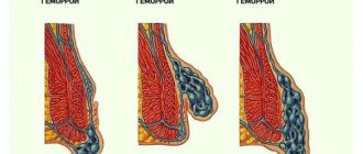
When choosing a method of treating patients with hemorrhoids, it is advisable to use a classification that subdivides chronic hemorrhoids into stage IV.
- I Art. Discharge of scarlet blood from the anus without prolapse of hemorrhoids.
- II Art. Prolapse of hemorrhoids with self-reduction into the anal canal (with or without bleeding).
- III Art. Periodic prolapse of hemorrhoids and the need for their manual reduction into the anal canal (with or without bleeding).
- IV Art. Constant prolapse of hemorrhoids along with the rectal mucosa, inability to reduce them into the anal canal using a manual aid (with or without bleeding).
Contraindications include: a combination of hemorrhoids with an anal fissure, rectal fistula, inflammatory diseases of the anal canal and perineum, acute hemorrhoids.
Ligation of hemorrhoids with latex rings is the most commonly used technique (32-82%), and sclerotherapy, due to the frequent development of complications (11-47%), is used less and less. Other minimally invasive treatment methods are used in less than 5% of cases.
Ligation of hemorrhoids with latex rings
Blaisdell first developed and used a tool for applying a circular latex ligature to the pedicle of a hemorrhoid in 1954. Subsequently, other, more advanced models of ligators were developed.
The use of this method is indicated for internal hemorrhoids II, sometimes III degree. Contraindications for performing ligation of hemorrhoids are: a combination of internal hemorrhoids with an anal fissure and rectal fistula; acute hemorrhoids; inflammatory diseases of the anal canal; treatment with anticoagulants.
Direct ligation (clamping) of hemorrhoids occurs using a latex ring with an internal diameter of 1 mm, which has good elasticity and provides uniform, constant compression of the tissue. Rejection of the hemorrhoid along with the ligature occurs 5 to 9 days after manipulation. During this period, as a rule, there is a slight discharge of scarlet blood from the anal canal, which does not require the prescription of medications, since it stops on its own. A connective tissue scar forms at the site of the rejected hemorrhoid.
There are two main methods for ligating hemorrhoids. The first is based on drawing cavernous tissue into the sleeve of a mechanical ligator using a special soft clamp, after which one or two ligatures are dropped from the instrument onto the stem of the hemorrhoid. The ring should only compress the pedicle of the node, without capturing tissue located below the anorectal line.
The essence of the second method is to use a vacuum ligator, which is connected to a suction device. The working part of the instrument should be pressed tightly against the hemorrhoid. After turning on the suction, negative pressure is created in the device cylinder, and the assembly is gradually drawn into the ligator coupling. When the pressure reaches 0.7 - 0.8 atmospheres, two latex rings are dropped from the instrument onto the stem of the hemorrhoid.
During the first session, ligation of one or two hemorrhoids is performed. The next stage of treatment is prescribed no earlier than 15 days later.
If the technique is followed correctly, the patient should not experience severe pain. After the manipulation, slight pain, a feeling of pressure, a feeling of a foreign body in the rectum, and tenesmus may appear, which may persist for 1 to 2 days. These sensations can be relieved by taking non-narcotic analgesics.
Complications of ligation of hemorrhoids are: pain (noted if the manipulation is performed incorrectly), thrombosis of external hemorrhoids (occurs in 2-3% of patients), bleeding (observed in 1% of patients). The effectiveness of the technique is more than 80%.
Sclerotherapy
For the first time, sclerotherapy as a method of treating hemorrhoids was used by I.I. Karpinsky (Russia) in 1870, using iron persulfate and phenol for these purposes. However, frequently developing complications after such sclerotherapy have led to the limitation of the use of this method. With the advent of new sclerosing drugs, anoscopes, and special needles, interest in this technique has increased again.
In the Russian Federation, preparations from the detergent group are approved for use. These include: polidocanol-ethoxysclerol, thrombovar, fibrowein, sodium morruate, sodium tetradecyl sulfate. Detergents are the most effective and safe phlebosclerosing chemicals. The mechanism of action of this group of drugs is based on the ability to cause coagulation of endothelial proteins and desquamation of the epithelium. Detergents have a local effect on vascular tissue and do not lead to systemic thrombus formation.
The indication for sclerotherapy is internal hemorrhoids of stages I-II; continued bleeding from hemorrhoids.
Contraindications to this method include: external hemorrhoids, paraproctitis, thrombosis of hemorrhoids, ulceration of the mucous membrane, anal fissure.
The essence of the sclerotherapy technique is to introduce the drug into the thickness of the hemorrhoidal node, using a specially curved needle with a limiter. Depending on the size of the hemorrhoid, from 0.5 to 2.0 ml of detergent is administered.
On the first day after the procedure, a tissue reaction to chemical coagulation occurs and pain may develop. A pronounced pain reaction may be associated with the introduction of the drug not into the thickness of the hemorrhoid, but into the muscular layer of the intestine, as well as with the introduction of a concentrated sclerosing drug in a larger volume. In this case, thrombosis and necrosis of the mucous membrane of the hemorrhoidal node may occur.
In order to prevent the development of pain and inflammation, it is advisable to perform sclerosis of no more than two hemorrhoids in one session. The second stage of treatment is prescribed no earlier than after 2 weeks. During a follow-up examination on days 12-14 after the procedure, a flat, round, painless, sclerotic area of cavernous tissue with unchanged mucosa is determined in the anal canal.
Sclerotherapy for hemorrhoids is most effective for stage I hemorrhoids. As the stage of the disease increases, the number of good results decreases and the number of relapses increases. Sclerosing therapy does not allow achieving a radical cure of patients from the manifestations of hemorrhoidal disease, and good long-term treatment results are observed in only 20% of patients.
Infrared photocoagulation
With the beginning of the use of ultraviolet and infrared radiation in medicine, A. Neiger in 1978 proposed a technique for infrared photocoagulation of hemorrhoids. The method is distinguished by its simplicity and short exposure time. It is used in the initial stages of internal hemorrhoids, as well as to stop hemorrhoidal bleeding. Contraindications are external hemorrhoids, thrombosis of internal hemorrhoids, combination with paraproctitis and anal fissure.
The principle of operation of the photocoagulator is that the infrared light flux is focused and directed through the light guide into the cavernous tissue. The tip of the light guide transmits infrared light, which, penetrating the hemorrhoidal node, is converted into thermal energy. As a result, coagulation of submucosal structures occurs with the development of necrobiotic processes in the vascular endothelium, which leads to a decrease in blood supply to the cavernous tissue. The depth of necrosis depends on the duration of exposure.
The technique is carried out as follows. A photocoagulator tube is inserted into the anal canal through an anoscope. The tip presses the mucous-submucosal layer against the muscle layer and photocoagulation is performed. This effect is carried out at 3-4 points, at the stem of the hemorrhoid, leaving gaps between the coagulation zones. In this case, a defect with a diameter of 4-5 mm is formed on the mucosa with a zone of local coagulation necrosis, the depth of which extends to no more than 5 mm. A week after photocoagulation, a scab forms at the site of exposure, which is gradually replaced by connective tissue to form a scar.
It is advisable to carry out coagulation of no more than two hemorrhoids in one stage. The procedure is repeated after 2 weeks. Repeated courses of photocoagulation are possible.
Observation of patients and analysis of treatment results showed that this method is most appropriate to use in stage I of chronic hemorrhoids, as well as to stop hemorrhoidal bleeding.
Cryosurgical treatment
One of the minimally invasive methods of treating hemorrhoids is cold destruction. Cryotherapy is based on the rapid freezing of hemorrhoids with liquid nitrogen. The disadvantages of this method are: pronounced, uncontrolled swelling of the perianal tissues, a feeling of discomfort in the anal canal, pain, a weeping wound, as well as long recovery times. These manifestations are observed in more than 50% of patients. The limited use of this method is also due to the difficulty of controlling the boundary of the spread of cryotherapy, the danger of deep tissue necrosis, and the possibility of bleeding. In this regard, cryotherapy, as a method of treating hemorrhoids, has practically not been used in recent years.
Suture ligation of hemorrhoidal arteries under ultrasound Doppler control
A relatively new minimally invasive technique, which has not yet become widespread in our service market, is suture ligation of hemorrhoidal arteries under the control of Doppler ultrasound. This method is attractive due to its ease of implementation and targeted impact on the etiological factor in the development of hemorrhoids.
The method is based on the identification of hemorrhoidal arteries using Doppler ultrasound, followed by suturing and ligating them with a regular thread. This method was developed and proposed by the Japanese surgeon Morigana R. (1996)
For diagnostic Doppler testing, an ultrasonic surgical device with a sound transducer and an anoscope with an ultrasound sensor built into it are used. After installing this sensor over the hemorrhoidal artery, a light and sound signal is heard on the device. Through the incisura in the anoscope, above the internal hemorrhoidal node, the distal branch of the hemorrhoidal artery is sutured and ligated with a figure-of-eight suture. The criterion for correct ligation of the artery is the disappearance of sound and light signals. In the same way, hemorrhoidal arteries are ligated around the entire circumference of the rectum. This leads to interruption of excess blood supply to internal hemorrhoids and their fixation in the anal canal. This technique is most effective for stages I-III of hemorrhoids.
Contraindications are external hemorrhoids, thrombosis of hemorrhoids, inflammatory diseases of the anal canal, combination with paraproctitis and anal fissure.
A small number of complications include short-term retention of urination, a feeling of discomfort in the anal canal for 2-3 days after the procedure. However, it should be remembered that if the ligature is excessively tightened, the hemorrhoidal artery may erupt with the development of massive arterial bleeding. To prevent delayed arterial bleeding, it is advisable to stitch no more than 2 hemorrhoidal arteries in one session. Subsequent sessions are carried out 2 weeks after the first procedure. Suture ligation of hemorrhoidal arteries under Doppler ultrasound control may be a promising minimally invasive method for the treatment of hemorrhoids. However, to assess the effectiveness of this method, like other techniques, it is necessary to study the long-term results of treatment.
Electrocoagulation of hemorrhoids
One of the modern minimally invasive techniques is electrocoagulation of hemorrhoids. This method of treating patients with hemorrhoids was first proposed by A. Gain in 1939. The literature reports on coagulation of hemorrhoids using various devices such as AKM, Bicap, Ultroid, WD-II. All these devices are based on the principle of diathermic action of electric current, by conducting it through a conductor to the mucous-submucosal structures. Through thermal and chemical effects on cavernous tissue, necrosis occurs, followed by fibrosis and the formation of scar connective tissue.
The manipulation technique is quite simple. Depending on the type of device used, application electrocoagulation of the mucous membrane near the stem of the hemorrhoidal node (Bicap device) is performed, similar to the photocoagulation method. When using the WD-II device, the mucous membrane of the hemorrhoidal pedicle is pierced with the two-point electrode included in the kit to a depth of 0.5 cm, and when the device is activated, electrocoagulation of the cavernous tissue gradually occurs. The current strength is adjusted individually. The disadvantage of this method when using this device is the long exposure time of the electrode in one hemorrhoidal node (10-15 minutes). During this period of treatment, both the patient in the corresponding position and the doctor performing the procedure become tired. Therefore, in one session, it is possible to coagulate only one hemorrhoid.
Indications for this technique are internal hemorrhoids of stages I - II, and contraindications are acute hemorrhoids, paraproctitis, anal fissure.
According to domestic and foreign researchers, treatment of hemorrhoids using electrocoagulation allows one to obtain good results only in patients with stages I - II of hemorrhoids.
In conclusion of this section, it should be said that the accumulated personal experience of using various minimally invasive techniques, observation of patients and analysis of long-term results of treatment of such patients has shown that these techniques are most effective in the initial stages of hemorrhoids. In stages IV and III of the disease, it is advisable to use a surgical treatment method. Minimally invasive techniques in late stages of hemorrhoids can be used to stop hemorrhoidal bleeding, which can be the first stage of further radical treatment of such patients, as well as in elderly, somatically burdened patients with palliative purposes. In our opinion, no more than 10-15% of patients diagnosed with chronic hemorrhoids can be radically cured using minimally invasive methods. However, the combination of various methods makes it possible to expand the indications for their use. Of course, the positive side of minimally invasive techniques is their ease of use, a small number of complications, low trauma, good tolerability of the procedure, and the possibility of using them on an outpatient basis, which is economically beneficial in modern conditions of insurance medicine.
Surgery
Currently in Russia, the most common method of treating hemorrhoids is hemorrhoidectomy. Most coloproctologists and surgeons in our country use a technique aimed at excision of the main collectors of cavernous tissue, proposed by Milligan E. and Morgan G. in 1937. This operation is used in two modifications. Some doctors use closed hemorrhoidectomy, when after excision of the hemorrhoid, suturing and ligation of the vascular pedicle, the mucous membrane is sutured tightly. Other coloproctologists use an open technique, without restoring the integrity of the rectal mucosa, leaving a solid mucocutaneous strip of tissue between the excised hemorrhoids. Each modification has its own advantages and disadvantages. In connection with the development of new technologies and the development of modern devices, they began to be used when performing hemorrhoidectomy, in order to reduce the number of postoperative complications and reduce the recovery time for patients after surgery. The most commonly used are the ultrasonic harmonic scalpel, the LigaSure electrothermal system, and the radio wave scalpel. In recent years, the method of circular resection of a section of the muco-submucosal layer of the distal rectum using a circular stapler (Longo method) has become widespread.
Ultrasonic harmonic scalpel
This method in our country began to be used in the practice of surgical treatment of hemorrhoids relatively recently, but immediately attracted attention. The operating principle of a harmonic scalpel differs from other electrosurgical devices in that it is based on a high frequency of vibration of the working blade in the longitudinal direction. This allows for simultaneous coagulation and dissection of tissue through mechanical cutting, cavitation and temperature effects. The installation allows you to reliably coagulate vessels up to 5 mm in diameter. It is important to note that in this case, a strictly targeted effect on tissue occurs, and the depth of thermal damage to adjacent structures does not exceed 1.5 mm, which distinguishes this device from electrocoagulators.
When performing hemorrhoidectomy using an ultrasonic scalpel, the first stage is dissection of the perianal skin using an electrocoagulator and separation of the external hemorrhoid from the fibers of the subcutaneous portion of the external sphincter. Then, using the coagulation and cutting mode, the external and internal hemorrhoids are excised en bloc. The vascular pedicle is treated only in coagulation mode. The remaining hemorrhoids are removed in a similar way. The wounds are not sutured, but left open.
Reliable coagulation and virtually bloodless excision of hemorrhoids allows reducing the time of surgical intervention. Shallow thermal damage to tissue leads to a decrease in pain response in the postoperative period. All this has a positive effect on the incidence of dysuric disorders and reduces the postoperative rehabilitation period for patients.
Hardware-controlled bipolar electrocoagulation
Designed for bipolar electrocoagulation and vascular division, the LigaSure electrothermal system delivers controlled energy to the jaws of the clamp. As a result, denaturation of collagen and elastin occurs in the tissues with the formation of a zone of coagulative necrosis. In addition, the clamp mechanically compresses the tissues, to which electric current is dosed. The strength of the treatment zone, consisting of partially denatured protein, is comparable to the strength of stitched fabric. In this regard, there is no need to isolate and additionally ligate the vascular pedicle of the hemorrhoid. The whole process takes about 5 seconds. The device allows you to coagulate vessels up to 7 mm in diameter. The depth of thermal effect on tissue, according to the characteristics, is 2 mm.
The LigaSure electrothermal system allows hemorrhoidectomy to be performed almost bloodlessly, without using suture material. At the same time, the operation time is significantly reduced. However, in some patients, in the immediate postoperative period, a fairly intense pain reaction develops, which may be associated with a deep thermal effect on the tissue of the anal canal. In this regard, this device, in our opinion, is most appropriate to use for hemorrhoidectomy of large hemorrhoids.
Radio wave scalpel
Some researchers suggest using a radio wave scalpel for hemorrhoidectomy, which has proven itself in cosmetic surgery.
The device emits a radio wave, which causes the formation of heat in the tissues, under the influence of which the breakdown of cellular structures occurs and tissue separation occurs. In this regard, the device has good dissection properties. Thermal damage to tissue is minimal, which creates optimal conditions for wound healing. However, the hemostatic properties of a radio wave scalpel are low, especially in the presence of biological fluids, which does not allow the use of this device alone (without an electrocoagulator) to perform hemorrhoidectomy.
Operation Longo
This operation differs from other methods of surgical treatment of patients with hemorrhoids in that the hemorrhoids are not removed. Due to circular excision of a section of the mucous membrane of the distal rectum, using a circular stapler, the hemorrhoids are proximally tightened and fixed in the anal canal. In this case, the terminal branches of the hemorrhoidal arteries are crossed, which leads to a significant decrease in the blood supply to the cavernous plexuses. All this determines the relief of clinical manifestations of hemorrhoids after this operation. The method was proposed in 1998. Italian surgeon A. Longo.
Indications for this type of surgical intervention are grade III-IV hemorrhoids. with loss of nodes, but without a pronounced external component, as well as relapse of the disease. Contraindications: inflammatory diseases of the anal canal and perineum, rectal fistula, prolapse of only one hemorrhoid.
The advantage of hardware hemorrhoidopexy is low trauma and short duration of the operation, mild postoperative pain syndrome, and short rehabilitation time for patients. However, it should be mentioned that in some patients, intense hemorrhage was noted in the postoperative period, requiring reoperation.
Despite the widespread use of this technique abroad, in Russia this operation is performed relatively rarely. Limiting factors are the high cost of the device and the lack of data on long-term treatment results. Of course, it is necessary to have information about what happens to the remaining hemorrhoids in long-term follow-up. It remains unclear whether revascularization of the remaining cavernous tissue occurs after a few years, whether the fixation of hemorrhoids in the anal canal is reliable, or whether the clinical manifestations of the disease will return in the long-term follow-up period.
So, the modern possibilities of surgical treatment of patients with chronic hemorrhoids are significant. The arsenal of methods for treating this common disease is large. It is impossible to adapt any one treatment method available in the clinic to all stages of hemorrhoids. It is necessary to skillfully determine the indications for treatment and, depending on the stage of the disease, choose the most appropriate method. It should be remembered that minimally invasive treatment methods, which patients readily agree to, especially those used on an outpatient basis, are most effective in the initial stages of hemorrhoids. When the stage of the disease increases, as well as when hemorrhoids are combined with other diseases of the anal canal and pararectal tissue, surgical treatment is indicated.