Minor pain in the leg muscles is quite common. Problems with legs most often arise when playing sports, when working with loads on the legs or doing household chores. In addition, injuries are a common cause of leg pain. Problems in the legs can be minor or, on the contrary, serious and manifest themselves with symptoms such as pain, swelling, cramps, numbness, tingling, muscle weakness or changes in temperature and skin color. Symptoms often appear after exercise, daily activities and excessive stress. People in the older age group are more susceptible to developing leg problems as muscle loss occurs over the years. In childhood, leg problems can be caused by similar factors as in adults, or they can be specific. The causes of pain in the legs are divided into two groups: traumatic and non-traumatic. By localization - pain in the thigh, buttocks, pain in the lower leg and pain in the foot. The main and most common causes of leg pain.
- Injuries associated with overuse or repetitive motion (bursitis, tendinitis, plantar fasciitis).
- Muscle cramps.
- Schlatter–Osgood disease
- Juvenile idiopathic arthritis
- Growing pains
- Medicines (side effects)
- Restless legs syndrome
- Transient ischemic attack or stroke
- Obliterating peripheral artery disease
- Venous thrombosis.
- Pregnancy
- Crick
- Muscle rupture
- Injury to a muscle or lower limb
- Bone lesions
- Rupture of popliteal cyst (Baker's cyst)
- Joint diseases (osteoarthritis, SLE, rheumatoid arthritis, arthrosis)
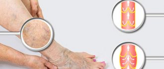
- Varicose veins
- Obesity
- Night crumpies
- Fibromyalgia
- Infections
- Neurogenic pain (disc herniation, stenosis, disc protrusion, lumbosacral radiculitis)
- Diabetic polyneuropathy
Muscle cramps
Muscle cramps (cramps) are strong, painful contractions and tightening of muscles that occur suddenly, lasting from a few seconds to several minutes. Muscle soreness may persist for several hours after the clenching (cramp) episode ends. They usually go away on their own and do not require special treatment.
The cause of seizures cannot always be determined. Muscle cramps can occur under various conditions or types of physical activity such as:
- Exercise, injury or overuse of muscles. When a muscle is overfatigued, has poor elasticity or an incorrect load vector, it can cramp.
- Dehydration associated with exercise during hot weather can cause heat cramps. In addition, various conditions (vomiting, diarrhea or lack of water) can also cause cramps.
- Pregnancy. Cramps during pregnancy can occur due to a decrease in the amount of minerals in the body, such as calcium and magnesium, especially in the last months of pregnancy (when the formation of the fetal musculoskeletal system is completed).
- Exposure to low temperatures, especially when immersed in cold water.
- Some diseases (peripheral artery occlusive disease, kidney disease, thyroid disease), multiple sclerosis, etc.
- Medicines - some drugs can cause seizures (neuroleptics, diuretics). In any case, if there are recurring episodes of seizures, you should consult a doctor.
Horseshoes on the heels
The formation of horseshoes around the heels is often called a sign of joint problems. Corns in the form of horseshoes are formed in cases where the load falls on the marginal parts of the heel, as a rule, associated with deformation of the knee and hip joints, problems in the lumbar spine. Often these deformations are associated with autoimmune processes occurring in the body.
The heels are smooth. How to help “your two” Read more
Osgood-Schlatter disease
Osgood-Schlatter disease (sometimes also called Schlatter disease or osteochondrosis of the tibial tuberosity) is an inflammation in the upper part of the tibia, where the patellar tendons attach to the tubercle called the tibial tuberosity. This disease is often the cause of knee pain in adolescence (from 10 to 15 years), is associated with the growth of the musculoskeletal system, and as growth completes, the symptoms may disappear.
Osgood-Schlatter disease can cause pain, swelling, and tenderness in the area of the front of the knee, below the kneecap. As a rule, one knee is affected (sometimes it happens on both sides). Symptoms worsen when the tendon attached to the tibial tuberosity is stressed (for example, when jumping). The diagnosis is made on the basis of radiography. The main goal of treatment is aimed at preventing symptoms (reducing certain types of stress, immobilization during exacerbation, physiotherapy). Surgical treatment methods are rarely used.
4.Bemer therapy
In our medical practice, Bemer therapy has proven to be the most successful way to combat any type of pain. BEMER therapy is electromagnetic physiotherapy from Switzerland, the main goal of which is to improve blood circulation.
The Bemer device consists of three elements: an induction mattress aimed at general health improvement, a reinforced applicator - a device that allows you to influence a specific place, and a laser magnet, which has the strongest effect on the pain point
read more about Bemer therapy by following the link. If you want to get a consultation or make an appointment, you can do this using the feedback form or by phone: +7-495-212-08-85
Sign up for a consultation
Juvenile idiopathic arthritis
Juvenile idiopathic arthritis, often called juvenile rheumatoid arthritis or juvenile chronic arthritis, is a childhood disease characterized by inflammation of the joints, which become swollen and painful (and therefore impair joint mobility). It manifests itself in children as pain in the joints, impaired gait, due to decreased mobility in the joints and stiffness in the joints, usually in the first half of the day, with regression within 1-1.5 hours and after normal physical activity. The causes of this disease are not entirely clear. Most researchers believe that the disease is caused by a combination of factors that cause excessive activation of the immune system. There are several types of juvenile arthritis. The division into types is based on the number of joints involved in the process during the first 6 months from the onset of the disease, the area of the body where the disease manifested itself, and the presence of other symptoms. Types of juvenile arthritis: oligoarthritis (affects no more than 4 joints), polyarthritis (affects 5 or more joints), rheumatic-positive and rheumatic-negative (depending on the presence of rheumatic factor in the blood), systemic (accompanied by rashes on the body, fever and lesions). eye).
Flat feet
According to RNIMU (Russian National Research Medical Institute) named after. Pirogov, from 40 to 60% of the Russian population are susceptible to flat feet . Of these , there are 4 times more women than men .
According to medical statistics from ROSZDRAVA: - by the age of two, 24% of children show the first signs of flat feet, - by the age of four - in 32%, - by the age of six - in 40%, - by the age of twelve - 50% (every second teenager is diagnosed with flat feet ), - by the age of twenty - 60% .
Flat feet can be either congenital or acquired.
Congenital flatfoot in its “pure” form is rarely observed - in approximately 5% of cases . Acquired flatfoot is more common .
Of all cases of acquired flatfoot, transverse flatfoot is in first place in frequency ; it is diagnosed in approximately 65% of cases. In second place in the frequency of detection (about 33% of cases) is longitudinal flatfoot with concomitant flatness of the forefoot. Longitudinal flatfoot is usually combined with hallux valgus . The frequency of this pathology is about 11% of all foot diseases.
Growing pains
Pain in the legs occurs with growing pains and is often found in children and adolescents. Most likely, pain manifestations are associated with uneven growth of muscles, bones and soft tissues. The pain is usually localized in the thighs or calf muscles and usually appears in the afternoon. The duration of the pain can be 1-2 hours. Growing pain is not accompanied by other symptoms (fever, swelling). Over time, the symptoms disappear on their own. It is possible to use massage and sometimes paracetamol. Aspirin is not recommended before age 20 due to the possibility of developing Reye's syndrome.
Foot injuries
Characteristic primarily in sports (running, football, tennis, figure skating, etc.). Statistics on occupational injuries for individual parts of the body (feet) are not kept.
Every tenth fracture and every third closed fracture occurs in the foot (for military personnel this figure is slightly higher and amounts to 13.8% in peacetime).
The most common foot fractures are:
- talus - less than 1%, of which about 30% of cases lead to disability;
- heel - 4%, of which 83% - as a result of a jump on straight legs from a great height;
— cuboid — 2.5%;
— scaphoid — 2.3%;
- metatarsal - the most common type of injury to the bone of the foot.
Moreover, for athletes, a fracture of the fifth metatarsal bone is typical under excessive loads, and for people experiencing unusual excessive loads, often in uncomfortable shoes, a fracture of the second, sometimes 3 or 4, and rarely 1 or 5.
The average duration of disability for a toe injury is 19 days.
A fracture of the foot bones can occur for several reasons:
- heavy objects falling on the foot;
- jump (fall) from a great height and land on your feet;
- when kicked;
- when hit on the leg;
- with subluxation of the foot due to walking on uneven surfaces.
Medicines
Some medications can cause leg problems due to side effects. Combination with smoking or drinking alcohol increases the risk of drug side effects. The main groups of drugs that can cause leg problems:
- Drugs that cause blood clots (for example, contraceptives)
- Neuroleptics (aminazine, haloperidol)
- Beta-2 receptor agonists (terbutaline or albuterol)
- Antihypertensive drugs (nifedipine, amlodipine or nicardipine)
- Statins are drugs that lower blood cholesterol levels (simvastatin or atorvastatin).
- Estrogens
- Lithium preparations
- Diuretics (Lasix and others)
- Opiates
- Steroids
If you are taking medications (especially long-term), you should inform your doctor about the occurrence of pain or cramps in the legs.
Restless legs syndrome
Restless legs syndrome is a condition in which the patient experiences discomfort from the position of the legs and, sometimes, an irresistible urge to move the legs. This syndrome can lead to sleep disturbances and interfere with daily life.
Patients describe their sensations as the touch of needles in their legs, and sometimes as nagging pain. Changing the position of the legs brings temporary relief. Most often, these sensations occur in the evening, when a person is trying to relax or go to bed. The cause of this syndrome is not known. Certain medications (such as antidepressants), pregnancy, or iron deficiency (anemia) may cause this syndrome. Restless legs syndrome is treated with drugs that increase dopamine levels in the brain (levodopa) or anticonvulsants (gabapentin) to control leg movements and improve sleep. Opiates are extremely rarely used.
Transient ischemic attacks
Transient ischemic attacks are a temporary disruption of the blood supply to the brain. They are harbingers of a stroke. The only difference from a stroke is that the symptoms gradually disappear. The following symptoms should alert you:
- Sudden numbness, tingling and weakness in half the body or limb
- Gait disturbance
- Impaired perception of reality
- Speech Impairment
- Strong headache.
If these symptoms occur, seek emergency medical attention
Obliterating peripheral artery disease
PADS is a permanent process of narrowing or blockage of the arteries that supply oxygen-rich blood to various organs, including the lower extremities.
The main cause of occlusive lesions of the peripheral vessels of the lower extremities is the accumulation of cholesterol, calcium and other substances on the walls of the arteries (atherosclerosis). The narrowing of the artery lumen leads to a decrease in the supply of arterial blood to the leg muscles, which is especially evident during physical activity. When the muscle is at rest, the blood supply may be adequate.
The main symptom of obliterating disease of the arteries of the legs is pain in the calf muscles, thighs and buttocks, which appear after physical activity (such as climbing a hill or stairs, running, walking). This pain is called intermittent claudication. This usually occurs after a certain amount of movement and disappears after rest. As the disease progresses, symptoms appear with minimal physical activity. In addition to pain, patients are often concerned about: decreased muscle strength in the legs and balance when standing, numbness in one or two legs, and the appearance of ulcers that heal slowly. Treatment is usually conservative. In case of severe disruption of blood supply, surgical treatment methods are indicated.
Why is it dangerous?
— Decreased quality of life
– A chronic infectious disease that steadily destroys nails and threatens to spread to surrounding people, especially family members of the patient.
- Untreated mycosis of the nails and mycosis of the feet are considered as an entry point for other infectious diseases - for example, bacterial - erysipelas.
— Complicates the course of diabetes mellitus and can lead to serious complications.
— Allergization of the human body with a fungal infection is the formation of hypersensitivity to the fungus as an allergen, that is, fungal allergy.
— Development or worsening of diseases such as bronchial asthma, allergic dermatitis, various skin rashes and reactions.
- In rare cases, against the background of immunodeficiency, untreated nail fungus led to the development of deep mycosis - germination or penetration of the fungus with blood into the internal organs, which caused death.
Venous thrombosis
The occurrence of pain during thrombosis (blockage of a vein with a blood clot) is promoted by inflammation of the vessel wall or surrounding tissue. In addition, impaired outflow can lead to swelling and discoloration of the skin (pallor, cyanosis, redness). Pallor can be detected in the early stages of thrombosis of the veins of the iliofemoral region. Cyanosis is caused by congestive hypoxia and, as a rule, occurs with obstruction of the proximal veins of the leg. Pain during thrombosis can be aching, sharp or dull, intense and moderate. Often the pain intensifies when walking or carrying heavy objects. Pain may be relieved by lying down with your legs elevated. With superficial vein thrombosis, diagnosis is not difficult. If there is a suspicion of deep vein thrombosis, then objective research methods are necessary.
Bunion
This is an inflammatory process near the joint capsule on the big toe (bursa). According to statistics, most women who wear uncomfortable shoes . The statistics are such that every 7th inhabitant of planet Earth faces inflammation of the foot joint every day. As a rule, these are elderly people, athletes and ballerinas. Women are more susceptible to inflammation of the foot joints.
Under pressure, deformation of the phalanx of the finger occurs; with constant wearing of uncomfortable shoes, the joint takes on the shape in which it is most often found. This deformation disrupts blood circulation and injures soft tissues. The same thing happens with injuries or bruises.
Pregnancy
Pain and tenderness in the lower leg (less often in the thigh) can occur during pregnancy. In pregnant women, pain may be accompanied by swelling of the lower leg and thigh (both one and both limbs). The causes of pain in the legs in pregnant women are not entirely clear. On the one hand, this is explained by a violation of the outflow from the venous network of the lower extremities due to the fetus. On the other hand, a decrease in electrolytes in the blood contributes to the appearance of muscle cramps, which serve as one of the sources of pain. Unilateral swelling and pain in the limb may be associated with compression of the iliac artery by an enlarged uterus.
Gangrene
The development of gangrene marks stage 4 of HUNC. Between the third stage and the appearance of gangrene, a phase of critical limb ischemia has recently been distinguished, which is characterized by intense pain at rest with the formation of superficial distal necrosis and trophic ulcers.
Gangrene manifests itself by the appearance of bluish lesions on the toes or heels, which subsequently become black. The lesions tend to spread, merge, and involve the proximal parts of the foot and lower leg.
Traditionally, dry and wet gangrene are distinguished. Their main difference is the delimitation (demarcation) of the area of necrosis from other tissues. With dry gangrene, there is an area of black skin, clearly demarcated from the surrounding unchanged tissue, with no tendency to spread. The general condition of the patients does not suffer (except for persistent pain), there are no signs of intoxication, and there is no hyperthermia. This type of gangrene with a small affected area (for example, dry gangrene of the distal phalanx of the toe) can be managed conservatively for a long time, without giving indications for surgery; in some cases, self-rejection of the necrotic area is possible. Haste with surgery in such a situation, due to surgical trauma, can cause progression of the necrotic process.
With wet gangrene, there is no demarcation, there are areas of both black and bluish color on the foot, the skin proximal to the focus of necrosis is hyperemic, and from under the necrosis there is a purulent discharge with an unpleasant odor. There are signs of intoxication (thirst, tachycardia, etc.), hyperthermia to subfebrile and febrile levels. The wet process is characterized by rapid progression, with necrosis spreading in the proximal direction.
In stage 4, some authors distinguish between stage 4A - when there is a prospect of preserving the supporting function of the limb (for example, if it is possible to perform amputation according to Sharp or Chopart while maintaining the supporting function of the heel) and 4B - when the patient is shown a high amputation at the level of the thigh or lower leg.
The presence of concomitant atrial fibrillation in a patient can cause a rapid transition from one stage of arterial insufficiency to another. With atrial fibrillation, in many patients, thrombotic masses accumulate in the left ventricle of the heart, the separation of which and migration in a large circle to the lower extremities can aggravate the existing arterial stenosis with transition to a more severe stage of ischemia, up to the development of gangrene.
Some tests can help in diagnosing limb ischemia, for example, the finger pressing symptom, Oppel's plantar ischemia symptom, Goldflam's test, Panchenko's knee phenomenon, etc.
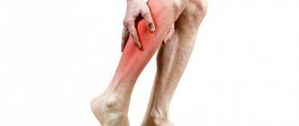
Crick
Muscle pain can appear after fast walking, running, playing sports, or walking in poorly fitting shoes. As a rule, discomfort occurs immediately after exercise or after 12-24 hours. Most often, pain occurs in the legs, sometimes in the hips. Possible pain on palpation. Sometimes swelling of the lower leg occurs. If there is a clear connection with physical activity and the pain is bilateral, then the diagnosis is beyond doubt. In cases where the pain is one-sided, it is necessary to exclude possible venous thrombosis using objective diagnostic methods.
Muscle rupture
Most often, muscle ruptures occur in the lower leg (possibly in the thigh). A rupture occurs as a result of a sudden stretch or contraction of a muscle. Typically, a small section of muscle is torn where it connects to the tendon. But there are also large ruptures, up to separation of the muscle from the tendon or separation of the tendon. Muscle rupture occurs mainly when the limb is sharply flexed in the direction opposite to the acting force. The rupture is accompanied by acute pain immediately after excessive stress. The pain may subside for a while, but as the hematoma grows, the pain will gradually intensify. Swelling occurs in the area of the damaged muscle and range of motion is limited.
Foot fasciitis (heel spur)
In 90% of cases, a heel spur develops against the background of flat feet. With this disease, the correct distribution of the load on the foot changes, the tendons become deformed and the places where the tendon is attached to the bone begin to become inflamed. Against the background of plantar fasciitis, the formation of marginal bone growths is possible - they are called “heel spurs”.
The consequence of the disease is acute pain in the heel, which may disappear, but occurs or intensifies with intense exercise. The causes of unpleasant pain lie in inflammatory and degenerative changes in the plantar fascia. The plantar fascia is a layer of tough fibrous tissue that runs along the perimeter of the bottom surface of the foot. In function it resembles a cable that connects the heel bone with the metatarsal bones and acts as a support for the longitudinal arch of the foot. The plantar fascia is the longest and strongest ligament in the human body.
Plantar fasciitis can be caused by injuries to the surrounding soft tissue . In other words, the plantar ligaments at the bottom of the foot, located between the heel and the ball of the foot, are pulled into an awkward position and the plantar fascia becomes deformed. Popularly, plantar fasciitis is often called heel spurs, but bone growth is a consequence of plantar fasciitis. Such spurs can appear along with pain in one leg, or in both legs together or alternately.
Problems associated with heel spurs usually appear after the age of forty. The likelihood of occurrence increases in people experiencing spinal diseases, who have arthritis, disease of the large joints of the legs, gout, or there is a serious violation of blood circulation in the legs. Foot fasciitis often occurs in athletes who have constant or prolonged stress on the legs.
According to statistics, every tenth person experiences problems caused by plantar fasciitis. Sometimes such acute pain occurs even in completely healthy people who do not complain at all about problems with their legs.
Several reasons can cause the onset of a disease such as heel fasciitis. The first of these is the development of flat feet . Due to flat feet, the arch of the foot becomes denser, while the plantar fascia becomes tense and micro-tears occur on it. Inflammation occurs in the area of the heel bone because the fascia is attached to it in this place.
Incorrectly selected shoes and constant wearing of them can provoke the development of plantar fasciitis.
The second possible cause of fasciitis is when the plantar fascia is regularly subjected to excessive stress . This can happen if the patient is overweight or if the person has increased their usual strength loads, for example, some kind of heavy work has arisen or sports have become too intense.
Bone damage
Damages to the bone tissue of the lower extremities (tumors, subperiosteal hematomas, fractures) may be accompanied by pain, tenderness to the touch, and swelling of the limb. Clinical manifestations may be similar to venous thrombosis. It can be especially difficult to diagnose a hip fracture in older people (the fracture is often accompanied by swelling of the lower limb). Considering the involutional changes in the central nervous system (cerebral vascular sclerosis), it is sometimes difficult to collect an anamnesis in this category of patients. In addition, elderly people often have a combination of a femoral neck fracture with venous thrombosis.
Pathomorphology
The main changes develop in the intima of the arteries. There are 5 morphological stages of atherosclerosis:
- Prelipidic – characterized by increased endothelial permeability, destruction of the basement membrane, destruction of elastic and collagen fibers.
- Stage of lipoidosis - focal infiltration of the arterial intima with lipids occurs.
- Stage of liposclerosis - a fibrous plaque forms in the intima of the artery.
- Stage of atheromatosis - destruction of the plaque occurs with the formation of an ulcer.
- Stage of atherocalcinosis – plaque calcification occurs.
Based on the type of damage to the vascular bed, segmental and diffuse atherosclerosis are distinguished. In the first case, the process develops in a limited area of the vessel from single plaques to complete occlusion of the lumen. This type is more favorable in terms of the potential for performing bypass reconstructive operations on blood vessels. The diffuse type involves widespread atherosclerotic lesions predominantly in the distal bed, leaving no “window” for the surgeon to apply a shunt or prosthesis. The destiny of such patients is conservative therapy in order to delay the onset of gangrene as much as possible.
Rupture of popliteal cyst (Baker's cyst)
The fluid contained in a Baker's cyst, when ruptured (in the case of injury, arthritis or surgery in the knee joint), can flow down into the intermuscular space and cause an inflammatory reaction. This may manifest itself as pain, swelling in the lower leg, tenderness on palpation and resemble the picture of venous thrombosis. Rupture of a Baker's cyst can occur without provoking reasons. The diagnosis can be made based on radiography. But the possible combination of cyst rupture and venous thrombosis requires first of all the exclusion of thrombosis.
Diagnostics
First of all, to diagnose the cause of pain, the doctor needs to get answers to a number of questions:
- Localization of pain
- Presence of pain in one or two limbs
- Nature of pain (sharp, dull)
- Dependence of pain on time of day
- Dependence of pain intensity on physical activity
- Possibility of reducing pain when raising legs
- The presence of other symptoms (fever, weakness, lower back pain).
In addition, the doctor will conduct a physical examination (visual examination, palpation, various tests). Instrumental examination methods:
- Laboratory research
- Ultrasound of the vessels of the lower extremities (Dopplerography of veins and arteries)
- Arteriogram
- Scintigraphy
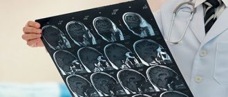
- MRI
- CT
- Radiography
At-risk groups
Patients with the following problems are most susceptible to vascular diseases of the lower extremities:
· Long history of smoking;
· Diabetes mellitus types 1 and 2;
· Alcohol abuse;
· Prolonged physical inactivity;
· High blood pressure;
· Hypercholesterolemia (increased concentration of cholesterol and triglycerides in the blood);
· High level of non-proteinogenic amino acid homocysteine in the blood;
· Obesity;
· Severe hormonal imbalance.
Pathologies of blood vessels and arteries of the legs mainly affect people who have crossed the threshold of fifty years of age, but in the last few years their active spread has also been observed among the young population. Men are more susceptible to such diseases than women.
It should be emphasized that most vascular dysfunctions are of a psychological nature, and people with a stressful type of character are most susceptible to them.
It is important to have a family history of disorders. This is especially true for atherosclerosis and varicose veins.
Treatment
Treatment of leg pain directly depends on the disease and can be either conservative (drug treatment, intra-articular injections, physiotherapy, acupuncture, exercise therapy, orthotics) or surgical. The following recommendations can be preventive:
- No smoking
- Don't abuse alcohol
- Monitor blood sugar levels (if you have diabetes)
- Control cholesterol levels
- When exercising, a mandatory warm-up before and sufficient rest after exercise
- Drinking enough fluids during exercise.
Author: V.I. Dikul