3.1. Risk levels of VTEC
The classification of a patient into a low, moderate or high risk group is carried out using a number of systems for assessing the patient's status and taking into account the nature of the upcoming treatment.
One of the most convenient tools for determining the risk of VTEC in surgery is the Caprini scale (Table 13). Table 13. Clinical characteristics scoring scale (according to Caprini)
| 3 points | 5 points | ||
| 41-60 years | 61-74 years | >74 years old | Stroke ( |
| Minor operation | Arthroscopic surgery | History of VTE | Major joint replacement |
| BMI >25 kg/m2 | Major open surgery (>50min) | Family history of VTE | Fracture of the femur, pelvis, tibia |
| Edema of the lower extremities | Laparoscopic surgery (>45 min) | Leiden mutation | Spinal cord injury ( |
| Phlebeurysm | Oncology | Mutation in the prothrombin gene | — |
| Pregnancy or postpartum period | Bed rest (>3 days) | Lupus anticoagulant | — |
| History of miscarriage (2-3 trimester) | Gypsum bandage | Antibodies to cardiolipin | — |
| Taking estrogens/gestagens | Catheter in the central vein | Increased plasma homocysteine levels | — |
| Sepsis ( | — | Heparin-induced thrombocytopenia | — |
| Severe lung disease, including pneumonia ( | — | Other thrombophilias | — |
| Respiratory dysfunction | — | — | — |
| Acute myocardial infarction | — | — | — |
| Congestive heart failure ( | — | — | — |
| History of inflammatory bowel disease | — | — | — |
| Therapy patient on bed rest | — | — | — |
Depending on the amount of points obtained during the collection of anamnesis and examination of the patient, he is assigned to one or another risk group.
It is advisable to distribute patients into groups according to the principle proposed in the recommendations of the American College of Chest Physicians as amended in 2012. Patients are divided into groups of very low risk, low, moderate and high risk depending on the number of points, as well as from the surgical option. The probability of developing VTEC in the absence of prophylaxis in these groups is respectively less than 0.5, 1.5, 3 and 6%. (Table 14). Table 14. Risk levels depending on the number of points or type of surgical intervention (according to Caprini as modified by ACCP)
| Risk | General surgery, including gastrointestinal tract, urology, vascular, thoracic, endocrine (thyroid) | Plastic and reconstructive surgery | Operation |
| Very low | 0 | 0-2 | Most "one day surgery" patients |
| Short | 1-2 | 3-4 | Surgeries on the spinal column (not oncology) |
| Moderate | 3-4 | 5-6 | Gynecological surgeries (non-oncology), cardiac surgery, thoracic surgery (most interventions), spinal column surgeries (oncology) |
| High | 5 and > | 7-8 | Bariatric surgery, gynecological surgery (oncology), pneumonectomy, craniotomy, traumatic brain injury, spinal cord surgery, severe trauma |
Due to the absence of fundamental differences in the frequency of VTEC and the tactics of management and prevention of patients with low and very low risks in clinical practice, it is advisable to combine them into one group with a low probability of VTEC.
Along with the Caprini scale, the risk assessment system using the volume, duration of surgical treatment and the patient’s age as the main factors is of great practical importance (Table 15).
Table 15. Risk levels of VTE
| Risk level | Patient characteristics |
| Moderate | Major operations* in patients 40-60 years old without additional risk factors |
| Major gynecological operations* in patients under 40 years of age during estrogen therapy | |
| Minor surgeries* in patients over 60 years of age | |
| Minor surgeries* in patients aged 40–60 years with risk factors | |
| Isolated shin bone fractures | |
| Rupture of ligaments and tendons of the leg | |
| Hospitalization with chronic heart failure III-IV functional class according to NYHA | |
| Acute infectious or inflammatory diseases requiring bed rest | |
| Acute lung diseases with respiratory failure that do not require artificial ventilation | |
| Burns up to 20% of the body surface | |
| Thermal inhalation injury, stage II | |
| High | Sepsis |
| Presence of verified thrombophilia | |
| Major operations* in patients over 60 years of age | |
| Major operations* in patients 40-60 years old who have additional risk factors for VTEC (history of VTEC, cancer, thrombophilia, etc.) | |
| Endoprosthetics of the hip and knee joints, operations for hip fractures | |
| Severe and combined trauma, spinal cord damage | |
| Burns over 20% of the body surface | |
| Thermal inhalation injury, stage III | |
| Complications of burn disease | |
| Chemotherapy, x-ray or radiotherapy in patients with cancer | |
| Ischemic stroke with paralysis | |
| Acute lung disease with respiratory failure requiring mechanical ventilation |
Note.
In all cases, the risk of VTEC increases with the presence of additional risk factors. * - see Appendix 2. Non-surgical patients. An approach to assessing the risk of VTEC in non-surgical patients is presented in Appendix 3.
5.6. Prevention of thromboembolic complications in surgical patients
The stable number of thromboembolic complications is due to many reasons, and only some of them are objective. For example, the improvement of surgical techniques and anesthetic management methods naturally leads to an increase in the number of elderly patients undergoing surgery, with a large number of concomitant diseases and, accordingly, a higher risk of pulmonary embolism. The number of people in the population with obvious or hidden hemostasis defects is also growing. These trends are certainly important, but the main problem of preventing VTEC is not the increase in the number of patients at high risk and not the lack of effective means of prevention, but the lack of proper organization of the prevention process, the absence or insufficient attention to this problem. Unfortunately, as the results of numerous studies show, in different countries, prevention is provided to no more than 40-60% of those who need it. In other patients, prophylaxis is either not carried out at all, or is insufficient in scope or duration. As a result, there is a large gap between the developed technologies for the prevention of VTEC and the implementation of these technologies in practical healthcare. And it is this gap that is currently considered as the main reason for the stable incidence of postoperative thrombosis and embolism. The main reasons for this gap are as follows.
Inadequate assessment of the danger of pulmonary embolism is primarily due to the peculiarities of the course of deep vein thrombosis, which in 80-88% of cases is asymptomatic. Among patients who died from massive pulmonary embolism after hip replacement, only 10-12% showed signs of thrombosis during their lifetime. Another factor leading to insufficient concern among hospital doctors about this problem is the late development of pulmonary embolism. Among patients with venous thrombosis, almost 70% develop this complication after discharge from the hospital. These patients do not often see the operating surgeon, but are usually treated by other specialists. Therefore, the prevalence of overt pulmonary embolism in hospitals is considered relatively rare, and its danger is lower than the risk of postoperative bleeding during prophylactic administration of anticoagulants. As a result, most surgeons have a negative attitude towards the prescription of anticoagulants in the preoperative period and the first administration of the drug is performed no earlier than 6-8 hours after the end of surgery. Meanwhile, with this approach, the effectiveness of prevention is reduced by 40%. This position can be changed only by systematically monitoring the patency of the deep veins in the immediate postoperative period and monitoring the future fate of the patient, creating good feedback between patients, clinics and surgical hospitals, when each case of pulmonary embolism and its clinical and economic results will become known to the operating surgeons.
The next reason for the irrational and insufficient use of preventive measures is a rather complex system for determining individual risk and choosing prevention methods. And if for operations such as hip or knee replacement this issue has been resolved in principle, since all patients are at high risk, then in many other situations it can be difficult to make such a choice, and all currently existing recommendations are only general and probabilistic character. As a result, the solution to key issues in the prevention of pulmonary embolism often turns out to be controversial, questionable and dependent not only on objective factors, but also on the competence of the doctor and his knowledge in this area.
The most significant examples of how “human factors” in this area affect patient safety were published in 1999 in a report by the Institute of Medicine of the National Academy of the United States, entitled: “To Err Is Human: Building a Safer Health Care System.” Human: Building a Safer Health System). The authors of this report, analyzing 37 million hospitalizations and 323,993 deaths, concluded that hospitals are one of the most dangerous places, where about 100 thousand people die every year from medical errors in this country alone. Taking into account the total number of doctors in the United States, this meant that on average, one in 10 doctors each year made an error that led to the death of a patient. None of the patients could consider themselves protected from such errors: on average, 1 out of 30 hospitalized patients developed a life-threatening complication, which, in principle, was not associated with his disease and general condition, but was the result of a violation of the usual algorithms for diagnosis, prevention, treatment, improper use of medications. In the structure of these complications, VTEC occupied one of the leading places, and PE turned out to be “the most common cause of death among those that can be prevented.” “Each of us,” wrote William Richardson, director of this project, “at some stage in our lives can become a patient in the health care system and face this problem to which we are so defenseless. I hope that our report will be a signal for action.” These actions began; the principles formulated in the report for building a patient safety system became the basis of a new government strategy in the healthcare of many world powers. For example, in order to change the attitude of the heads of medical institutions to this problem, in April 2008 in the United States a decision was made according to which, within the framework of the public health insurance system Medicare, payment for the treatment of patients with VTEC, as well as some other complications, was stopped, such as transfusion of incompatible blood, deep bedsores, iatrogenic pneumothorax, or a napkin forgotten in the wound. So far, such measures are in effect only in certain countries and insurance companies and apply to VTECs that arise, for example, after prosthetic surgery for large joints. But the general position is clear - now PE can no longer be considered a fatal and rare complication, the development of which is difficult to predict and almost impossible to prevent. Today, the prevention of TEC is one of the main elements of national programs to improve patient safety, and the frequency of these complications is a criterion for the quality of medical care in a hospital.
How to organize the prevention of VTEC in a hospital? In our opinion, the answer to this question is most revealing in the study by D. Levy et al. (1998), who studied the quality of TEC prevention during an inspection of intensive care units in three hospitals (1830 case histories). According to the authors, in a hospital where this problem was not paid attention to, prophylaxis was carried out in 38% of patients who needed it. If doctors were trained in prevention, i.e., additional lectures, classes, and clinical discussions were conducted, then the frequency of adequate prevention increased almost 2 times (62%). And where the prophylaxis process was determined by a special Protocol, almost all patients (97%) received the prophylaxis that met the standards.
The low effectiveness of lectures, conversations, and seminars as ways to attract the attention of doctors to the problem of pulmonary embolism and the high effectiveness of the local Prevention Protocol were also noted in the latest edition of the ASSR recommendations, based on data from “evidence-based medicine” (2008).
The presented data indicate that the main step towards creating an effective and safe system for the prevention of VTEC is the creation of a protocol in each hospital. The main goal of creating this protocol is to maximally adapt and specify the main provisions of industry standards, national and international recommendations to the specifics of the work of a given hospital, taking into account its own organizational, diagnostic, clinical and financial capabilities. Creating such a protocol in practice is not an easy and very responsible task, since the resulting document at the hospital level will become a mandatory order. This protocol should define algorithms for assessing the degree of risk of TEC, identify groups of patients in need of various methods of prevention, determine the procedure for prescribing and recording means of mechanical and specific prevention, methods of laboratory and instrumental monitoring of its effectiveness.
The methodological basis of all provisions of the Protocol should be modern ideas about the mechanisms of thrombogenesis, the reasons for their implementation in patients with critical conditions and pathogenetically justified ways to prevent these complications. Understanding by doctors of the essence of the changes taking place will, in turn, facilitate the implementation of the remaining provisions of the Protocol.
Pathogenetic mechanisms of venous thrombosis
The processes of hemostasis and thrombosis in any part of the vascular bed can have different meanings in human life. When a vessel is damaged, the formation of a hemostatic plug is a normal reaction that prevents blood loss. At the same time, in contrast to hemostasis, aimed at preserving the integrity of the circulatory system, the processes of thrombus formation, i.e. the formation of a blood clot in the lumen of a vessel, are pathological in nature, lead to disruption of the blood supply to organs and can not only cause various morphofunctional disorders, but also lead to to the sudden death of a person.
The list of diseases in the genesis of which thrombus formation is important includes most nosological forms known today, but their effect on hemostasis in all cases is realized in the form of three main components: changes in the thrombogenic potential of the vascular wall, increased activity of the coagulation system (thrombophilia) and disturbances of regional or systemic blood flow. The importance of these three mechanisms in the pathogenesis of venous thrombosis was proven in 1859 by Rudolf Virchow, and his “triad” retains its significance to this day. At the same time, knowledge of only this triad for the prevention and treatment of DVT in surgical patients is clearly not enough; a more detailed understanding of the mechanisms of venous thrombus formation, the processes that initiate or prevent the formation of a blood clot in critical conditions is necessary.
These mechanisms are very complex, they involve hundreds of substances produced by various organs and tissues, the interaction and interaction of which provides the main beneficial result - maintaining the blood in a liquid state with constant readiness for the formation of a hemostatic plug at the site of vessel damage. In the absence of vascular damage or inflammation, platelets are not capable of adhesion to the endothelium, since a normally functioning endothelium has no receptors for unstimulated platelets and because the endothelium synthesizes and secretes various biologically active substances - prostacyclin, nitric oxide, thrombomodulin, tissue plasminogen activator, endothelin, thromboplastin , heparin cofactors, protease inhibitors and other metabolites, the combined action of which ensures the atrombogenicity of the vascular intima. When the endothelium is damaged, platelets interact with subendothelial ligands, for which they have specific receptors. The first of these contacts between circulating platelets and the subendothelial layer is mediated by the platelet glycoprotein (GP) complex Ib-IX-V, which binds to the subendothelial von Willebrand factor (VWF). Through this interaction, platelets find themselves close to the endothelial surface, lose speed, and begin to roll over the surface, which provides conditions for closer interaction with subendothelial proteins of other platelet receptors. Once adhesion has occurred, transmembrane signals triggered by ligand receptors activate platelets, leading to conformational activation of integrins, most likely α2β3 and αIIbβ3. As a result, calcium release is activated, which leads to the release of granules with the secretion of a variety of procoagulant molecules, such as factor (F) V, VWF and fibrinogen, as well as transformation of the platelet membrane, on the surface of which phosphatidylserine appears. Phosphatidylserine is a matrix for the formation of complexes from plasma coagulation factors, which ultimately leads to the formation of thrombin and fibrin clot. Histopathological studies of arterial thrombi are fully consistent with this model, since the base of the arterial thrombus consists almost exclusively of platelets directly associated with the vessel wall at the site of injury and surrounded by dense fibrin masses above and below the attachment site with single red blood cells embedded in the structure of the thrombus. This model is also supported by the clinical results of the use of antiplatelet agents, effective means of preventing arterial thrombosis.
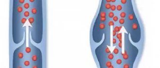
The sequence of events in venous thrombosis is much less known, but it is clear that, unlike arterial thrombosis, damage to the walls of the deep veins is not usually the cause of DVT. Such damage is extremely rarely found in autopsies of patients with DVT and TEC: moreover, the structure of venous thrombi also does not correspond to the arterial model. Most venous thrombi consist of two parts: the first, consisting mainly of fibrin and red blood cells (red thrombus), and the second, aggregated platelets (white thrombus). Since it is the first, fibrin-rich part that ensures the attachment of the thrombus to the vascular wall, it can be assumed that in the initiation of venous thrombus formation, the activation of plasma coagulation factors occurs before the activation of platelets.
Of key importance in understanding how the initiation of the coagulation cascade can occur in vivo in the absence of endothelial damage was numerous facts that confirmed the constant circulation in the blood of a type I transmembrane protein, the so-called tissue factor (TF). Previously, it was believed that TF is primarily contained only outside the lumen of the vessel and appears in the bloodstream only at the site of endothelial damage, where it forms a complex with FVIIa, which leads to the activation of factor X, triggering coagulation mechanisms. In turn, Xa combines with cofactor Va, forming a prothrombinase complex on the phosphatidylserine-rich surface of activated platelets, which converts prothrombin into thrombin.
In recent years, convincing evidence has been obtained that TF circulates in normal plasma, both in the form of microbubbles formed by cell membranes and in soluble form. It has been established that endogenous microvesicles containing TF lead to experimental thrombosis through the binding of platelets at the site of injury through mechanisms including the interaction of specific receptors (PSGL-1) on microvesicles and P-selectin on activated platelets. Microvesicles not only attach activated platelets, they adhere to them via PSGL-1 and phosphatidylserine and transfer TF and other proteins to the platelet membrane, accordingly increasing the activity of the prothrombinase complex, stimulating the formation of thrombin and fibrin. The evidence base is constantly growing that microvesicles containing TF can play an important role in the pathogenesis of DVT, increasing the risk of its development and increasing the volume of thrombotic masses. A significant increase in the concentration of microvesicles with TF was noted in sepsis, common oncological processes, severe autoimmune diseases, acute stroke and other conditions accompanied by stimulation of the systemic inflammatory response.
It is important to emphasize that microvesicles containing TF can interact with intact but activated endothelial cells. Like platelets, endothelial cells contain P-selectin and phosphatidylserine in intracellular granules, which appear on the cell surface when activated. From this moment on, the surface of the endothelial cell is able not only to interact with TF from microvesicles, but also to act as a matrix, a catalytic surface for the formation of the prothrombinase complex and the launch of the coagulation cascade, replacing platelets in this role. Platelets can be involved in the fibrin bundle for the second time, through adhesion with the help of GPIb-IX-V αIIbβ3 receptors, and then ensure the growth of the thrombus. This hypothesis is confirmed by the fact that in venous thrombi platelets are usually concentrated far from the site of fixation, and, secondly, the data that antiplatelet agents, in particular aspirin, cause, although insignificant and not comparable with anticoagulants in effectiveness, but statistically significant reduction in the risk of DVT.
The diagram presented above is still largely hypothetical. But already now it allows us to draw at least two conclusions that are very important for the prevention of feasibility studies.
Firstly, for the development of these complications, the presence of trauma or surgical impact on the vein wall is not necessary. In many severe diseases, in critical conditions, especially those accompanied by activation of the inflammatory response, the concentration of TF can increase significantly without additional injury to the vessel wall, providing an increased, almost pathological readiness of the circulating blood for thrombus formation. Therefore, the problem of preventing TEC is much broader; it concerns every patient who is or has experienced a critical condition, an infectious complication, and the absence of a history of surgery or trauma to the venous vessels in such a patient cannot serve as a sufficient basis for refusing to carry out preventive measures. This conclusion is supported by the fact that currently only one out of every five patients who die from TEC is a “surgical” patient.
Secondly, knowledge of the mechanisms of venous thrombus formation suggests that the main efforts to prevent these complications should be aimed at preventing endothelial activation. There is already TF in the blood, but it can only interact with activated endothelial cells, cells that have changed their properties and have lost their atrombogenicity.
Among the factors that cause a change in the functional properties of the vascular wall and a violation of its thromboresistance, there are “external” ones associated with mechanical damage to the vessel; physical - radiation exposure, hyper- and hypothermia; chemical - exotoxins, some medications (glucocorticoids, hormonal contraceptives, chemotherapeutic agents), which always increase the risk of thromboembolic complications and require targeted prevention. “Internal” factors are no less important, primarily ischemic, hypoxic, reperfusion and toxic damage to endothelial cells, which occurs and becomes systemic in any acute violation of the vital functions of the body. The entire system of intensive care is designed to prevent these disorders, including measures aimed at protecting and quickly restoring central regulatory mechanisms (multi-level anesthesia, minimally sufficient and depth-controlled sedation, early activation), maintaining oxygen balance, rational infusion and cardiotropic therapy, preventing hyper - and hypodynamia of blood circulation, satisfaction of metabolic and energy needs, sanitation of infectious foci). It is these measures, and not artificial hypocoagulation, that should form the basis for the prevention of TEC in every patient. Specific prevention only complements this complex, being essentially the price that has to be paid for the inability to prevent pathological activation of the endothelium in many conditions.
In addition, the entire complex of intensive care measures also limits the effect of the remaining components of Virchow’s triad - venous stasis and hypercoagulation. Almost every patient in the hospital also has the conditions for implementing these elements. In particular, the presence of venous stasis is a factor that determines the highest probability of localization of thrombosis in the deep veins of the leg. These veins contain a large number of valves, the petals of which are covered with endothelium, which differs in structure from the endothelial lining of other parts of the venous wall in its higher density and lack of orientation to the direction of blood flow. The walls of the valves, unlike the walls of the veins, do not have their own blood supply (their own vasa vasorum) and are therefore most sensitive to changes in oxygen tension in the venous blood.
There are several factors that determine the particular predisposition of valve flaps to the formation of blood clots. First, it is the endothelial cells of the venous sinuses that are primarily affected by the reduced blood flow and shear rates. In particular, during immobilization, this is where the maximum decrease in blood flow velocity was noted.
Secondly, in this zone the blood is least saturated with oxygen. For example, in one study, the oxygen tension in the deepest part of the sinus was 5-70% of its value in the central part of the vascular bed. Thirdly, it is in the venous sinuses that there is a tendency towards a constant accumulation of leukocytes and platelets.
The main background for the occurrence of thrombosis of these veins in surgical patients or patients in critical condition in the ICU is usually local venodilation, leading to a decrease in the volumetric velocity of blood flow in the muscular-venous sinuses and the occurrence of stasis, as well as, probably, local accumulation of activated factors coagulation. Of the most significant causes of venodilation, two are usually identified: the entry into the bloodstream of toxins and inflammatory mediators, causing venodilation and pharmacological myoplegia caused by the use of muscle relaxants. Maximum varicose veins occur in the presence of venous hypertension due to mechanical ventilation or the addition of heart failure.
In second place in terms of the frequency of non-traumatic thrombus formation are the common iliac vein and tributaries of the internal iliac vein. Of all thrombosis of the main veins of the lower extremities, primary thrombus formation in this area occurs in 10-15% (up to 49%) of all observations. Unlike the muscular veins of the leg, in the iliac vein basin, where there is intense volumetric blood flow, a simple disturbance of regional hemodynamics, even against the background of hypercoagulation, is not enough for spontaneous thrombus formation. In this area, the main reason for the formation of blood clots is anatomical changes in the common iliac vein, caused by the abnormal structure of the vascular bed, intravascular changes or external compression of the vein, for example, against the background of an aneurysm of the common iliac or internal iliac artery, fibrosis of the retroperitoneal tissue, structural changes in the spine. These changes are often accompanied by a violation of the venous outflow from the underlying segments, which is the necessary condition under which venous thrombosis of this or underlying areas of the venous system subsequently develops. The trigger for thrombus formation in this segment is most often an increase in outflow resistance (childbirth, pregnancy) or local hypercoagulation (inflammation or surgery on the pelvic organs).
The third place in the frequency of primary thrombus formation, amounting to no more than 5%, is occupied by other localizations, the most common of which are the popliteal and femoral vein in the popliteal and iliopectineal fossa at the level of their lymphatic collectors. Primary thrombus formation in these places is closely related to regional inflammatory foci, installation of venous catheters, and improper use of elastic compression.
A special place in the structure of thromboembolic complications is occupied by venous thrombosis caused by various coagulopathies, including thrombosis due to heparin-induced thrombocytopenia. The sites of primary thrombus formation in these cases are the same segments of the vascular bed, and such thromboses are distinguished by their significant prevalence, tendency to relapse, severity of damage and resistance to treatment.
Among the causes of acquired thrombophilias, first of all, it is necessary to highlight violations of the rheological properties of blood (changes in blood viscosity, deformability and aggregation of erythrocytes, their hydrodynamic strength), usually caused by hemoconcentration, dehydration, endogenous intoxication, hypercatecholaminemia, blood transfusions and other well-known components of critical conditions that can be and needs to be diagnosed and eliminated in a timely manner.
It is more difficult to identify thrombophilias caused by congenital or acquired deficiency of natural coagulation inhibitors, disorders of the fibrinolytic system (dysfibrinogenemia, changes in the activity of fibrinolysis activators or inhibitors), homocysteinemia and other reasons. In intensive care practice, the most common is acquired antithrombin deficiency, which is realized clinically in the form of a lack of effect from heparin prophylaxis or heparin therapy. For this reason, in critically ill patients, it is advisable to determine the antithrombin concentration before prescribing heparin. And if this is not possible, the use of anticoagulants should be started with an infusion of UFH under APTT control. In this case, the development of hypocoagulation will make it possible to clinically exclude antithrombin deficiency and switch to the use of low molecular weight heparins (LMWH).
More rarely seen in clinical practice are thrombophilias caused by hereditary or acquired resistance of factor V to activated protein C (RAPS), deficiency or disorder of the structure of protein C and protein S, antiphospholipid syndrome and other reasons. The prevalence of these thrombophilias remains unknown, but they should be kept in mind, especially in the presence of an atypical course of thrombotic complications or in the presence of a relevant medical history.
Considering the wide variety of causes that can provoke pathological thrombus formation, the number of conditions and diseases that increase the risk of thrombosis, it is obvious that every patient in a hospital may be at risk of developing VTEC. This conclusion determines the content of the VTE Prevention Protocol.
Protocol for the prevention of thromboembolic complications in patients in a multidisciplinary hospital
The main principle of creating a standard or protocol for treating a patient is that this document should make it as difficult as possible for the doctor to make the wrong decision and make it easier for the doctor to make the right decision. Therefore, the Protocol for the prevention of VTEC in patients in a multidisciplinary hospital should contain general algorithms for assessing the degree of risk of VTEC, determine the procedure for prescribing and recording means of mechanical and specific prevention, methods of laboratory and instrumental monitoring of its effectiveness. At the same time, the document must be accessible to the practitioner, simple and easy to implement.
In accordance with the Protocol in force at the National Medical and Chemical Center named after. N.I. Pirogov, carrying out the prevention of feasibility studies includes the following five stages.
3.2. Methods for preventing VTE
Antiplatelet agents (acetylsalicylic acid). According to some data, acetylsalicylic acid helps prevent VTEC. However, evidence of its preventive effectiveness is limited and not as convincing as that of anticoagulants. In addition, there is reason to believe that acetylsalicylic acid has no safety advantage over anticoagulants. Therefore, although the use of acetylsalicylic acid for the prevention of VTEC can be discussed in individual patients, in most cases anticoagulants should be preferred.
Anticoagulants have well-proven preventive efficacy in various clinical situations; they reduce the risk of venous thrombosis and pulmonary thromboembolism by approximately half and should be used in all patients with an increased risk of venous thrombosis who do not have contraindications.
Subcutaneous prophylactic doses of UFH, LMWH, fondaparinux sodium, or VKA should be used. In addition, in traumatology and orthopedics, oral administration of NOACs (apixaban, dabigatran, etexilate or rivaroxaban) is possible. It is advisable to use mechanical methods of preventing DVT in patients of any risk level.
Mechanical methods are of particular importance when the use of anticoagulants is impossible due to the high risk of bleeding. In patients at high risk of DVT, it is reasonable to combine them with anticoagulants. Elastic stockings should be used to ensure optimal distribution of compression on the lower extremities (compression hosiery - see Appendix 3), or instrumental methods of prevention (alternating pneumatic compression of the lower extremities, and in some cases a venous pump for the foot, electromuscular stimulation, systems that provide flexion- extension movements in the ankle joints).
Early cessation of bed rest is one of the conditions for successful prevention of venous thrombosis and should be practiced in all cases where possible. However, in patients with an increased risk of venous thrombosis, this approach should not be the only method of prevention, since it does not provide sufficient protection.
Overdose of anticoagulants
In case of bleeding, doctors administer a 1% solution of protamine sulfate intravenously. Heparin is used as follows: to neutralize 100 units of heparin, 0.1 ml of a 1% solution of protamine sulfate is administered. The use of low molecular weight heparins for these purposes is relevant: 0.6 ml of a 1% solution of protamine sulfate neutralizes 0.1 ml of low molecular weight heparin.
Selected anticoagulants
Heparin
Heparin sodium is prescribed at a dose of 15,000 IU for 24 hours, and if the patient’s body weight is less than 50 kg, the daily dose of heparin is reduced to 10,000 IU. In elective surgery, the first injection is carried out 2 hours before surgery, the interval between injections is 8 hours
Low molecular weight heparins
There are two approaches to prescribing drugs: based on calculations and on the results of clinical studies. Dose calculation: from 4000 to 6000 units of anti-Xa per day. Injections are given 1 or 2 times a day. In emergency surgery, it is possible to start heparin prophylaxis after surgery, but no later than 12 hours after its completion.
Dalteparin (Fragmin) daily dose 5000 IU, for body weight above 120 kg daily dose 7500 IU. In clinical studies, a daily dose of 5000 IU has been studied.
Nadroparin calcium (Fraxiparin) daily dose 5750 IU (0.6 ml), for body weight above 120 kg daily dose 7550 IU (0.8 ml). Clinical studies have shown higher efficacy of the 0.3 dose (2875 units) than unfractionated heparin at a dose of 15,000 units per day; in one study, 0.6 ml of nadroparin calcium (5750 units) was used in patients weighing over 70 kg.
Enoxyparine (Clexane) daily dose 4000 IU (40 mg), for body weight above 120 kg daily dose 6000 IU (60 mg). Studies have generally shown the effectiveness of 30-40 mg of enoxaparin, and there are reports that a dose of 20 mg is not statistically different in effectiveness from 15,000 units of unfractionated heparin per day.
Factor Xa inhibitors
Rivaroxaban (Xarelto) is a direct factor Xa inhibitor (available in tablets), prescribed 10 mg once a day. Contraindicated in pregnancy and severe renal failure (creatinine clearance ≤15 ml/min).
Fondaparinux (Arixtra), a synthetic factor Xa inhibitor. The drug is administered no earlier than 6 hours after completion of the operation, 2.5 mg 1 time per day subcutaneously. In patients whose age is more than 75 years, and/or body weight is less than 50 kilograms, and/or who have moderately impaired renal function (creatinine clearance ≤ 3050 ml/min), it is better to refrain from administering the drug.