The cause of acute coronary syndrome (ACS) is almost always a sharp decrease in coronary blood flow caused by occlusion or thrombosis of a coronary artery. Clinical manifestations and consequences of ACS depend on the location of the obstruction, severity and duration of myocardial ischemia.
Of great importance in the treatment of ACS is the identification of individuals at high risk of developing cardiac complications, such as recurrent or repeated acute myocardial infarction (AMI) or death.
Stratification models (scales) make it possible to identify risk groups and, based on the degree of risk, to plan a particular treatment method, which help to more accurately determine the risk in patients with ACS, compared with the expert assessment of experienced doctors.
Risk stratification is considered one of the indicators of the quality of care for patients with ACS, and for its implementation (risk calculation) in patients with ACS, stratification models (scales) TIMI , GRACE, PURSUIT (for patients with ACS without ST-segment elevation), TIMI II (for patients with ACS and ST segment elevation); CADILLAC (when referring patients for percutaneous coronary interventions) and others.
Differentiated use of risk stratification models (scales) for the development of adverse outcomes helps in choosing a treatment strategy for patients with ACS (early invasive or initially conservative), thereby reducing mortality and the frequency of re-hospitalization for myocardial infarction or unstable angina.
Indications for reperfusion therapy
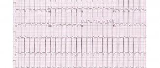
Reperfusion therapy should be carried out if no more than 12 hours have passed since the onset of an anginal attack, and the electrocardiogram shows ST segment elevation ≥0.1 mV in at least two consecutive chest leads or in 2 limb leads, or blockade appears LNPG.
The administration of thrombolytics or PCI should be done at the same time when ECG signs of true posterior myocardial infarction (high R waves in the right precordial leads and ST segment depression in leads V1-V4 with an upward T wave). It is recommended to consider the feasibility/possibility of reperfusion even if more than 12 hours have passed since the onset of symptoms (according to the patient), but there is clinical and/or electrocardiographic evidence of ongoing myocardial ischemia. Performing PCI on a completely occluded infarct-causing artery after 24 hours from the onset of clinical symptoms in stable patients is not recommended.
Emergency PCI
What is the cha2ds2-vasc scale and its interpretation
Emergency PCI, performed within the first two hours from the moment of admission to the hospital, is indicated in the following group of patients with acute coronary syndrome without ST-segment elevation:
1. The presence of ongoing or recurrent myocardial ischemia;
2. Dynamic changes in the ST segment (depression more than 1 mm or transient elevation (less than 30 minutes) more than 1 mm from the isoline);
3. The presence of deep ST segment depression in leads V2-V4, indicating ongoing transmural damage to the posterior myocardium of the left ventricle;
4. Acute heart failure (III-IV class according to Killip);
5. The presence of life-threatening arrhythmias (ventricular fibrillation, ventricular
tachycardia);
GRACE score explained
Causes of hypoglycemic coma, its symptoms and emergency care
The GRACE risk score stratifies mortality risk (6 mo – 3 y) from myocardial infarction (ST- elevation and non-ST elevation) in patients suffering from acute coronary syndrome.
GRACE comes from the Global Registry of Acute Coronary Events, an international ACS database and is calculated at hospital admission and at discharge.
There are eight variables taken into account: patient age, heart rate (HR), systolic blood pressure (SBP), serum creatinine, Killip heart failure class, the existence or not of cardiac arrest at admission, any deviations of the ST segment and cardiac enzyme levels.
The values introduced for the first four and the presence of the other four, are weighted differently in the final score.
The scoring guidelines are introduced in the following tables:
| 1) Age | Pts | 2) Heart Rate (bpm) | Pts |
| 30 — 39 | 8 | 50 — 69 | 3 |
| 40 — 49 | 25 | 70 — 89 | 9 |
| 50 — 59 | 41 | 90 — 109 | 15 |
| 60 — 69 | 58 | 110 — 149 | 24 |
| 70 — 79 | 75 | 150 — 199 | 38 |
| 80 — 89 | 91 | ≥200 | 46 |
| ≥90 | 100 | ||
| 3) Systolic BP (mmHg) | Pts | 4) Creatinine Level (mg/dL) | Pts |
| 58 | 0 — 0.39 | 1 | |
| 80 — 99 | 53 | 0.40 — 0.79 | 4 |
| 100 — 119 | 43 | 0.8 — 1.19 | 7 |
| 120 — 139 | 34 | 1.20 — 1.59 | 10 |
| 140 — 159 | 24 | 1.6 — 1.99 | 13 |
| 160 — 199 | 10 | 2.0 — 3.99 | 21 |
| ≥200 | ≥4 | 28 | |
| 5) Killip classification of prior or current congestive heart failure | |||
| Cardiogenic shock | 59 | ||
| Acute pulmonary edema | 39 | ||
| Rales and/or jugular venous distension | 20 | ||
| No CHF | |||
| 6) Cardiac arrest at admission | 39 | ||
| 7) ST segment deviation | 28 | ||
 Abnormal cardiac enzymes Abnormal cardiac enzymes | 14 |
There are other mortality prognosis tools for patients with ACS, such as the TIMI score (Thrombolysis in Myocardial Infarction).
Abstract
Purpose:
Short-term outcomes have been well characterized in acute coronary syndromes; however, longer-term follow-up for the entire spectrum of these patients, including ST-segment-elevation myocardial infarction, non-ST-segment-elevation myocardial infarction, and unstable angina, is more limited. Therefore, we describe the longer-term outcomes, procedures, and medication use in the Global Registry of Acute Coronary Events (GRACE) hospital survivors undergoing 6-month and 2-year follow-up, and the performance of the discharge GRACE risk score in predicting 2-year mortality.
Methods:
Between 1999 and 2007, 70,395 patients with a suspected acute coronary syndrome were enrolled. In 2004, 2-year prospective follow-up was undertaken in those with a discharge acute coronary syndrome diagnosis in 57 sites.
Results:
From 2004 to 2007, 19,122 (87.2%) patients received follow-up; by 2 years postdischarge, 14.3% underwent angiography, 8.7% percutaneous coronary intervention, 2.0% coronary bypass surgery, and 24.2% were re-hospitalized. In patients with 2-year follow-up, acetylsalicylic acid (88.7%), beta-blocker (80.4%), renin-angiotensin system inhibitor (69.8%), and statin (80.2%) therapy was used. Heart failure occurred in 6.3%, (re)infarction in 4.4%, and death in 7.1%. Discharge-to-6-month GRACE risk score was highly predictive of all-cause mortality at 2 years (c-statistic 0.80).
Conclusion:
In this large multinational cohort of acute coronary syndrome patients, there were important later adverse consequences, including frequent morbidity and mortality. These findings were seen in the context of additional coronary procedures and despite continued use of evidence-based therapies in a high proportion of patients. The discriminative accuracy of the GRACE risk score in hospital survivors for predicting longer-term mortality was maintained.
About this calculator
Cardiovascular disease risk score calculator
Prediction of mortality in ICU patients according to the APACHE II scale (Acute Physiological Disorders and Chronic Disorders Evaluation Scale II)
Formula
Sum of points according to the following criteria:
| Criterion | Number of points |
| Age, years | |
| 45-54 | 2 |
| 55-64 | 3 |
| 65-74 | 5 |
| >74 | 6 |
| History of severe organ dysfunction or immunosuppression | |
| No | |
| Yes, elective surgery patients | 2 |
| Yes, non-operated patients operated on for emergency reasons | 5 |
| Rectal temperature, °C | |
| >40.9 | 4 |
| 39-40.9 | 3 |
| 38.5-38.9 | 1 |
| 36-38.4 | |
| 34-35.9 | 1 |
| 32-33.9 | 2 |
| 30-31.9 | 3 |
| 4 | |
| Mean arterial pressure, mm Hg | |
| >159 | 4 |
| 130-159 | 3 |
| 110-129 | 2 |
| 70-109 | |
| 50-69 | 2 |
| 4 | |
| Heart rate, beats/min | |
| >179 | 4 |
| 140-179 | 3 |
| 110-139 | 2 |
| 70-109 | |
| 55-69 | 2 |
| 40-54 | 3 |
| 4 | |
| Respiratory rate, breaths/min | |
| >49 | 4 |
| 35-49 | 3 |
| 25-34 | 1 |
| 12-24 | |
| 10-11 | 1 |
| 6-9 | 2 |
| 4 | |
| Oxygenation (if FiO22, mm Hg; if >= 0.5 - Aa - gradient, mm Hg) | |
| Aa - gradient >499 | 4 |
| Aa - gradient 350-499 | 3 |
| Aa - gradient 200-349 | 2 |
| Aa - gradient 2 > 0.49) or PaO2 >70 (if FiO2 | |
| PaO2 61-70 | 1 |
| PaO2 55-60 | 3 |
| PaO2 | 4 |
| Arterial blood pH | |
| >7.69 | 4 |
| 7.60-7.69 | 3 |
| 7.50-7.59 | 1 |
| 7.33-7.49 | |
| 7.25-7.32 | 2 |
| 7.15-7.24 | 3 |
| 4 | |
| Serum sodium, mmol/l | |
| >179 | 4 |
| 160-179 | 3 |
| 155-159 | 2 |
| 150-154 | 1 |
| 130-149 | |
| 120-129 | 2 |
| 111-119 | 3 |
| 4 | |
| Serum potassium, mmol/l | |
| >6.9 | 4 |
| 6-6.9 | 3 |
| 5.5-5.9 | 1 |
| 3.5-5.4 | |
| 3-3.4 | 1 |
| 2.5-2.9 | 2 |
| 4 | |
| Serum creatinine, µmol/l | |
| >300.56 and surge arrester | 8 |
| 176.8-300.56 and surge arrester | 6 |
| >300.56 and chronic renal failure | 4 |
| 132.6-176.7 and surge arrester | 4 |
| 176.8-300.56 and chronic renal failure | 3 |
| 132.6-176.7 and chronic renal failure | 2 |
| 53.04-132.5 | |
| 2 | |
| Hematocrit, % | |
| >59.9 | 4 |
| 50-59.9 | 2 |
| 46-49.9 | 1 |
| 30-45.9 | |
| 20-29.9 | 2 |
| 4 | |
| Leukocytes, *109/l | |
| >39.9 | 4 |
| 20-39.9 | 2 |
| 15-19.9 | 1 |
| 3.0-14.9 | |
| 1.0-2.9 | 2 |
| 4 | |
| Glasgow Coma Scale | 15 - Glasgow coma score |
| Bicarbonate, mmol/l. Used when it is impossible to assess blood gas composition in patients with normal oxygenation. | |
| >52 | 4 |
| 41-52 | 3 |
| 32-40.9 | 1 |
| 22-31.9 | |
| 18-21.9 | 2 |
| 15-17.9 | 3 |
| 4 |
Severe organ dysfunction implies (historical history, before the current hospitalization):
Liver
- Biopsy-proven cirrhosis
- Confirmed portal hypertension (PH)
- GI bleeding associated with PG
- Hepatic encephalopathy, coma
Heart and blood vessels
Heart failure NYHA IV
Respiratory system
- Chronic restrictive, obstructive or vascular disease leading to significant limitation of physical activity - inability to climb stairs or inability to manage housework.
- Documented chronic hypoxia, hypercapnia, secondary polycythemia, severe pulmonary hypertension (>40 mm Hg), ventilator dependence.
Kidneys
Program hemodialysis
Immunosuppression
The patient received immunosuppressive therapy, which reduced his resistance to infection: chemotherapy, radiation, high doses of steroids. Diseases that reduce resistance to infection: leukemia, lymphoma, HIV, etc.
additional information
The APACHE III and APACHE IV scales, despite their thorough development and high predictive value, are not widely used due to the protection of their statistical methods by copyright.
The APACHE II scale can be used to inform the patient's relatives about the likely outcome of treatment and the aggressiveness of therapy.
All scales used in the ICU must be periodically recalibrated to accommodate constantly changing treatment approaches and demographics.
Literature
Acute physiology and chronic health evaluation (APACHE II) and Medicare reimbursement, DP Wagner, EA Draper - Health Care Financing Review, Nov. 1984
For USA use only
Consider FDA-approved medical therapies in postmenopausal women and men aged 50 years and older, based on the following:
- A hip or vertebral (clinical or morphometric) fracture
- T-score ≤ -2.5 at the femoral neck or spine after appropriate evaluation to exclude secondary causes
- Low bone mass (T-score between -1.0 and -2.5 at the femoral neck or spine) and a 10-year probability of a hip fracture ≥ 3% or a 10-year probability of a major osteoporosis-related fracture ≥ 20% based on the US-adapted WHO algorithm
- Clinicians judgment and/or patient preferences may indicate treatment for people with 10-year fracture probabilities above or below these levels
From the editor: Problems after inflammatory processes in the chest
RECORD
Based on data obtained in the ACS RECORD registry, which was carried out in domestic hospitals, a prognostic scale of the same name was proposed. This suggests its significance specifically for Russia, although it does not exclude the possibility of use in other countries.
The RECORD register was conducted from November 2007 to February 2008. 18 hospitals from 13 cities participated in it. A total of 796 patients were included in the register. The condition of 550 of them at admission was regarded as PD ST ACS. Condition 246 was assessed as ACS P ST.
According to the registry, factors independently associated with death were identified (Killip class >11, ST segment elevation on the initial electrocardiogram >1 mm, systolic blood pressure on admission
These factors are combined as equivalent (1 point) into a system (scale) for assessing the risk of developing relevant events. The separation point between high and low risk of death in hospital during ACS was determined (2 points).
According to the results obtained, the proposed scale for assessing the risk of adverse events in patients with ACS has a fairly high prognostic power in relation to both fatal outcome and death or MI during the patients’ hospital stay.
In terms of prognostic value, the RECORD scale was not inferior to the GRACE scale, the use of which is recommended by modern guidelines and which, in patients with PD ACS, in this regard is superior to the TIMI and PURSUIT scales.
However, this result was obtained on a cohort of patients in whom independent prognostic factors for adverse outcomes were identified and included in the RECORD scale. Convincing data can only be obtained by comparing prognosis estimates using different scales on large independent samples of patients with ACS.
The main disadvantage of the RECORD register can be considered its limitations, i.e. small number of participating institutions. Among the advantages of the RECORD scale, one should note its simplicity (even in comparison with the rather simple TIMI scale), as well as the ability to quickly determine its components. To definitively determine the clinical significance of the scale obtained from the RECORD registry, its verification in large independent groups of patients with ACS is required.
Thus, the presented scales demonstrate high predictive accuracy when assessing the risk of death and development (new or recurrent) MI at different times from the moment of hospitalization of patients with ACS.
Risk gradation in the diagnosis of acute coronary syndrome
Various parameters for detecting the level of risk are summed up to estimate the risk.
For this purpose, several scales have been developed and studied on various patient samples.
Although the contribution of individual risk factors is difficult to assess at the bedside, the GRACE and TIMI scores provide simple methods for assessing risk in patients with acute MI and guide the management of these patients.
The GRACE risk score is based on a large sample from a registry of patients with ACS.
Ease of assessment - age, heart rate, systolic blood pressure, creatinine level, presence of ST segment deviation and biochemical markers, as well as the presence of cardiac arrest are included in this scale.
CADILLAC
The CADILLAC risk score was developed based on data from the CADILLAC study of the same name (the Controlled Abciximab and Device Investigation to Lower Late Angioplasty Complications), which included 2082 patients with MI without cardiogenic shock. All underwent primary (but not salvage) PCI with randomization to angioplasty or stenting with or without abciximab.
The resulting criteria were then tested in another independent study, the Stent-Primary Angioplasty in Myocardial Infarction (Stent-PAMI), which included similar patients with MI (900 in total) and performed similar interventions, with the exception of less frequent use of abciximab (5% vs 53%). in CADILLAC). However, 1-month and 1-year mortality rates did not differ between the two studies.
7 predictors were selected that showed their independence in predicting both annual and monthly mortality. They turned out to be:
- left ventricular ejection fraction less than 40% (strength 4 points),
- renal failure - glomerular filtration rate less than 60 ml/min (3 points),
- MI severity class according to T. Killip 2/3 (3 points),
- post-procedural blood flow on the TIMI scale 0-2 (2 points),
- age over 65 years (2 points),
- anemia (2 points),
- three-vessel coronary artery disease (2 points).
To simplify risk stratification, 3 groups were identified: low risk (sum from 0 to 2 points), intermediate risk (3-5 points) and high risk of death (>6 points), which showed high predictive accuracy for both 1-month and for 1-year survival. At the same time, the developed CADILLAC scale turned out to be better than other models used for this purpose (TIMI ST-segment elevation, 2000; PAMI, 2004).
T. Palmerini et al. A comparative study of stratification scales in patients with NSTE-ACS who underwent PCI was conducted, which found that stratification scales, including clinical and angiographic variables, have the highest predictive accuracy for a wide range of endpoints - cardiac mortality, myocardial infarction, target vessel revascularization and stent thrombosis in patients with ACS undergoing PCI.
During annual follow-up, attempts were made to study the prognostic accuracy of models for assessing short- and long-term risk in patients with ACS in the Russian population.
The GRACE prognostic score was ineffective in predicting deaths and cumulative deaths/MI during hospitalization. At the 6-month follow-up period, the scale demonstrated an average level of prognostic significance. Similar performance was found for the TIMI model in predicting death/MI/refractory ischemia at 14-day and 1-year follow-up.
The PURSUIT risk model showed a very good level of predictive value at 30-day and 1-year follow-up. When comparing the GRACE and PURSUIT models for all-cause death and death/MI composites at 12-month follow-up, the PURSUIT scale had better predictive accuracy. One of the features of this study is the small sample size.
A slightly larger study showed higher predictive accuracy of the GRACE score compared with the TIMI score.
According to the Canadian ACS Registry, which is significantly larger in scale than both previous studies, the GRACE and PURSUIT models had comparable predictive power for predicting mortality during hospitalization and within a year after the onset of ACS, superior to the TIMI model. According to other data, the GRACE scale is superior in accuracy to TIMI and PURSUIT.
It is believed that the prognostic value of the GRACE score needs to be tested in patient populations from countries that did not participate in the registry.
TIMI scale
The TIMI (Thrombolisis In Myocardial Infarction) scale proposed by Antman et al. in 2000, based on data from two well-known large-scale studies, TIMI-11B and ESSENCE, comparing unfractionated heparin and enoxaparin in PD-ACS.
The ESSENCE study followed 3171 patients from 176 centers in the United States, Canada, South America and Europe, randomized within 24 hours of the onset of acute myocardial ischemia without ST-segment elevation on the ECG that occurred at rest. The end point was the sum of deaths, AMI and recurrent angina.
The TIMI 11B (Thrombolysis in Myocardial Infarction 11B Trial) study is the largest comparative clinical trial of low molecular weight heparins conducted. The study involved 3910 patients with unstable angina or myocardial infarction; randomization was performed within the first 24 hours of the onset of the disease. Moreover, the majority of patients (81%) also had ischemic changes on the ECG and/or elevated levels of markers of myocardial necrosis in the blood. The end point was the sum of cases of death, MI (including repeated ones) and emergency myocardial revascularization performed due to resumption of angina.
The result of the TIMI 11B study was similar to the ESSENCE study - the reduction in the risk of the sum of adverse outcomes in those receiving enoxaparin was about 15% and persisted until at least 43 days after the start of treatment. At the same time, a significant advantage of enoxaparin was noted already 48 hours after the start of treatment and during this period was maximum during the entire observation period.
E. M. Antman et al. performed a meta-analysis of the effectiveness of enoxaparin in comparison with conventional heparin according to the ESSENCE and TIMI 11B studies, on the basis of which a comprehensive approach was developed to determine the risk of death, MI, recurrent angina and/or the need for urgent revascularization in patients with ACS without ST-segment elevation.
TIMI scale for ACS without ST elevation
The TIMI scale for PD ACS allows you to determine the risk of adverse events within 14 days after their occurrence.
The scale takes into account seven main risk factors:
- age over 65 years;
- the presence of at least three risk factors for IHD (hypercholesterolemia, family history of IHD, diabetes, hypertension);
- previously identified 50% or more stenosis of the coronary artery;
- ST segment deviation;
- two or more attacks of angina in the previous 24 hours;
- taking aspirin within the last seven days;
- elevated serum levels of cardiac biomarkers.
The basic level of risk in the absence of these factors is 4.7%. Each risk factor adds one point, to a maximum of 7 points, which increases the risk level to 40.9%, that is, almost 10 times.
A high TIMI score in PD ST ACS indicates a high risk of death, MI, and recurrent ischemia requiring revascularization. The high-risk group includes patients whose score exceeds 4.
TIMI scale for ST-elevation ACS
The TIMI scale for P ST ACS is based on the EARLY II study and allows you to determine the risk of death of a patient within 30 days after the onset of the disease.
It must be remembered that the scale was created on the basis of clinical studies that included patients admitted in the first 6 hours of acute MI with ST segment elevation and treated with thrombolytic drugs - tissue plasminogen activators (alteplase, lanoteplase).
The scale takes into account the following risk factors:
- age over 65 years;
- the presence of diabetes mellitus, hypertension or angina pectoris;
- systolic blood pressure
- heart rate >100 per minute;
- symptoms of acute heart failure (T. Killip II-IV class);
- body mass
- anterior localization of MI or blockade of the left branch of the His bundle;
- initiation of reperfusion therapy >4 hours.
The basic level of risk in the absence of these factors is 0.8%. Each factor adds points, from a maximum of 9 to 14, which increases the risk level to 36%, i.e. almost 40 times. In addition, the possibility of long-term (3 years) prediction of an increased risk of death, the development of chronic heart failure, and their combination has been shown.
PURSUIT
The PURSUIT risk assessment model is known, which is based on the design and results of the corresponding study (Platelet Glycoprotein Ilb-IIIa in Unstable Angina: Receptor Suppression Using Integrilin Therapy, 2000).
The protocol for this study was designed to examine the effectiveness of eptifibatide (Integrilin) given in addition to required standard therapy in patients with ACS.
A total of 10,948 patients participated in the study and were randomized within 24 hours of the onset of symptoms of unstable angina or non-Q wave myocardial infarction. Inclusion criteria also included ischemic changes in the terminal portion of the ventricular ECG complex without persistent ST segment elevation.
Changes in the level of serum MB fraction of creatine kinase were taken into account. The end points of the study were death and MI within 30 days of follow-up. The number of end points was also analyzed after 96 hours from the start of treatment.
The scale takes into account criteria such as age, increased heart rate, increased systolic blood pressure, ST segment depression, signs of heart failure, and increased cardiac enzymes in the blood serum. These criteria are associated with an increased mortality and risk of MI (including recurrent MI) within a 30-day period.
Also in the section
| Acute heart failure Acute heart failure is a clinical condition that either acutely debuts or is a worsening of the course of heart failure, which... |
| Acute coronary syndrome: treatment Treatment, which will be discussed below, is carried out for all patients with ACS - with myocardial infarction with ST-segment elevation, non-ST-segment elevation myocardial infarction, unstable ... |
| Variant angina Variant angina is a type of angina in which pain occurs at rest, with a transient rise in the 5G segment. This is explained by a transient spasm... |
| Heart attack and unstable angina What is a heart attack? A heart attack occurs when blood flow to the heart is blocked. Without blood and the oxygen it supplies, part of the heart begins... |
| Sinus arrhythmia Sinus arrhythmia is an abnormal sinus rhythm characterized by periods of gradual acceleration and deceleration of the rhythm. The most common sinus... |
| Diagnosis of hypertension Arterial hypertension, as defined by the WHO Expert Committee, is persistently elevated systolic and/or diastolic pressure. Essential… |
| What medications increase blood pressure? Medicines that increase blood pressure are of interest to either hypotensive patients who need to get rid of a symptom, or hypertensive patients who want to prevent further... |
| Constrictive Pericarditis Constrictive pericarditis is named after a Latin word that translates to “compression.” The disease is characterized by thickening and fusion… |
| Cor pulmonale “Cor pulmonale” is a secondary lesion of the heart, manifested by hypertrophy and/or dilation of the right ventricle due to pulmonary hypertension,… |
| Infectious endocarditis Infective endocarditis is the name of the disease that refers to damage to the myocardium caused by microbes or fungi, which leads to… |
What is the test for?
The GRACE scale for ACS allows you to assess the overall risk of developing certain complications or pathological conditions that lead to the death of the patient. This indicator takes into account many factors and, based on them, a variation model is built, measured as a percentage ratio.
Thanks to advances in technology, anyone can perform a GRACE analysis using their home computer or phone. Despite the fact that the indicator in automatic research is measured as a percentage, an incorrect conclusion is practically excluded.
Due to the fact that the automatic calculation of the indicator using a calculator eliminates the human factor, this GRACE calculation makes it possible to quickly and effectively assess the main risk of death.
Differentiated use of the scale allows you to choose the best treatment method in case of acute development of ACS. Most therapeutic courses show high effectiveness in early or conservative stages of the disease. Using the scale when making a diagnosis significantly reduces mortality, based on numerous studies, by 50-70%.
Acute coronary syndrome
ACS is the name given to a group of cardiovascular conditions in which the muscular function of the heart is impaired or completely ceases due to a decrease in coronary artery blood flow.
These include: myocardial infarction, non Q wave myocardial infarction or unstable angina. Their most common symptom is chest pain which radiates towards the left arm.
Conditions pertaining to ACS require emergency hospital admission and are associated with different degrees of mortality risk.
Differential diagnosis is supported by Electrocardiogram EKG investigation and determination of myocardial markers (Troponin I or T, among others).
When an accompanying pulmonary embolism is suspected, a D-dimer test is also ordered.
Similar articles
- Global Registry of Acute Coronary Events (GRACE) hospital discharge risk score accurately predicts long-term mortality post acute coronary syndrome.
Tang EW, Wong CK, Herbison P. Tang EW, et al. Am Heart J 2007 Jan;153(1):29-35. doi: 10.1016/j.ahj.2006.10.004. Am Heart J. 2007.
PMID: 17174633
- Risk-prediction model for ischemic stroke in patients hospitalized with an acute coronary syndrome (from the global registry of acute coronary events).
Park KL, Budaj A, Goldberg RJ, Anderson FA Jr, Agnelli G, Kennelly BM, Gurfinkel EP, Fitzgerald G, Gore JM; Grace Investigators. Park KL, et al. Am J Cardiol. 2012 Sep 1;110(5):628-35. doi: 10.1016/j.amjcard.2012.04.040. Epub 2012 May 19. Am J Cardiol. 2012.
PMID: 22608950
Clinical trial.
- Management patterns of non-ST segment elevation acute coronary syndromes in relation to prior coronary revascularization.
Elbarasi E, Goodman SG, Yan RT, Welsh RC, Kornder J, Wong GC, Déry JP, Anderson F, Gore JM, Fox KA, Yan AT; Canadian Acute Coronary Syndrome Registries I and II (ACS I and ACS II); Canadian Global Registry of Acute Coronary Events (GRACE/expanded-GRACE) Investigators. Elbarasi E, et al. Am Heart J 2010 Jan;159(1):40-6. doi: 10.1016/j.ahj.2009.09.019. Am Heart J. 2010.
PMID: 20102865
- Long-term outcome of a routine versus selective invasive strategy in patients with non-ST-segment elevation acute coronary syndrome a meta-analysis of individual patient data.
Fox KA, Clayton TC, Damman P, Pocock SJ, de Winter RJ, Tijssen JG, Lagerqvist B, Wallentin L; FIR Collaboration. Fox KA, et al. J Am Coll Cardiol. 2010 Jun 1;55(22):2435-45. doi: 10.1016/j.jacc.2010.03.007. Epub 2010 Mar 30. J Am Coll Cardiol. 2010.
PMID: 20359842
Review.
- Acute coronary syndromes: Diagnosis and management, part II.
Kumar A, Cannon CP. Kumar A, et al. Mayo Clin Proc. 2009 Nov;84(11):1021-36. doi: 10.1016/S0025-6196(11)60674-5. Mayo Clin Proc. 2009.
PMID: 19880693 Free PMC article.
Review.
Unstable angina
Unstable angina (UA) is acute myocardial ischemia due to decreased coronary blood flow, the severity and duration of which is insufficient for the development of myocardial necrosis. But at any time, NS can transform into myocardial infarction. Based on symptoms and ECG data, early treatment clinicians are often unable to distinguish between UA and non-ST-segment elevation MI.
With NS, attacks of coronary pain increase in frequency, duration and intensity, exercise tolerance sharply decreases, and the effectiveness of nitroglycerin decreases. Along with this, ECG changes appear that were previously unnoted. NS also includes post-infarction (recurrent) angina (PS) - the occurrence or increase in frequency of angina attacks within 24 hours and up to 8 weeks after the development of MI.
It is divided into early (up to two weeks) and late post-infarction angina. In the presence of early PS, patient mortality; after myocardial infarction, increases from 2 to 17-50% over the course of 1 year. The main complication directly related to PSC is the expansion of the necrosis zone, observed in 20-40% of such patients.
ECG signs of unstable angina recorded during an attack include depression of the ST segment, less often - its rise above the isoelectric line, the appearance of tall T waves in the chest leads, their inversion, or a combination of these changes. Signs of ischemia are unstable and disappear either soon after the cessation of the attack of pain, less often - over the next 2-3 days.
In fairly common cases, the ECG remains within normal limits. In fact, angina pectoris differs from non-ST segment elevation myocardial infarction (NSTEI) only by the absence of an increase in blood biomarkers of myocardial necrosis in quantities sufficient for the diagnosis of myocardial infarction.
Recently, with the advent of highly sensitive tests (troponins, H-FABP) for markers of myocardial necrosis, the frequency of diagnosing non-ST segment elevation myocardial infarction has increased significantly. This suggests that the difference between these two forms of ACS is quite relative. Treatment of UA and non-ST segment elevation MI, at least with medication, should be the same.
About the study
GRACE score was developed by Fox et al. in 2006 following a study on a population of 43,810 patients from 94 worldwide hospitals (21,688 in derivation set and 22,122 in validation set).
The main outcome measures considered were death and myocardial infarction. The research used multivariable regression to develop a final predictive model, with prospective and external validation.
The factors found to have independent predictability were included in the score. It was found that GRACE is a reliable risk prediction tool for cumulative six-month risk of death or myocardial infarction across the spectrum of patients with ACS.
Abstract
Purpose:
Short-term outcomes have been well characterized in acute coronary syndromes; however, longer-term follow-up for the entire spectrum of these patients, including ST-segment-elevation myocardial infarction, non-ST-segment-elevation myocardial infarction, and unstable angina, is more limited. Therefore, we describe the longer-term outcomes, procedures, and medication use in the Global Registry of Acute Coronary Events (GRACE) hospital survivors undergoing 6-month and 2-year follow-up, and the performance of the discharge GRACE risk score in predicting 2-year mortality.
Methods:
Between 1999 and 2007, 70,395 patients with a suspected acute coronary syndrome were enrolled. In 2004, 2-year prospective follow-up was undertaken in those with a discharge acute coronary syndrome diagnosis in 57 sites.
Results:
From 2004 to 2007, 19,122 (87.2%) patients received follow-up; by 2 years postdischarge, 14.3% underwent angiography, 8.7% percutaneous coronary intervention, 2.0% coronary bypass surgery, and 24.2% were re-hospitalized. In patients with 2-year follow-up, acetylsalicylic acid (88.7%), beta-blocker (80.4%), renin-angiotensin system inhibitor (69.8%), and statin (80.2%) therapy was used. Heart failure occurred in 6.3%, (re)infarction in 4.4%, and death in 7.1%. Discharge-to-6-month GRACE risk score was highly predictive of all-cause mortality at 2 years (c-statistic 0.80).
Conclusion:
In this large multinational cohort of acute coronary syndrome patients, there were important later adverse consequences, including frequent morbidity and mortality. These findings were seen in the context of additional coronary procedures and despite continued use of evidence-based therapies in a high proportion of patients. The discriminative accuracy of the GRACE risk score in hospital survivors for predicting longer-term mortality was maintained.
Later PCI
Late PCI, which is performed within the first 72 hours from the patient’s admission to the hospital, is indicated in the following group of patients with acute coronary syndrome without ST-segment elevation:
- GRACE score 108 (if calculated manually), estimated mortality rate less than 3% but >1% (if calculated using an automatic calculator);
- Patients without multiple other high-risk criteria who experience relapse of symptoms during intensive drug therapy or who experience the appearance of induced myocardial ischemia during stress testing.
GRACE
The most common model for assessing the risk of ACS is the GRACE scale. The scale is based on relevant research (Global Registry of Acute Coronary Events); verified in the GRACE and GUSTO .
The registry included data from 43,810 unselected patients with ACS (with and without ST-segment elevation) admitted to 94 hospitals in 14 countries in Europe, North and South America, Australia and New Zealand from April 1999 to September 2005. The end points were all-cause mortality. and all-cause mortality + MI during hospitalization and for 6 months after discharge.
The performance of the scale was tested prospectively on patients from the GRACE registry admitted from October 1, 2002 to September 30, 2005, and on an external population of patients with MI included in the GUSTO study.
Using this scale, you can assess the risk of in-hospital mortality, mortality and the development of MI, as well as death and the development of MI within six months (including after discharge from the hospital); determine the most appropriate treatment method and intensity for a given patient with ACS.
GRACE uses 8 criteria:
- age;
- class of acute heart failure according to T. Killip;
- increased systolic blood pressure;
- ST segment change;
- heart failure;
- increase in serum creatinine concentration;
- positive cardiac biomarkers;
- tachycardia.
Scoring on the GRACE scale can be done either manually or automatically using an electronic calculator, available both on the website and for installation on a personal computer or mobile device (smartphone, communicator).
The risk assessed according to the GRACE scale is usually interpreted as follows:
- low risk - mortality less than 1% (when calculated using an automatic calculator), the number of points (when performing manual calculations) is less than 109;
- average risk - mortality from 1% to 3% (when calculated using an automatic calculator); number of points (when performing manual calculations) from 109 to 140;
- high risk - mortality rate more than 3% (when calculated using an automatic calculator); the number of points (when performing manual calculations) is more than 140.
Interpreting the results
GRACE scores provide mortality risk stratification for ACS and vary for hospital mortality and 6 months prognosis and also according to ST for non ST elevation and ST elevation.
| ST elevation - Acute coronary syndrome | |||
| Setting | Score | Risk | Mortality |
| In hospital | Low | ||
| In hospital | 126 — 154 | Intermediate | 2 — 5% |
| In hospital | >154 | High | >5% |
| 6 months | Low | ||
| 6 months | 100 — 127 | Intermediate | 4.5 — 11% |
| 6 months | >127 | High | >11% |
| non ST elevation — Acute coronary syndrome | |||
| Setting | Score | Risk | Mortality |
| In hospital | Low | ||
| In hospital | 109 — 140 | Intermediate | 1 — 3% |
| In hospital | >140 | High | >3% |
| 6 months | Low | ||
| 6 months | 89 — 118 | Intermediate | 3 — 8% |
| 6 months | >118 | High | >8% |
Similar articles
- Global Registry of Acute Coronary Events (GRACE) hospital discharge risk score accurately predicts long-term mortality post acute coronary syndrome.
Tang EW, Wong CK, Herbison P. Tang EW, et al. Am Heart J 2007 Jan;153(1):29-35. doi: 10.1016/j.ahj.2006.10.004. Am Heart J. 2007.
PMID: 17174633
- Risk-prediction model for ischemic stroke in patients hospitalized with an acute coronary syndrome (from the global registry of acute coronary events).
Park KL, Budaj A, Goldberg RJ, Anderson FA Jr, Agnelli G, Kennelly BM, Gurfinkel EP, Fitzgerald G, Gore JM; Grace Investigators. Park KL, et al. Am J Cardiol. 2012 Sep 1;110(5):628-35. doi: 10.1016/j.amjcard.2012.04.040. Epub 2012 May 19. Am J Cardiol. 2012.
PMID: 22608950
Clinical trial.
- Management patterns of non-ST segment elevation acute coronary syndromes in relation to prior coronary revascularization.
Elbarasi E, Goodman SG, Yan RT, Welsh RC, Kornder J, Wong GC, Déry JP, Anderson F, Gore JM, Fox KA, Yan AT; Canadian Acute Coronary Syndrome Registries I and II (ACS I and ACS II); Canadian Global Registry of Acute Coronary Events (GRACE/expanded-GRACE) Investigators. Elbarasi E, et al. Am Heart J 2010 Jan;159(1):40-6. doi: 10.1016/j.ahj.2009.09.019. Am Heart J. 2010.
PMID: 20102865
- Long-term outcome of a routine versus selective invasive strategy in patients with non-ST-segment elevation acute coronary syndrome a meta-analysis of individual patient data.
Fox KA, Clayton TC, Damman P, Pocock SJ, de Winter RJ, Tijssen JG, Lagerqvist B, Wallentin L; FIR Collaboration. Fox KA, et al. J Am Coll Cardiol. 2010 Jun 1;55(22):2435-45. doi: 10.1016/j.jacc.2010.03.007. Epub 2010 Mar 30. J Am Coll Cardiol. 2010.
PMID: 20359842
Review.
- Acute coronary syndromes: Diagnosis and management, part II.
Kumar A, Cannon CP. Kumar A, et al. Mayo Clin Proc. 2009 Nov;84(11):1021-36. doi: 10.1016/S0025-6196(11)60674-5. Mayo Clin Proc. 2009.
PMID: 19880693 Free PMC article.
Review.
From the editor: Riboxin tablets: instructions for use, indications, composition, side effects and contraindications
Previous fracture
A special situation in the case of a previous spinal fracture. If the fracture was detected only by X-ray examination (morphometric vertebral fracture), then it is taken as a previous fracture. However, a particularly strong risk factor is a previous clinically manifest fracture of the spine, along with a fracture of the femoral neck. Therefore, in this case, the calculated probability of fracture may even be artificially low. In addition, the calculated probability of fracture will be artificially low if you have previously suffered multiple fractures.
GRACE score for ACS
These are the scores for each of the eight variables in the scoring system. According to the particular situation of the patient, the corresponding points will be summed and will provide the result which will then be interpreted based on the model guidelines.
| Age | Pts | Heart Rate (bpm) | Pts |
| 30 — 39 | 8 | 50 — 69 | 3 |
| 40 — 49 | 25 | 70 — 89 | 9 |
| 50 — 59 | 41 | 90 — 109 | 15 |
| 60 — 69 | 58 | 110 — 149 | 24 |
| 70 — 79 | 75 | 150 — 199 | 38 |
| 80 — 89 | 91 | ≥200 | 46 |
| ≥90 | 100 | ||
| Systolic BP (mmHg) | Pts | Creatinine Level (mg/dL) | Pts |
| 58 | 0 — 0.39 | 1 | |
| 80 — 99 | 53 | 0.40 — 0.79 | 4 |
| 100 — 119 | 43 | 0.8 — 1.19 | 7 |
| 120 — 139 | 34 | 1.20 — 1.59 | 10 |
| 140 — 159 | 24 | 1.6 — 1.99 | 13 |
| 160 — 199 | 10 | 2.0 — 3.99 | 21 |
| ≥200 | ≥4 | 28 |
| Killip classification of prior or current congestive heart failure | |
| Cardiogenic shock | 59 |
| Acute pulmonary edema | 39 |
| Rales and/or jugular venous distension | 20 |
| No CHF |
| Cardiac arrest at admission | 39 |
| ST segment deviation | 28 |
| Abnormal cardiac enzymes | 14 |
Cited by 13 articles
- Good Practices in the Clinical Management of Patients with Acute Coronary Syndrome: Retrospective Analysis in a Third-Level Hospital in Mexico.
Flores-Salinas HE, Casillas-Muñoz F, Valle Y, Guzmán-Sánchez CM, Padilla-Gutiérrez JR. Flores-Salinas HE, et al. Cardiol Res Pract. 2020 Jul 6;2020:9624283. doi: 10.1155/2020/9624283. eCollection 2020. Cardiol Res Pract. 2021.
PMID: 32695506 Free PMC article.
- The Role of Clopidogrel in 2021: A Reappraisal.
Patti G, Micieli G, Cimminiello C, Bolognese L. Patti G, et al. Cardiovasc Ther. 2021 Mar 16;2020:8703627. doi: 10.1155/2020/8703627. eCollection 2021. Cardiovasc Ther. 2021.
PMID: 32284734 Free PMC article.
Review.
- Evaluation of the impact of the GRACE risk score on the management and outcome of patients hospitalized with non-ST elevation acute coronary syndrome in the UK: protocol of the UKGRIS cluster-randomised registry-based trial.
Everett CC, Fox KA, Reynolds C, Fernandez C, Sharples L, Stocken DD, Carruthers K, Hemingway H, Yan AT, Goodman SG, Brieger D, Chew DP, Gale CP. Everett CC, et al. BMJ Open. 2021 Sep 5;9(9):e032165. doi: 10.1136/bmjopen-2019-032165. BMJ Open. 2021.
PMID: 31492797 Free PMC article.
- Long-Term Survival after Acute Myocardial Infarction in Lithuania during Transitional Period (1996-2015): Data from Population-Based Kaunas Ischemic Heart Disease Register.
Radisauskas R, Kirvaitiene J, Bernotiene G, Virviciutė D, Ustinaviciene R, Tamosiunas A. Radisauskas R, et al. Medicina (Kaunas). 2021 Jul 9;55(7):357. doi: 10.3390/medicina55070357. Medicina (Kaunas). 2021.
PMID: 31324034 Free PMC article.
- Development of New Antithrombotic Regimens for Patients with Acute Coronary Syndrome.
George S, Onwordi ENC, Gamal A, Zaman A. George S, et al. Clin Drug Investig. 2021 Jun;39(6):495-502. doi:10.1007/s40261-019-00769-6. Clin Drug Investig. 2021.
PMID: 30972665 Free PMC article.
Review.