Home — For the public
- Map of medical organizations
- Vaccination
- Clinical examination
- Fluorography
- Addresses and opening hours of clinics
- Emergency rooms
- Oncology
- Where to take an HIV test
- Healthy child's office
- Services
- Prevention of CVD
- Disease Prevention
- World Patient Safety Day
- Newspaper "Medical News"
- specialist
- School of Health
— Disease prevention
- HIV infection
- All about vaccination
- All about proper nutrition
- Hepatitis
- Flu
- Dementia
- Schoolchildren's health
- STD
- Tick-borne encephalitis
- Whooping cough
- Measles
- Legionellosis
- Meningococcal infection
- Oncology
- Acute intestinal infection
- Pediculosis
- First aid
- Pneumococcal infection
- Pneumonia
- Prevention of rabies
- Dependency Prevention
- Rotavirus infection
- Diabetes
- Cardiovascular diseases
- Injuries
- Tuberculosis
- Tularemia
- Physical activity
- Obstructive pulmonary disease
- Exotic infections
- Ecology
- Why is swimming in ponds dangerous?
— Cardiovascular diseases — Pregnancy and cardiovascular diseases
Pregnancy is a special period in a woman’s life. Along with pleasant worries and joyful expectations, this is a time of serious hormonal changes and increased stress on all body systems. Newly diagnosed or chronic cardiovascular diseases in pregnant women can lead to the loss of the baby or severe complications for the mother.
Diagnostics
Decoding electrocardiogram data takes 5-15 minutes . Its data can reveal:
- Size and depth of ischemic lesion;
- Localization of myocardial infarction, how long ago it occurred in the patient;
- Electrolyte metabolism disorders;
- Enlarged heart cavities;
- Thickening of the walls of the heart muscle;
- Intracardiac conduction disorders;
- Heart rhythm disturbances;
- Toxic damage to the myocardium.
Features of diagnosis for various myocardial pathologies:
- myocarditis – the cardiogram data clearly shows a decrease in the waves in all leads, a violation of the heart rhythm, the result of a general blood test shows the presence of an inflammatory process in the body;
- myocardial dystrophy - ECG indicators are identical to the data obtained for myocarditis; this diagnosis can only be differentiated using laboratory test data (blood biochemistry);
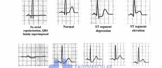
- myocardial ischemia - ECG data show changes in the amplitude, polarity and shape of the T wave in those leads that are associated with the ischemic zone;
- acute myocardial infarction – horizontal displacement of the ST segment upward from the isoline, trough-shaped displacement of this segment;
- necrosis of the heart muscle - irreversible death of myocardial cells is reflected on the ECG graph as a pathological Q wave;
- transmural necrosis - this is an irreversible damage to the entire thickness of the heart muscle wall, expressed in the cardiogram data as the disappearance of the R wave and the acquisition of the QS type by the ventricular complex.
When making a diagnosis, you should additionally pay attention to the symptoms of concomitant diseases . This may include pain in the heart with myocardial ischemia, swelling of the legs and arms with cardiosclerotic changes, signs of heart failure as a result of a heart attack suffered on the legs, hand tremors, sudden weight loss and exophthalmos with hyperthyroidism, weakness and dizziness with anemia.
The combination of such symptoms with diffuse changes detected on the ECG requires an in-depth examination .
Heart rhythm and conduction disturbances in pregnant women. Clinical observation
MM. MANGUSHEVA, T.V. RUDNEVA, S.P. YAKUPOVA, N.G. SHAMSUTDINOVA, L.S. SHAMEEVA
Kazan State Medical University
Republican Clinical Hospital of the Ministry of Health of the Republic of Tatarstan, Kazan
Shamsutdinova Nailya Gumerovna -
Candidate of Medical Sciences, Assistant at the Department of Hospital Therapy of KSMU
420049, Kazan, st. Butlerova, 49, tel. 7 (904) 7638372, e-mail
Rhythm disturbances during pregnancy pose a special medical problem because they can cause disturbances in the functioning of the fetus. The article describes current issues in the diagnosis of heart rhythm disturbances during pregnancy, the possibility of drug and non-drug correction. A demonstration of clinical observation is provided.
Keywords
:
heart rhythm disturbances, pregnancy.
M. _ M. _
MANGUSHEVA , T. _ V. _ RUDNEVA , S. _ P. _ YAKUPOVA , N. _ G. _ SHAMSUTDINOVA , L. _ S. _ SHAMEEVA
KazanStateMedicalUniversity,
Republican Clinical Hospital of the Ministry of Health of the Republic of Tatarstan, Kazan
Disturbances of a heart rhythm and conductivity of pregnant. Clinical study
Rhythm disturbances during pregnancy are the special medical problem as they may cause fetus life activity disturbances. The article describes the current problems of heart rhythm disturbances of pregnant, possibilities of drug and non-drug correction. Is demonstrated a clinical study.
Key words:
heart rhythm disturbances, pregnancy.
Cardiac arrhythmia (CHD) is a change in the basic electrophysiological properties of the heart (automaticity, excitability, conductivity), leading to a disruption in the coordinated contraction of the entire heart or its parts and manifested by a change in frequency, rhythm regularity and conduction of excitation through the conduction system of the heart. Quite often (from 5.1% to 38.7%), practically healthy pregnant women may experience various rhythm disturbances. They represent a serious medical problem for a number of reasons. Firstly, arrhythmias themselves can pose a threat to the health and life of a pregnant woman and fetus. Secondly, the frequency of arrhythmias during pregnancy increases, which is due to significant physiological changes in the mother’s body. Pregnancy itself can act as a proarrhythmogenic factor [1].
The mechanisms of development of rhythm disturbances in pregnant women are associated with functional, hormonal and hemodynamic changes that occur during pregnancy. In pregnant women, the level of estrogen and human chorionic gonadotropin increases several times. In addition, high levels of catecholamines in the blood plasma and increased sensitivity of adrenergic receptors lead to excessive activation of the sympathetic nervous system. All these changes in pregnant women create favorable conditions for the development of arrhythmias.
Diagnosis of cardiac arrhythmias and follow-up during pregnancy do not differ significantly from those in non-pregnant women. Pregnant women with complaints of palpitations, “interruptions” in the work of the heart, as well as healthy pregnant women with asymptomatic arrhythmias detected on the electrocardiogram, should undergo a thorough examination, including 24-hour Holter monitoring of the ECG and, if necessary, an electrophysiological study (transesophageal for the purpose of diagnosis, clarification of the mechanism of tachycardia, it is possible to provide relief therapy).
It is advisable to carry out Holter monitoring over time (at 28-30 weeks, before birth and 2 months after birth). When rhythm disturbances are detected in healthy pregnant women, a more detailed examination is required to exclude, first of all, organic heart pathology. Heart rhythm disturbances most often accompany heart defects, in addition to pathologies of the bronchopulmonary system, thyroid dysfunction, electrolyte disturbances and other pathological conditions.
Of course, analysis of the course of previous pregnancies is important. To diagnose heart rhythm disturbances, as well as the causes that cause them, in the first half of pregnancy, patients should be sent for examination to the cardiology department of a therapeutic hospital, and in the second half of pregnancy - to the pathology department of pregnant maternity hospitals. Pregnant women with a history of arrhythmias, as well as patients in whom rhythm disturbances were detected in previous pregnancies, should be under clinical observation by antenatal clinic therapists.
NRS are found in almost 20% of pregnant women, and most often, according to various authors, supraventricular extrasystoles (SVES) (in 28-67% of cases) and ventricular extrasystoles (VES) (in 16-59% of cases) are detected. NRS is clinically more often asymptomatic and is detected only during routine ECG registration or ECG Holter monitoring [2]. In the vast majority of cases, extrasystolic heart rhythm disturbance is not a contraindication to natural childbirth and does not require drug treatment.
The prescription of antiarrhythmic drugs, primarily cardioselective β-blockers, is indicated for poor subjective tolerability of extrasystole and in pregnant women with high grade extrasystole, prognostically unfavorable.
Reciprocal supraventricular tachycardia is the most common type of tachycardia found in women of childbearing age. This type of heart rhythm disturbances often occurs in young women, is cyclical in nature and is often associated with premenstrual changes in the body. In these women, SVT is likely due to a period of low estrogen levels in the blood.
Regarding the incidence of SVT during pregnancy, data are conflicting. According to the literature, the risk of primary occurrence of SVT during pregnancy increases by 34%, and the risk of developing recurrent SVT by 29%. On the other hand, in women with accessory pathways, episodes of tachycardia are significantly more common compared to AV reentrant tachycardia [3].
The management tactics for pregnant patients with paroxysmal AV tachycardia are the same as for non-pregnant patients. Vagal tests should be used initially. If paroxysm is successfully relieved using vagal tests, no additional antiarrhythmic therapy is required. If tachycardia continues, according to the recommendations of the American Heart Association, the method of choice is intravenous ATP. A retrospective analysis showed the safety and effectiveness of this drug in the second and third trimester of pregnancy. The effectiveness and safety of ATP in the first trimester have not been studied.
In order to timely detect bradycardia, fetal cardiac monitoring is recommended. It is not advisable to administer ATP to pregnant women with WPW syndrome (possible development of atrial fibrillation with a high frequency of ventricular excitations). If ATP is ineffective, intravenous propranolol and metoprolol can be used. Verapamil should be avoided due to its long-term hypotensive effect.
If the paroxysm cannot be controlled with medication, or the patient experiences hemodynamic instability, the method of choice is synchronizing electropulse therapy. To relieve antidromic WPW tachycardia (with wide QRS complexes), it is preferable to use antiarrhythmic drugs that can impair conduction through accessory pathways (propafenone, procainamide).
Radiofrequency ablation is indicated for the treatment of episodes of SVT during pregnancy. Radiofrequency ablation is also the treatment of choice in patients with SVT who are refractory to medical therapy. The operation is recommended to be performed in the second trimester of pregnancy.
Atrial fibrillation and flutter are rarely observed during pregnancy in the absence of organic heart disease or any endocrine disorders. If a woman develops atrial fibrillation or flutter during pregnancy and does not have a history of these episodes, the possibility of congenital heart disease, rheumatic heart disease, or hyperthyroidism should be excluded.
Currently, the number of pregnant women with congenital heart defects is growing. It should be noted that women with congenital heart defects are several times more likely to experience episodes of AF. In most cases of AF paroxysms, women had a history of surgical interventions involving the atria.
Atrial fibrillation often occurs in patients with atrial septal defect and scarring of the atria due to surgical interventions. Management of an episode of AF should be aimed at reducing the heart rate with beta-blockers or digoxin. In many cases, restoration of sinus rhythm occurs spontaneously. If spontaneous rhythm restoration does not occur quickly, electrical cardioversion should be performed within 48 hours. After this period, the issue of prescribing anticoagulant therapy should be decided.
EIT is also indicated for patients with unstable hemodynamics. Fetal monitoring should be performed during and immediately after EIT. Antiarrhythmic therapy with drugs IA- (disopyramide), IC- (propafenone), III- (sotalol) classes is indicated for patients with recurrent atrial fibrillation and flutter to prevent relapses of AF, as well as for patients with hemodynamic disorders.
In addition, patients with chronic AF should be prescribed anticoagulant therapy to prevent cardioembolic complications. However, the teratogenic effect of warfarin during the first trimester of pregnancy has been proven. Unlike warfarin, heparin does not cross the placenta and is the drug of choice in such patients.
For atrial flutter, drug therapy is less effective, and transesophageal pacing (TEPP) or EIT is often required to restore sinus rhythm. For severe paroxysms of AF and AFL that are refractory to drug treatment, it is possible to use RFA, which is most effective for typical atrial flutter.
Ventricular tachycardia (VT) is rare during pregnancy. Most often these are paroxysms that have arisen for the first time.
VT from the right ventricular outflow tract occurs in patients without structural heart disease and is benign. Most episodes of tachycardia occur due to exposure to stress factors and respond well to therapy with beta-blockers. The occurrence of episodes of VT or group ventricular extrasystoles does not depend on the timing of pregnancy. More often, ventricular tachycardia is monomorphic, and 73% of cases originate from the outflow tract.
The number of paroxysms of ventricular tachycardia decreases significantly in the postpartum period in most women. It is assumed that hemodynamic and neurohormonal changes during pregnancy play a large role in the occurrence of VT.
A more serious prognosis for patients in whom ventricular tachycardia occurs against the background of organic heart damage, long QT interval syndrome, Brugada syndrome.
Relief of hemodynamically unstable VT should include the entire range of measures for electrical cardioversion. In patients with episodes of VT not accompanied by hemodynamic instability, lidocaine and procainamide can be used.
The drugs of choice for catecholamine-sensitive tachycardias are β-blockers. Patients with long QT syndrome have a high risk of developing a life-threatening cardiac arrhythmia - torsades de pointes. As previously stated, such patients are advised to prescribe β-blockers during pregnancy and the postpartum period. In some cases, especially to prevent sudden cardiac death, a cardioverter-defibrillator (ICD) may be required. ICVD can be performed both before pregnancy and at any stage of pregnancy using means of maximum fetal protection [1].
The authors observed supraventricular tachycardia in a pregnant woman at 30 weeks' gestation.
Patient M., 27 years old, was delivered on December 30, 2011 via air ambulance to the cardiology department of the Republican Clinical Hospital at 30 weeks of pregnancy with complaints of a feeling of rapid heartbeat. This is the second pregnancy. The first pregnancy and childbirth proceeded without complications. Previously, she was not registered with a cardiologist. According to previous ECG data, the patient had sinus arrhythmia and mild tachycardia.
The feeling of pronounced palpitations appeared two days before admission. At an appointment at the antenatal clinic, supraventricular tachycardia was recorded. The ATP injection did not restore the rhythm. The patient was hospitalized in the cardiology department of the emergency hospital in Nab Chelnov. Conservative therapy was carried out: verapamil 80 mg 3 times a day, polarizing mixture. During the therapy, the rhythm was not restored, and therefore the patient was transferred to the Republican Clinical Hospital.
Upon admission, the patient's ECG showed supraventricular tachycardia with a heart rate of 180 beats. per minute Hemodynamics were stable. After examination and examination, the diagnosis was made: “Impaired heart rhythm: Paroxysmal form of left atrial tachycardia with orthograde blockade with a conduction ratio of 1:1, 3:1.” According to Echo-CS, no organic pathology of the heart was detected.
On 12/30/11, due to the ineffectiveness of drug therapy, radiofrequency ablation surgery was performed. After surgical treatment, sinus rhythm was restored.
On January 3, 2012, the patient had a relapse of tachycardia, the heart rate was in the range of 120—140—180 beats per minute. Then the patient was under observation in the cardiology department and was regularly examined by obstetricians and gynecologists. Attacks of tachycardia with a frequency of 180 per minute were accompanied by shortness of breath and were stopped by infusions of a potassium-magnesium mixture.
At 37-38 weeks of pregnancy, the patient was transferred to the department of pathology of pregnant women for delivery by cesarean section. On the second day after delivery, sinus rhythm was restored, and no recurrence of tachycardia paroxysms was observed.
Conduction disorders (slowdown or complete cessation of the conduction of excitation from the sinus node through the conduction system of the heart) during pregnancy usually do not pose a danger. Even with complete atrioventricular block, especially if it is caused by a congenital or acquired heart defect in childhood, pregnancy and childbirth can proceed normally. A contraindication to pregnancy should be considered complete atrioventricular block with a ventricular contraction rate of less than 40 per minute, as well as the presence of Morgagni-Adams-Stokes syndrome.
A particular problem is solving the issue of the possibility of pregnancy and childbirth in women with an implanted artificial pacemaker (APM). IVR with a constant pacing rate does not allow the heart to flexibly respond to changing hemodynamic conditions during pregnancy, which in some conditions complicates the situation. Implantation of an IVR with an adjustable frequency of stimulation allows you to maintain pregnancy, subject to frequent monitoring of the operation of the IVR.
LITERATURE
1. Oganov R. G., Mamedov M. N. National clinical guidelines for cardiology. - M., 2009. - 392 p.
2. Akselrod A. S., Chomakhidze P. Sh., Syrkin A. L. Holter monitoring of ECG. Opportunities, difficulties, mistakes / ed. A. L. Syrkina. - M.: Medical Information Agency LLC, 2007. - 192 p.
3. Tak T., Berkseth L., Malzer R. A case of supraventricular tachycardia associated with Wolff-Parkinson-White syndrome and pregnancy. —Wisconsin Medical Journal. - 2012. - V. 111 (5). - P. 228-232.
Non-drug treatment
Therapy for any diseases of the heart and blood vessels should begin with lifestyle modification and the elimination of bad habits. The basic principles are:
- Rational nutrition and diet. Diet therapy in the presence of diffuse changes requires excluding fatty, fried, smoked foods from the diet, minimizing animal fats and increasing the consumption of vegetable fats. Refusal of pickled foods and overly seasoned dishes. Particular attention should be paid to the diet of people with high blood cholesterol levels and chronic coronary heart disease.
- Dosed physical activity. Physical activity is very important for the normal functioning of the heart muscle. However, it should be remembered that weightlifting and physical overload cause hypertrophy of the heart muscle, which increases its need for oxygen. Therefore, physical activity should be moderate. Walking in the fresh air, a short jog, and exercise in the morning are enough.
- Quitting alcohol and smoking.
- Correction of blood pressure when it increases by limiting salt and water intake.
- Weight correction for obesity under the strict supervision of a nutritionist.
- Normalization of work and rest schedules, full 8-hour sleep.
Why does the heart contract?
The heart is “started” by an electrical impulse. It is formed from multiple electrical currents from the junction of heart cells.
Every living cell has its own negative electrical discharge inside. The difference between the external and internal voltage on the two sides of the cell membrane is 80-90 mV. This is the transmembrane potential. It does not change throughout life and is characteristic of each type of cell.
But cardiac cells are characterized by a change in potential due to the movement of ions (charged particles of sodium, potassium, calcium) through open tubules. Thanks to them, an electric current arises. It is also called the action potential.
Therapy
The treatment method is determined by a cardiologist after determining the diagnosis. This may be drug therapy or surgery. In addition, traditional medicine can be used as auxiliary medicines.
Treatment must be comprehensive and carried out with an individual approach . It is aimed at the main cause of the development of pathology. In addition, therapy requires restoration of myocardial function.
A heart disease specialist prescribes the necessary vitamin and mineral complexes to the patient.
- Carrying out an ECG for angina pectoris and interpreting the results
When treating a patient, it is necessary to protect him from conflict situations and stress. It is also important to establish proper nutrition.
Drug therapy
If diffuse damage is caused by heart disease, then the following groups of medications may be prescribed:
- beta-blockers (Coriol, Bisoprolol, Corvitol, Nebilet);
- glycosides (Corglicon or Digoxin);
- aldosterone receptor antagonists (Veroshpiron, Eplerenone);
- potassium-based products (Asparkam or Panangin);
- sartans (Mikardis, Kandesar);
- ACE inhibitors (Captopril, Ramipril, Enap).
Medicines are used that normalize the conductivity of the organ, eliminate vasospasm, and ensure the flow of energy to the heart.
Only a specialist has the right to prescribe these medications. Self-medication can lead to aggravation of the situation and deterioration of health.
Other methods
In severe cases, cardiac surgery may be prescribed.
Physiotherapy is also used to treat pathology. Such patients are indicated for sanatorium-resort treatment.
The patient must adhere to a diet.
What is “depolarization” and “repolarization”
The occurrence of an impulse (electric current or action potential) in heart cells goes through two main periods:
- Depolarization - sodium and calcium ions enter the cell and the charge changes to positive. At a certain speed, the depolarization wave is transmitted to neighboring cells and covers the entire muscle. Actin combines with myosin and the heart contracts. The speed of wave propagation depends on the presence of healthy or altered cells (ischemic or scar tissue) in the path of the impulse.
- Myocardial repolarization is a longer period, it is necessary to restore the negative intracellular charge, the flow of potassium ions must leave the cells. This phase determines the accumulation of energy in the heart muscle and preparation for the next contraction. Visible rest actually includes all the biochemical mechanisms of energy production, enzymes and oxygen from the blood are consumed. Until complete recovery is completed, the heart is not able to contract.
The most important mechanism ensuring sufficient action potential is the sodium-potassium pump.
Diagram of depolarization (right) of a cell membrane
Violation of myocardial repolarization can be detected during an electrocardiographic examination by determining the repolarization time.
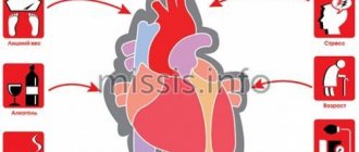
Causes of pathology
The reasons may lie in the following factors:
- Aching pain in the heart: causes and treatment, possible diseases
- myocarditis (inflammatory process in the heart muscle);
- cardiosclerosis;
- angina pectoris;
- cardiac ischemia;
- heart failure;
- a disrupted metabolic process in the organ, which can be provoked by various factors;
- autoimmune neuropathy;
- water-salt imbalance in the body;
- hormonal changes;
- dystrophic damage to the myocardium;
- chronic diseases;
- impaired function of the endothelium (layer of blood vessels);
- use of certain medications;
- alcohol abuse;
- use of narcotic substances.
In addition, the development of such a process can be caused by past diseases, for example, tonsillitis, influenza, typhoid, diphtheria, scarlet fever, measles and other infections.
Another reason for this condition is a deficiency of substances necessary for the body, as well as deviations from the norm in glucose levels. Slight changes in the heart muscles are also caused by advanced age.
Signs of SRS
There are no characteristic clinical signs of early ventricular repolarization syndrome. There are only specific changes on the ECG:
- ST segment and T wave changes;
- in a number of branches, the ST segment rises above the isoline by 1-2-3 mm;
- often ST segment elevation begins after the notch;
- the ST segment has a rounded shape and directly passes into a tall positive T-wave;
- the convexity of the ST segment is directed downwards;
- The base of the T wave is wide.
Basic diagnostic methods
After a thorough and scrupulous conversation with the patient, the doctor refers him to additional examination methods. The main methods to establish a diagnosis are as follows:
- Electrocardiography (ECG) involves recording the conduction of impulses in the heart muscle by applying electrodes.
- Holter monitoring is a 24-hour electrocardiogram, on which the patient, using a special button, can also record the time of occurrence of unpleasant sensations (shortness of breath, chest pain, etc.).
- ECG with bicycle ergometer is a method of recording an ECG with a load on a bicycle ergometer.
- Echocardiography is a study of the heart using ultrasound.
Kinds
There are two options for SRR:
- without damage to the cardiovascular and other systems;
- involving the cardiovascular and other systems.
From the point of view of the nature of the course, a distinction is made between transient and permanent SRGC.
Based on the localization of ECG signs, doctor A.M. Skorobogaty proposed the following classification:
- Type 1 – with a predominance of signs in leads V1-V2;
- Type 2 – with a predominance in leads V4-V6;
- Type 3 (intermediate) – without a predominance of signs in any leads.