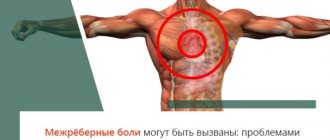
What diseases provoke a pressing condition in the chest in the middle?
Pain behind the sternum in the middle can be one of the symptoms of obesity, atherosclerosis, anemia and other diseases that, at first glance, have nothing to do with the thoracic area. Let's look at what diseases provoke pressing pain in the sternum.
Angina pectoris
This is a serious form of coronary heart disease. Pressing pain in the chest occurs as a result of decreased oxygen supply to the heart muscle. The condition is primarily caused by atherosclerosis of the coronary arteries that supply blood to the heart.
In case of narrowing of the artery due to the atherosclerotic process, during physical activity it does not allow enough blood to pass through, therefore, oxygen deficiency occurs. This causes a feeling of tightness in the chest.
Discomfort occurs during tension or physical activity. Does not last long, disappears 3-5 minutes after the end of physical activity. If the disorder is not treated, the narrowing continues, the pressure in the left chest worsens (pain occurs with less effort or even at rest).
At any stage of angina pectoris, a blood clot can form in an artery damaged by atherosclerosis, completely clogging the coronary vessel. Myocardial infarction occurs.
Pericarditis
Pericarditis is an inflammation of the pericardial sac, the pericardium. It is a membrane surrounding the heart. When inflammation of any etiology occurs (infection, lupus erythematosus, rheumatism, oncology, etc.), pressing pain occurs.
The pain is not as pronounced as with a heart attack, but it is often difficult to distinguish pericarditis from a heart attack. In differential diagnosis, ECG or echocardiography is used.
Pleurisy
When the pleura is inflamed, it also puts pressure on the chest. The typical pleural nature of the pain is pain associated with breathing, worsening with inspiration.
Inflammation of the endocardium and heart valves
Inflammation can affect different parts of the heart wall. When the internal mucous membrane, the endocardium, is inflamed, we are talking about endocarditis. The heart valves are often affected. Pain in the middle of the sternum is similar to discomfort with other inflammations (pericarditis, pleurisy), the pain syndrome is moderate.
Myocarditis
With inflammation of the heart muscle (myocarditis) of any etiology, pain occurs in the chest in the middle or on the left. Heart rhythm disturbances and signs of heart failure may also be present.
Reflux disease
With reflux, pain radiating into the chest is caused by the return of stomach contents to the esophagus (gastroesophageal reflux).
Typically, the esophagus is separated from the stomach by a muscular sphincter, allowing food to pass from the esophagus to the stomach, but not back. If the sphincter does not work properly, stomach acid can back up into the esophagus and damage the walls. This is accompanied by heartburn, sudden pain in the chest. Discomfort occurs after eating; antacids (alkaline substances that reduce the acidity of the stomach contents) help to cope with it.
Sometimes the soreness is “just” unpleasant, sometimes it is so severe that it resembles a myocardial infarction, including an unpleasant lump in the throat .
If a heart attack is suspected, it is recommended to seek immediate medical attention to rule out an acute condition.
Esophageal motility disorder
In case of muscle disorders of the esophagus, there is also often pressure on the chest. This symptom is typical of abnormally increased peristalsis.
Respiratory tract infections
Pain from inflammation of almost all respiratory tracts can radiate to the chest. Pain is often found with pneumonia and is accompanied by fever and cough (dry or productive).
The diagnosis is confirmed by chest x-ray and other examinations. An important blood test showing the presence of an inflammatory process in the body.
Spine pain
Pressing pain is a symptom of problems with the thoracic spine and its nerves. The peculiarity of the pain is a stabbing nature, often radiating from the back to the chest. She is not dangerous, but very unpleasant.
Abdominal pain
Some abdominal disorders present with pain in the middle of the chest. Such diseases include acute pancreatitis and biliary colic.
Shingles
Shingles is an infection caused by the chickenpox virus that affects the nerves leading from the skin and subcutaneous tissue.
The disease can affect almost any part of the body, including the scalp, face, and the typical location is the chest area. In the first stages of the disease, burning or pressing pain of varying intensity occurs.
Pneumothorax
This is a condition in which air enters the pleural cavities. There are two pleural cavities, one around each lung. These cavities are lined with a membrane, pleura, and have a vacuum. It is this vacuum that allows the lungs to open when inhaling.
Spontaneous pneumothorax is relatively common. It usually occurs in completely healthy young people. When part of the lungs bursts for a certain reason, air from them enters the pleural cavity.
A depressed lung cannot open during breathing. This condition is associated with sudden pain in the chest on the left or right, depending on the location of the lesion; the person has difficulty breathing and feels dizzy.
With extensive pneumothorax, in addition to pain in the chest on the left or right, shortness of breath is also present. The condition is determined by listening with a phonendoscope, when breathing is not heard above the pneumothorax (the lung does not contract), the final diagnosis is confirmed by an x-ray of the lungs.
While a smaller pneumothorax is not treated (air is absorbed on its own), a large one requires pressure on the pleural cavity.
Special, acute conditions in which there is pressure in the chest
The most common acute conditions are those in which there is pressure in the middle of the sternum.
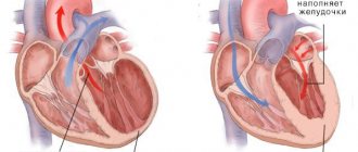
Myocardial infarction
A heart attack occurs when a vessel that supplies oxygen to the heart muscle suddenly becomes blocked, stopping the flow. Due to lack of oxygen, necrosis of part of the muscle occurs.
During a heart attack, pain manifests itself in the chest, in the area of the heart, persists for more than 20 minutes, and can radiate to the upper limbs, neck, lower jaw, back, epigastrium (abdominal area).
Pressing pain is accompanied by sweating, pallor, and anxiety. There may be shortness of breath, a lump in the throat, and rapid heartbeat.
The feeling of tightness during a heart attack, unlike angina, does not go away with rest and does not respond to taking nitroglycerin. If such pain occurs, you must immediately seek medical help and take care to minimize the load on the heart.
Aortic aneurysm
Sharp pain behind the breastbone in the middle, shooting into the lower part of the body, especially the stomach or back, may indicate an aortic aneurysm. This is a very serious and life-threatening condition. Requires immediate medical attention. The pain is intense, medications do not help.
Broken ribs
Fractures usually occur due to injuries to the thoracic area. The pain is very unpleasant and worsens with inhalation.
Pulmonary embolism
This is a condition in which a blood clot forms in a vessel (usually in the vessels of the lower extremities).
If a lump, an embolus, breaks off, it enters the lungs with the bloodstream, where it blocks the pulmonary artery and disrupts the flow of blood through part of the pulmonary bed.
A large clot that blocks a pulmonary vessel can cause sudden death. Smaller blockages of the pulmonary system may manifest as pain in the chest area associated with a dry cough and shortness of breath.
Diagnosis and treatment
The diagnosis is clarified using laboratory and instrumental research methods.
The diagnostic complex includes:
- X-ray with or without contrast.
- Endoscopic examination with collection of biopsy material.
- Fecal occult blood test.
- Esophageal manometry.
- pH-metry.
- Impendancemetry.
If damage to the esophagus is not confirmed, then an underlying disease of extraesophageal origin is sought. An ultrasound of the thyroid gland and heart is required. An ECG is taken. Blood is taken for markers of rheumatological pathology, as well as thyroid hormones.
After diagnosis, treatment is started. Treatment depends on the nature of the pathology. First of all, medications and diet are prescribed.
A healthy diet is an important part of treatment. The diet allows you to relieve the esophagus, reducing the functional load.
During treatment exclude:
- baked goods;
- fatty, hot, spicy;
- mushrooms;
- soda;
- smoked meats;
- canned foods;
- alcohol;
- tea and coffee.
You should eat in small portions. Watch your posture while eating. Each piece must be chewed thoroughly.
Drug therapy:
- Proton pump inhibitors, for example, Omez or Ezolong.
- Agents that stimulate peristalsis (Domrid).
- Antacids. These include Phosphalugel, Silicea-Gastrogel.
- H2 blockers. Kvamatel, Famotidine are used.
Medicines must be prescribed by a doctor. It is prohibited to treat a lump in the throat on your own.
Traditional medicine is used as an adjuvant therapy. Healing herbs have a strong anti-inflammatory and healing effect. To achieve the effect of such treatment, it is necessary to adhere to the frequency of administration and the rules for preparing medications.
Recipes for folk remedies:
- 1 tsp. St. John's wort, mint, chamomile flowers, pour a glass of boiling water and leave for 15 minutes. Drink 1/3 glass 3 times a day.
- 1 tbsp. dry nettle pour a glass of boiling water. Leave for 1 hour. Drink 1 tbsp. 5-6 times a day.
- 1 tsp. Drink sea buckthorn oil 30 minutes before meals 3 times a day.
- 1 tsp add aloe juice to 2 tbsp. liquid honey. Take 1 tsp. per day on an empty stomach.
To prevent gastrointestinal diseases, it is necessary to lead a healthy lifestyle and undergo regular medical examinations. A lump in the esophagus is a signal that you should take care of the health of your nervous system.
What to do, what examinations need to be completed
Basic methods of investigating the causes of chest pain, including a thorough history and physical examination, provide 90% of success towards the correct diagnosis.
Increased body temperature, accompanying a feeling of tightness, is common with pneumonia, acute myocardial infarction, pericarditis, and herpes zoster. Pain in the chest along with weakening of the pulse is characteristic of dissection of the aorta or aneurysm.
Enlarged jugular veins occur in congestive heart failure, sometimes occurring as a complication of an inferior heart attack or pulmonary embolism, when the blockage accounts for more than 60% of the blood supply to the lung.
Pain in the middle of the chest, palpable on palpation, indicates a musculoskeletal cause. Unilateral discomfort in the thoracic region is a sign of damage to one or more adjacent ones involving the spinal or peripheral nerve (herpes zoster).
Auscultation, in which the doctor listens to the chest, is an important part of the physical examination. Auscultation findings:
- weak one-sided breathing, in which pressure on the chest, raises suspicion of pneumothorax, local attacks of auditory detection - effusion;
- tubular breathing is heard over the inflamed area, sometimes over the effusion;
- limited areas with audible crepitus in the chest are present with prolonged lobar pneumonia, enlarged areas accompanied by pain in the heart indicate either multilobar inflammation or pulmonary edema due to left ventricular failure after acute myocardial infarction (or heart failure);
- with pericarditis and pleurisy, a rustling sound occurs (pericardial sounds are heard only when you hold your breath).
The main tests aimed at determining the cause of discomfort behind the sternum in the middle include a blood test. With pneumonia, leukocytosis is detected (changes in the cellular composition of the blood), which may be present to a lesser extent in acute myocardial infarction. Cardiac-specific markers increase within 6 hours after myocardial infarction.
Chest examinations that reveal pathologies that accompany pressing pain in the sternum:
| Diagnostic measures. | What is expected during the examination. |
| History, physical examination. | Musculoskeletal and skin diseases. |
| ECG, O2 saturation. | Cardiac arrhythmia, myocardial infarction, pulmonary embolism, hypoxic diseases. |
| Elevated troponin T. | Acute myocardial infarction, myocardial necrosis of another etiology. |
| D-dimer. | Rule out pulmonary embolism. |
| X-ray. | Pneumonia, pneumothorax, damage to the ribs and vertebrae. |
| Echocardiography. | Aortic dissection, delayed pulmonary embolism, delayed acute myocardial infarction, aortic stenosis. |
| CT, angio computed tomography | Pulmonary embolism, aortic dissection, pneumothorax, pneumonia, pleural disease, rib injury. |
| Stress tests, coronary angiography, gastroscopy, psychiatric examinations | Other causes (for example: cardiac neurosis, cardiac phobia) |
What else to read:
- Drug treatment of reflux esophagitis: drugs, treatment regimens Contents of the article:1 Antisecretory drugs1.1 Proton pump inhibitors1.2 H2-histamine receptor inhibitors1.3 Antacids and alginates2 Prokinetics3 Cytoprotectors4 Drugs for symptomatic treatment and vitamins5 How long does treatment last6 Treatment7 Treatment regimen......
- What to do if you have pain in the esophagus when swallowing? Contents of the article:1 Features of pain2 Causes of pain when swallowing2.1 Reflux esophagitis2.2 Traumatic injuries and burns2.3 Foreign bodies in the esophagus2.4 Peptic esophagitis2.5 Pregnancy2.6 Hernia2.7 Other possible causes of pain when swallowing3……
- Causes and treatment of fungal infections of the esophagus Contents of the article:1 What is candidiasis of the esophageal mucosa2 Causes and risk factors3 Degrees of severity and symptoms4 What is dangerous about the disease5 Diagnostic methods6 How to treat candidiasis of the esophagus6.1 Fighting the fungus……
Which doctor should I contact?
If you have pain behind the sternum in the middle, it is best to immediately consult a therapist. He will examine the chest and, in accordance with the initial findings, will refer you to a specialized specialist:
- cardiologist (suspected heart disorders or diseases);
- neurologist (neurological etiology of chest pain);
- psychotherapist (psychological reasons);
- gastroenterologist (digestive tract diseases);
- pulmonologist (pulmonary diseases);
- vertebrologist (vertebrogenic causes).