An electrocardiogram is the most important diagnostic technique that allows you to assess the functioning of the heart, as well as timely diagnose cardiac diseases. The key to reliable and comprehensive information is strict adherence to the algorithm for performing the procedure. This includes, first of all, the normal functioning of the device itself, the technique of placing electrodes, as well as the features of their application.
An electrocardiographic study records the electrical fields arising during the life of the heart on paper or a display, it depends on the type of device, a record is issued.
With the help of an ECG, the doctor can determine the further type of adequate therapy
There are three main components of an electrocardiograph:
- input devices, which include the electrodes themselves and the cable;
- amplifiers;
- sensor.
ECG recording algorithm
For correct diagnosis, making an accurate diagnosis and prescribing optimal treatment, it is important to adhere to the following algorithm of actions, namely:
- preparing the patient for the procedure;
- checking the operation of the device;
- application of electrical transmission gels and antiseptic liquids;
- adherence to clear rules on where to place electrodes depending on color;
- selection of lead registration scheme;
- directly recording.
In most cases, the electrocardiogram is recorded in a separate room in a medical facility, but sometimes it can even occur at home, in a ward, or in an ambulance. The room in which the procedure will be performed must be at a sufficient distance from possible electrical interference. As for the couch itself, on which the patient lies, it is located approximately two meters from the power supply.
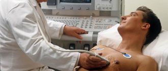
An ECG is an absolutely painless procedure that does not cause absolutely any negative emotions
The procedure is carried out in a lying position, with free access to the upper torso, legs and arms. If there are contraindications, then an ECG can be performed in a sitting position. Before an electrocardiogram, it is better not to engage in intense physical activity, and also not to take food or drinks that could activate the heart.
Influencing factors
Although the ECG technique has a fairly clear algorithm, some errors cannot be ruled out.
- The appearance of interference from inrush currents of the electrical network (irregular oscillations with a frequency of 50 Hz).
- Fluctuations caused by poor contact of the electrode with the skin.
- Record muscle tremors if the patient is anxious or cold.
- Incorrect application of electrodes. Mostly, the electrodes on the upper limbs are swapped and signals of unusual polarity are received.
- Record additional leads without switching the recorder from enhanced leads to lead V.
- Failure to insert a roll of thermal tape into the device, as a result of which the drawing mechanism can slow down and affect the width of the recorded complexes.
Rules for applying electrodes for ECG
I would like to immediately note that the electrodes for the ECG are located in different places. What is this connected with? This is the only way to catch all the heart impulses. There may be disturbances in heart rate and rhythm in different places.
The electrode application scheme includes the following:
- degreasing the skin with alcohol;
- abundant hair is treated with a soap solution;
- if the electrodes are disposable, then for high-quality recording of heart impulses it is necessary to remove the hair;
- Although some people use gauze, it is better to use conductive paste. As a last resort, you need to take saline solution.
ECG electrodes can be disposable or reusable
I would like to make a special mention regarding disposable electrodes. They are not included in all kits; most likely, they can be found in the latest brands of the device. According to experts, such electrodes are simple and easy to use, and also help reduce the preparation period for the patient before the procedure. They are usually used by private clinics. They are made of foil, which reduces the likelihood of disease transmission. In addition, disposable electrodes improve contact due to better conductivity. They are often used in cases where a cardiogram needs to be taken quickly, as well as for hyperhidrosis.
If we are talking about reusable electrodes, then we cannot fail to note their durability; of course, they are stronger. They are produced in the form of suction cups and clothespins. The signal passage is a little difficult, unlike the above mentioned electrodes for reusable use of a rougher design.
Stages of the ECG procedure
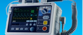
Electrocardiography is a research method in which the electrical fields of the heart are recorded, provoked by contractions of the heart muscle. As a result, the specialist receives a cardiogram - an image of curve graphs. They can be displayed on a monitor or recorded on a special paper tape. To carry out this type of diagnosis, you need professional medical equipment - a cardiograph. This device consists of three required elements.
- An input device including electrodes, lead cables and their switches.
- Signal amplifier - allows you to amplify them to the value necessary for analysis.
- Recording unit for direct recording of a cardiogram.
When conducting an ECG, a specialist works sequentially with all these blocks, following a clear algorithm. It, in turn, is divided into several stages:
- preparing the room, equipment and patient for ECG;
- application of electrodes and connection of leads according to a specific scheme;
- adjusting the gain to correctly display the signal on the cardiogram;
- direct recording of the electrocardiogram.
For informative and reliable research results, it is necessary to avoid errors at each of these stages. Therefore, let’s take a closer look at their algorithms and features.
Electrode locations
Devices must be applied to the following areas of the body:
- lower limbs: right and left leg (black and green electrode, respectively);
- upper limbs. Red on the right hand, yellow on the left;
- rib cage.
Chest electrodes are installed according to the following scheme:
- 1 lead. On the right side at the level of the fourth intercostal space;
- 2 lead. On the left side – the fourth intercostal space;
- 3 lead. Between the first and second;
- 4 lead. Fifth intercostal space on the left side;
- 5 lead. The same line goes, only along the anterior premuscular region;
- 6 lead. Corresponds to the left premuscular zone.
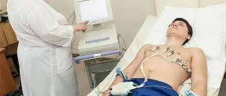
Despite the fact that an electrocardiogram seems to be a simple manipulation, to obtain correct results it is important to adhere to medical recommendations
In order to obtain reliable information about the functioning of the heart, you must adhere to the following recommendations:
- the day before you should have a good rest both physically and emotionally;
- Before the procedure, a light breakfast is allowed a maximum of two hours;
- strong coffee and tea, alcohol, smoking, energy drinks - all this is prohibited on the day of the study;
- if possible, it is necessary to reduce the amount of hygiene products, because they impair conductivity due to the formation of a fatty film;
- the room in which the study is carried out must be warm, the patient must be comfortable, but if physiological tremors are present, this will certainly affect the results.
Fixation of electrodes on the limbs
On the legs, a pair of electrodes are fixed on the inside of the lower leg at the very bottom. It must be remembered that a grounding sensor is attached to the right leg. On the hands, electrodes are attached to the forearms.
With standard leads, indicators are taken as follows:
- left and right hands - first position;
- right arm and left leg - second position;
- left leg and arm - third position.
Thus, an Einthoven triangle is obtained, each side of which corresponds to one and possible leads.
Enhanced leads involve collecting a signal using one electrode, which records the potential difference between the area where it is applied and the conventional electrical zero. They have the following designations:
- right hand - aVR;
- left hand - aVL;
- left leg - aVF.
Carrying out an ECG according to Slopak
The methodology is not very different from standard research. In this case, not six, but nine chest leads are used. The first six leads are also attached, and the next ones are presented below:
- 7 lead. Fifth hypochondrium, axillary region;
- 8 lead. Scapular line at the same level;
- 9 lead. Paravertebral line.
Electrocardiogram according to Slopak allows you to get a more detailed picture of the heart’s work
ECG by Sky
This type of research also differs from the standard technique in the placement of electrodes. The bottom line is that electrodes are placed on the chest area in three places, namely:
- 1 red lead is placed on the right side at the level of the second intercostal space;
- 2 yellow lead is installed on the left at the same level only along the posterior axillary line;
- Green lead 3 is the midclavicular line on the left side of the sternum.
As a result, a kind of triangle is created, the corners of which will serve as projection sites for the leads of the electrical activity of the heart. As for the indications, this technique is carried out in case of suspected heart attack, as well as for athletes during sports.
To increase the information content of the method, medication and stress tests are carried out. To identify areas of hidden ischemia, the test may be done after physical activity, such as squats, or after taking certain medications.
Sky leads will help identify coronary heart disease
Fixation of electrodes in chest leads
If a single-channel study is carried out, then only one electrode is used. But in modern ECGs, for more accurate diagnosis, in most cases, 6 are used. They are designated by the Latin letter V and are superimposed at the following points:
- V1 - at the right edge of the sternum in the 4th intercostal space, the red wire is connected;
- V2 - also in the 4th intercostal space, but on the left edge of the sternum, wire color - yellow;
- V3 - in the middle between the 4th and 5th intercostal spaces along the left line, connect the green cable;
- V4 - along the midclavicular line in the 5th intercostal space, wire color - brown;
- V5 - along the anterior axillary line in the 5th intercostal space, connected to a black cable;
- V6 - in the middle of the axillary line at the same level with V450m, the wire color is blue (possibly purple).
Long registration
Data on the electrical work of the heart can be recorded over several hours or even days. Why is this necessary? Thus, doctors can identify transient disturbances in heart rhythm, and also compare the identified deviations with the patient’s activity during the day and his complaints of pain.
Let's consider the indications for long-term monitoring:
- complaints from patients about interruptions in the functioning of the heart, as well as a sensation of short-term palpitations, the interpretation of which is difficult on a conventional electrocardiograph;
- complaints in which angina pectoris can neither be excluded nor confirmed;
- the occurrence of attacks of weakness, fainting, dizziness, the cause of which has not been established;
- monitoring the functioning of an artificial heart pacemaker;
- IHD, as a control and identification of a type of arrhythmia that is asymptomatic;
- as a monitoring of the effectiveness of drugs and to identify undesirable effects on cardiac activity.
Where to put children's electrodes?
An ECG of the heart helps to identify such pathological processes in children: defects, myocardial infarction, angina pectoris, etc. The study is easy to conduct and accessible. The manipulation can be carried out before planned surgical interventions, when heart murmurs are detected, before entering a sports section or school.
During the procedure, the child must behave calmly, so it is necessary to prepare him and explain that installation of the sensors is absolutely painless
The specialist installs nine to twelve electrodes on the child’s body: in the ankles, wrists and on the chest. Sensors of smaller sizes and special shapes are used, since children have more delicate skin. It is best to use electrodes for one-time use.
So, an electrocardiogram is an informative diagnostic method used for both adults and children. The procedure is simple, accessible, and does not cause any discomfort. An ECG will help identify serious cardiac disorders and determine the correct treatment. To obtain accurate results, it is important to adhere to the recommendations regarding preparation and the manipulation itself. Early diagnosis is the key to a speedy recovery!
Home visit
Modern devices for taking ECGs are compact enough to call a cardiologist to conduct an examination at home. Portable devices have the same accuracy class as stationary devices in diagnostic and treatment institutions. As a rule, disposable electrodes are used with the device. In addition, usually a doctor who arrives to a patient’s call can decipher the cardiogram on the spot, which is very convenient and sometimes vital for seriously ill patients with acute chest pain and children.
The portable electrocardiograph can be six or twelve-channel, which fully meets diagnostic needs
The electrocardiography method allows you to evaluate the activity and condition of each part of the heart. Determine the areas of ischemia and the threat of infarction, the area and localization of the lesion, atrial and ventricular rhythm disturbances. This is a painless and fairly simple method that has high informative value and high patient adherence.