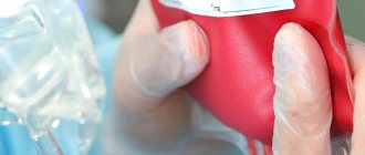
In medicine, blood transfusion is called blood transfusion. During this procedure, the patient is injected with blood or its components obtained from a donor or from the patient himself. This method is used today to treat many diseases and to save the lives of people in various pathological conditions.
People tried to transfuse the blood of healthy people to the sick back in ancient times. At that time there were few successful blood transfusions; more often such experiments ended tragically. It was only in the twentieth century, when blood groups (in 1901) and the Rh factor (in 1940) were discovered, that doctors were able to avoid deaths due to incompatibility. Since then, transfusion has not become as dangerous as before. The method of indirect blood transfusion was mastered after they learned how to store the material for future use. For this, sodium citrate was used, which prevented coagulation. This property of sodium citrate was discovered at the beginning of the last century.
Today, transfusiology has become an independent science and medical specialty.
In what cases is blood transfusion performed?
Whole blood cannot be transfused; it is possible to use only its components, such as fresh frozen red blood cells and platelet concentrate. One of the main indications for blood transfusion is a hemoglobin level of less than 70 g/l and a decrease in saturation (oxygen saturation of the blood) to 80%. The procedure is necessary for the disintegration of massive tumors; the disintegration process is accompanied by chronic blood loss. Usually these are tumors of the uterus, vagina, and cervix. And such an oncological problem as melanoma has a depressing effect on the red blood cell, in this case, in order to carry out chemotherapy, it is necessary to restore the normal level of red blood cells and hemoglobin, so a blood transfusion is performed.
The need for a transfusion of fresh frozen plasma is due to profuse edema and the presence of a state of suppressed hematopoiesis; this transfusion is also carried out to prevent the development of disseminated intravascular coagulation syndrome with a sharp inhibition of blood clotting.
Blood transfusion
The bulk of blood and plasma for transfusion and preparation of various therapeutic drugs is obtained from donors.
Donors are people who give their blood for medicinal purposes. In the USSR there is a unified national blood transfusion service.
Free donors are the most numerous category of donors who, due to their high consciousness, fulfilling their civic duty, voluntarily and freely give their blood. Donors can be healthy people aged 18 to 55 years. Persons suffering from infectious diseases, malignant tumors, heart defects, arrhythmias, gastric ulcers, acute rheumatism, disorders of the nervous and endocrine systems, and dystrophy cannot be donors.
In addition to donor blood, waste, placental, retroplacental, fibrinolysis (post-mortem) and autologous blood are used, i.e. the blood of the recipients themselves, which can be prepared several days before surgery or collected from sterile serous cavities of the body, where it is poured during some internal bleeding.
Recipients are people (sick people) who receive blood transfusions.
Most often, a transfusion of canned blood is performed, to which a stabilizing substance (sodium citrate, heparin) and a preservative mixture containing glucose, antiseptic substances and antibiotics are added.
Blood preserved with glucose-citrate mixtures can be stored at a temperature of +4 to +6 ° C for 2 weeks, maximum up to 3 weeks, after which it cannot be transfused, but can be used for the preparation of some medicinal preparations.
Erythrocyte mass can be stored for 7-23 days, leukocyte mass - up to 48 hours, platelet mass - only for 12-18 hours. Native plasma can be stored for no more than 5 days. Specially prepared and preserved blood can be stored at temperatures from -8 to -12°C for up to 100 days. Red blood cells freshly frozen at ultra-low temperatures (-196°C) can retain their properties for several years. Dried plasma, thrombin and fibrinogen are stored for 3-5 years.
There are absolute and relative indications for blood transfusion.
Absolute indications are: 1) life-threatening or fatal blood loss; 2) traumatic shock; 3) severe anemia not associated with blood loss.
Relative indications for blood transfusion are various conditions and diseases in which the administration of blood may be beneficial.
A deficiency in the patient’s blood of its individual parts serves as an indication for transfusion of various blood components (erythrocytes, leukocytes, plasma, protein, etc.).
Contraindications to blood transfusion can also be absolute or relative.
An absolute contraindication is intolerance to transfusion with sharply increased sensitivity to its components.
Relative contraindications are: 1) severe hepatitis and cirrhosis of the liver; 2) severe nephrosonephritis, amyloidosis, severe renal failure; 3) severe heart failure with symptoms of edema, ascites, liver enlargement, cyanosis of the skin and mucous membranes; 4) lung diseases with pronounced stagnation in the pulmonary circulation; 5) active tuberculosis in the infiltrate stage; 6) some allergic diseases, such as eczema.
If there are vital indications, blood transfusion should be performed, despite the existing relative contraindications.
Blood transfusion can be safe only when it is carried out taking into account compatibility by group, Rh factor and other factors. Therefore, it is necessary: 1) to establish the recipient’s blood group and Rh factor; 2) establish the group and Rh factor of the donor blood; 3) establish the suitability of blood for transfusion; 4) conduct a test for individual compatibility; 5) conduct a test for Rh compatibility; 6) conduct a biological test.
The above studies are carried out by a doctor, but the nurse must be sufficiently oriented in the technique of conducting them.
Blood groups and their compatibility
. It has been established that human tissue cells, including erythrocytes, may contain innate antigens - agglutinogens, designated A and B, and in blood plasma or serum - innate antibodies - agglutinins, designated α (alpha) and β (beta). It turned out that agglutinogen A has several varieties, and then it was discovered that there are a number of agglutinogens in erythrocytes. Depending on the constantly occurring combination of agglutinogens and agglutinins in the blood, all people are divided into 4 groups.
First group
blood is designated as 0(I). It does not contain agglutinogens in erythrocytes, and in plasma it contains both agglutinins α and β.
Second group
blood is designated as A (II). It contains agglutinogen A in erythrocytes, and agglutinin β in plasma.
Third group
blood is designated as B (III). It contains agglutinogen B in erythrocytes, and agglutinin α in plasma.
Fourth group
blood is designated as AB(IV). It contains both agglutinogens, A and B, in erythrocytes, and does not contain agglutinins in plasma.
Among the population of the USSR, 0(I) blood group occurs in 35.2%; A(II) - in 36.7% B(III) - in 20.2% and AB(IV) - in 7.9%.
When blood of different groups is mixed with related agglutinogens and agglutinins, i.e. A with α or B with β, agglutination occurs - the gluing of red blood cells into strong conglomerates, clearly visible to the eye in the form of grains or grains of sand. After agglutination, the red blood cells in such a conglomerate are destroyed and hemoglobin comes out of them, i.e., hemolysis occurs. The same thing happens in people's blood when they are transfused with incompatible blood. In this case, a severe complication develops - blood transfusion shock, which can result in death. This complication can only be avoided by transfusion of same-group blood or small amounts of other group (500-700 ml) compatible blood (the so-called Ottenberg rule, which is illustrated in Fig. 16). In this case, the agglutinins of the injected (donor) blood are highly diluted and therefore are not capable of causing dangerous agglutination of the recipient’s red blood cells. However, when transfusing a large amount of compatible blood of a different group (2000-3000 ml or more), agglutination of the recipient's red blood cells by agglutinins of the transfused blood may occur and transfusion shock may develop, so it is safe to transfuse only blood of the same group. Transfusion of compatible blood of a different group is possible only for health reasons in the absence of blood of the same group.
16. Scheme of compatible blood transfusions
Blood group determination
. Determining a blood group means establishing the presence or absence of agglutinins and agglutinogens in it using an agglutination reaction.
To determine agglutinogens, standard isohemagglutinating (hemagglutinating) sera are used, and standard isohemagglutinating (hemagglutinating) erythrocytes are used to determine agglutinins. More often, blood type is determined using standard hemagglutinating sera - a simple reaction, and if doubt arises, standard hemagglutinating erythrocytes are additionally used (crossover method).
Hemagglutinating serum must be stored at a temperature of +4 to +6 ° C and have a titer of at least 32, i.e., even being diluted 32 times, it must still give a clear agglutination of the erythrocytes of the corresponding blood. The serum of each group is colored in a specific color:
On a specially marked white plate (cassette or cup) the locations of serums of all four groups are indicated. After that. At each mark, 2 drops of hemagglutinating sera from two series of each group are applied. You must first ensure that the hemagglutinating serum is suitable for use. Do not use rotten, contaminated and dried serums, as well as serums that have expired. Serum is collected from ampoules or vials using eye pipettes. Each bottle should have its own pipette. It is important not to confuse or mix serums.
To determine group affiliation, blood is taken from the pulp of the nail phalanx of a finger or earlobe, which is first wiped with alcohol. Individual scarifiers or needles are used for injection. You cannot use the same needle on more than one person. The skin is punctured to a depth of about 1.5 mm. The released drop of blood is introduced into a drop of serum using the corner of a glass slide and lightly stirred. Each time, blood is added to the drops using different angles of the glass slide. To determine the group, you can obtain blood from a vein or a gauze ball soaked in it during surgery. The volume of a drop of blood should be 5-10 times less than a drop of serum. The results of the sample can be assessed no earlier than 5 minutes after mixing it with the serum. Determination of blood type should be carried out in good lighting and at an ambient temperature of +18 to +24°C. Depending on the blood group, the following results can be obtained (Table 6).
Table 6. Reaction results when mixing blood of different groups with standard hemagglutinating sera
Note
. The + sign indicates the presence of agglutination, the sign indicates its absence.
If the blood being tested belongs to group 0(I), then agglutination is not observed in any of the samples, since red blood cells of group 0(I) do not contain agglutinins, so agglutination is impossible.
If the blood belongs to group A (II), then agglutination occurs only in sera of groups 0 (I) and B (III), which contain agglutinin a, which is related to agglutinogen A introduced with erythrocytes.
If the blood belongs to group B (III), then agglutination will occur in the sera of groups 0 (I) and A (II), since they contain agglutinin β, related to agglutinogen B, administered together with red blood cells of group B (III).
If the blood belongs to group AB(IV), then agglutination is observed in the sera of three groups 0(I), A(II) and B(III). Only group AB(IV) serum will not exhibit agglutination since it does not contain the related agglutinins, while all other sera contain agglutinin α or β, or both.
When determining a blood group, errors are possible depending on technical errors, the use of low-quality sera, or the biological characteristics of the blood being tested. In this case, it is possible to mistakenly detect agglutination where it should not be, or vice versa - not to detect agglutination where it should occur.
Technical errors may include confusion and mixing of sera of different groups with each other, taking too large a drop of blood, non-compliance with the sample temperature, premature conclusion about the results of the reaction (earlier than 5 minutes), drying out of blood samples and hemolysis of red blood cells under the influence of a hypotonic solution.
Poor quality sera also lead to erroneous conclusions, since they either do not cause agglutination or can produce it with all samples. Whey may lose activity during long-term storage, overheating or the development of microorganisms in it.
The biological characteristics of the blood itself also lead to errors. There is a phenomenon of panagglutination, which consists in the appearance of agglutination in all samples. It is extremely rare in healthy people. The appearance of “coin columns” is possible - one of the variants of false agglutination. To exclude false agglutination, add 2-3 drops of isotonic sodium chloride solution to the sample and warm it to +22...+24°C. Under such conditions, only true agglutination can be preserved. Infected blood also produces agglutination in all samples.
Determining the blood group by a double reaction involves, in addition to determining it using standard hemagglutinating sera, a repeated determination using hemagglutinating erythrocytes of three groups: A (II), B (III) and AB (IV). In this case, a 5-10 times smaller drop of a suspension of standard erythrocytes is added to three large drops of the test serum applied to a plate. The reaction is carried out at room temperature and assessed 5 minutes after mixing. Add 1-2 drops of isotonic sodium chloride solution to each sample. Possible reactions depending on the group affiliation of the serum are presented in table. 7.
Table 7. Reaction results when mixing sera of different blood groups with standard hemagglutinating erythrocytes
Note
. The designations are the same as in the table. 6.
Rh factor and its definition
. Another special agglutinogen was discovered in human erythrocytes, which was called the Rh factor and is designated as “Rh”. It is found in the blood of only 86% of people who are called Rh positive (Rh+). The remaining 14% of people who lack it are called Rh negative (Rh-). There are no innate antibodies (agglutinins) to Rh factor agglutinogens, but they are formed in Rh-negative people after the first transfusion of Rh-positive blood. Repeated transfusions of Rh-positive blood to these people cause agglutination, hemolysis of red blood cells and transfusion shock.
Knowing the Rh affiliation of the donor and recipient allows you to correctly select blood based on this factor and avoid Rh conflict. Therefore, determining the Rh factor (Rh factor) is a mandatory step before blood transfusion.
To determine the Rh factor in the blood being tested, standard anti-Rh sera of all 4 blood groups are used. They are the blood sera of Rh-negative people immunized with the Rh factor, and therefore containing Rh antibodies, i.e. antibodies to the Rh factor.
The definition is that anti-Rhesus serum of the same group as the blood being tested is taken. 5 drops of anti-Rhesus serum are applied to a Petri dish, and a 12-15 times smaller drop of the blood being tested is added to each of them. After carefully mixing each sample, the Petri dish is placed in a water bath with a water temperature of +43...+45°C. In this case, the temperature of the sample itself will not exceed +37°C. If agglutination occurs, then the blood is Rh positive, since anti-Rh antibodies met the Rh factor and caused agglutination. If agglutination does not occur, then the blood being tested is Rh negative, that is, it does not contain the Rh factor.
Errors in determining the Rh factor may be associated with technical errors, poor-quality anti-Rh sera, or with the biological characteristics of the blood being tested.
Mechanical errors include incorrect selection and mixing of anti-Rhesus sera, incorrect ratio of serum volume to test red blood cells, non-compliance with temperature conditions and premature conclusion about the results of the reaction (sample).
Serum may lose its ability to agglutinate due to prolonged storage or the development of infection in it.
Determining the suitability of blood for transfusion
. Before blood transfusion, it is necessary to once again determine the recipient’s blood group and the donor’s blood group (in the vial), and also determine the recipient’s Rh status. The Rh factor of the donor blood is not determined again; it is established on the basis of the passport data (label) of the bottle. On bottles with Rh-positive blood, no information about the Rh factor is provided, but on bottles with Rh-negative blood there is a stamp “Rh(-)” and an additional label with the same stamp can be affixed. A bottle of blood of the required type must be carefully examined, paying attention to passport data (blood type, Rh status, date of collection, preservative, surname of the donor, doctor who collected the blood, and No.), closure and appearance of the blood. The closure must be intact and the glass bottle must not be damaged or cracked.
The blood that has settled in an ampoule or vial before shaking should be clearly divided into 2 layers: the lower one - red blood cells and the upper one - plasma with a preservative, and between them a thin whitish-gray layer of leukocytes, visible only from above through the transparent layer of plasma. Blood that has clots, cloudy plasma, with films and flakes, or plasma that is colored red as a result of hemolysis cannot be transfused. After establishing the suitability of blood for transfusion and control determination of its group affiliation, a test is performed to determine the individual compatibility of the donor blood with the recipient’s serum.
Individual compatibility test
(cold test). To do this, serum is sucked out of a test tube with the recipient's blood and applied in the form of 2 large drops to a Petri dish. A drop of donor blood that is 10 times smaller in volume is added to each drop. The test is carried out at room temperature (+18… +24°C). If after 5 minutes agglutination does not appear in the samples, then such blood is compatible and can be transfused, but it is first necessary to conduct a test for Rh compatibility and a biological test.
Rh compatibility test
(thermal test) consists of adding a drop of donor blood 10 times smaller in volume to the recipient’s blood serum. This test is carried out in 3 drops, which are applied to a Petri dish, and the dish itself is placed in a water bath at a temperature of +37°C for 10 minutes. If after 10 minutes no agglutination appears in the drops (inspect carefully, since agglutination can be very small), then such blood is compatible for the Rh factor and can be transfused after a biological test.
Biological sample
. This test is very sensitive, since it is carried out with the patient himself and allows one to identify compatibility not only by proven factors, but also by untested and even unknown ones. The test consists of 25 ml of blood being transfused intravenously from a bottle and over the next 3 minutes, careful observation of the patient’s subjective sensations and objective condition is carried out. If the recipient has no discomfort and no changes in pulse, blood pressure and respiration are noted, then another 25 ml of blood is injected. Wait another 3 minutes, also observing the patient. If there are no changes in his condition, then another 25 ml is injected and again wait 3 minutes. Only after this, if no alarming signs appear, can the rest of the blood be transfused.
Blood transfusion under anesthesia
. When performing major operations under anesthesia, blood transfusions are often necessary to replace blood loss. The amount of blood transfused can reach several liters, and some of the blood has to be transfused as a stream. Under these conditions, blood and blood-substituting solutions must be heated to +37°C. The most convenient way to do this is to place the bottles in a water bath for 8-10 minutes at a water temperature of +43... +45°C. Warming of blood and injected solutions is undertaken to prevent dangerous reactions to cold blood and preserve the body's energy reserves.
To reduce the negative impact on the recipient's body of substances contained in transfused blood, it is necessary to administer 40-50 ml of a 5% sodium bicarbonate solution and 5-10 ml of a 10% calcium chloride solution for every 500 ml. At the end of the blood transfusion, 500-1000 ml of saline solution and diuretics (furosemide - 20-40 mg or aminophylline - 10-20 ml of 2.4% solution) are administered.
Anesthesia is such a powerful anti-shock measure that even a transfusion of 500-1000 ml of incompatible blood does not cause changes in the body, and if they occur, they are very weakly expressed and can be mistakenly associated with the operation or anesthesia itself. Therefore, before a blood transfusion under anesthesia, all tests must be carried out especially carefully. After the end of the operation and anesthesia, the patient must release urine with a catheter and examine its color. The appearance of pink, red or brown coloring indicates hemolysis, which is most likely associated with transfusion of blood that is incompatible for any factors. Therefore, in parallel with carrying out vigorous therapeutic measures, it is necessary to once again carefully check the correctness of all actions associated with blood transfusion.
Methods and techniques of blood transfusion
. There are fresh blood transfusions (direct transfusions directly from person to person) and preserved blood transfusions (indirect transfusions of blood stored in vials, ampoules and soft plastic bags). In addition to direct transfusion of fresh blood, there is also a transfusion of warm blood, which consists of quickly transfusing the donor’s stabilized blood taken into a vial, without allowing it to cool, to the recipient. Direct blood transfusions are currently prohibited in the USSR due to the risk of infection of the donor, so transfusion of warm blood becomes important, since with this method the blood best retains its properties and is almost no different from that of direct transfusions. Blood can be transfused intravenously, intraarterially, and intraosseously (into cancellous bone).
Intravenous method
Blood transfusion is most widespread and is used in various ways. For this purpose, the saphenous veins of the upper and lower extremities, main veins (external and internal jugular, subclavian, superior and inferior vena cava), umbilical vein and fontanelle in newborns, cavernous bodies of the penis can be used. Blood is transfused using special systems that can be prepared by a medical institution, or using single-use plastic systems manufactured in a factory. Packages containing such sterile systems can be stored for several years.
Intra-arterial transfusion
blood is now rarely used. Its variety is intra-aortic transfusion, which is used for an open chest and massive bleeding during complex operations on the heart and great vessels.
Intraosseous transfusion
blood is indicated when it is impossible to use other routes of administration for transfusion. For such transfusion, conventional systems are used, but the cortical layer of the cancellous bone is pierced with a special Kassirsky needle.
Complications and reactions during blood transfusion
. Blood transfusions may cause reactions (pyrogenic, allergic and anaphylactic) and complications. Complications include: shock, hemolysis, acute cardiovascular failure, acute pulmonary failure, acute renal failure, acute liver failure, toxicosis and infectious complications.
The reasons causing these complications are the following: 1) incompatibility of transfused blood due to group factors, Rh factor, etc.; 2) poor quality of transfused blood (bacterial contamination, hemolysis, long periods and violation of the temperature regime of storage, overheating, etc.); 3) technical errors during transfusion (embolism, etc.); 4) massive amounts of blood transfused; 5) underestimation of the patient’s condition (presence of contraindications, increased reactivity); 6) blood transfusion containing pathogens of infectious diseases.
The most severe cases and require urgent treatment measures, and sometimes resuscitation, are acute, cardiac enlargement, embolism and thromboembolism, blood transfusion (post-transfusion), nitrate and anaphylactic shock.
Acute dilatation of the heart
is accompanied by the appearance of acute cardiac weakness and requires the cessation of blood transfusion, administration of cardiac drugs, calcium chloride or gluconate, and oxygen therapy.
Air embolism
occurs more often with intra-arterial injection of blood or when blood is pumped into a vein under pressure, as well as with a leaky system that sucks in air. In this case, squelching noises are heard over the heart and loss of consciousness may occur. It is necessary to stop the transfusion, lower the head end of the table, release air from the heart with a puncture, return the patient to a horizontal position and begin cardiopulmonary resuscitation.
Pulmonary embolism
may occur when using a system without filters. Pain in the chest, a feeling of suffocation, cyanosis and swelling of the neck veins develop. It is necessary to stop the blood transfusion, begin transfusion of a blood replacement solution, administer painkillers, as well as substances that increase blood pressure and dilate the vessels of the pulmonary circulation. In case of massive embolism, a special technique is used to catheterize the pulmonary artery and “wash out” the embolus and thrombus with fibrinolysin and heparin.
Blood transfusion shock
develops after the onset of severe intravascular hemolysis and is accompanied by sharp pain in the chest, lower back, abdomen, nausea and vomiting, facial hyperemia, alternating with pallor or cyanosis. In especially severe cases, severe tachycardia, a drop in blood pressure and loss of consciousness develop.
In this case, it is necessary to stop the blood transfusion, replacing it with a low molecular weight plasma-substituting solution, administer cardiac and painkillers, or even give anesthesia; transfuse 250-400 ml of 5% sodium bicarbonate solution, 200-250 ml of 15% mannitol solution and perform an exchange blood transfusion. The blood plasma and urine of these patients have a pronounced pink or brown color. From the very first day, they may experience acute renal failure, in which the amount of fluid administered must be sharply limited. In the following days, while maintaining satisfactory renal function, it is necessary to continue the administration of sodium bicarbonate and forced diuresis. Sometimes you have to resort to hemodialysis.
Transfusion shock during the transfusion of infected or toxically altered blood due to overheating develops 20-120 minutes after the transfusion and is accompanied by stunning chills, headache, blackouts and an increase in body temperature to 40°C. In this case, a decrease in blood pressure, the appearance of paleness of the face, cyanosis, and yellowness of the sclera are noted. Oliguria is often associated. Treatment consists of administering cardiac medications, glucose, calcium chloride, pipolfen or diphenhydramine, as well as detoxifying and antiseptic solutions and diuretics.
Citrate shock
develops towards the end of massive canned blood transfusions and is accompanied by pallor, increased heart rate and decreased blood pressure. In the prevention and treatment of this complication, the administration of calcium chloride or gluconate (10 ml of a 10% solution for every 500 ml of preserved blood) is of great importance.
Allergic shock
develops at the very beginning of blood transfusion and is accompanied by the appearance of bronchospasm, allergic edema and tremendous chills with an increase in body temperature to 39... 40°C and above. At the same time, tachycardia occurs and blood pressure drops sharply. It is necessary to stop the blood transfusion and begin the administration of hemodez or rheopolyglucin, simultaneously administer intravenously 50 mg of diphenhydramine or pipolfen, 10 ml of a 10% solution of calcium chloride or gluconate and 5-10 ml of a 5-10% solution of ascorbic acid, pantopon, adrenaline and atropine (the latter are administered mainly subcutaneously 1 ml, but 0.2-0.3 ml should be administered intravenously), as well as glucocorticoid hormones.
Blood transfusion procedure
Upon admission to the hospital, each patient's blood group according to the ABO system and antigens are checked. If a patient has a negative KO (Kell system), then he is allowed to use only blood with similar characteristics for transfusion. If this rule is not followed, hemolysis will occur and the red blood cells will be destroyed by the patient's own antibodies.
Also, before each blood transfusion procedure, the blood group according to the ABO system and the Rh factor, a compatibility test between the patient and the donor, and a biological test must be carried out (20-25 ml of blood is administered and the patient is monitored for 15 minutes). If the Rh factors, blood group match, the compatibility test is positive and there are no abnormalities in the biological test, further blood transfusion is carried out.
It is possible to individually select various blood components for patients with existing Rh conflict, hemolytic anemia and multiple previous transfusions. For such patients, a gel test is performed in a specially equipped blood bank laboratory.
Transfusion media
The main blood transfusion media include the following.
Blood preserved
For the preparation, special solutions are used, which includes the preservative itself (for example, sucrose, dextrose, etc.); a stabilizer (usually sodium citrate), which prevents blood clotting and binds calcium ions; antibiotics. The preservative solution is present in the blood in a ratio of 1 to 4. Depending on the type of preservative, the product can be stored for up to 36 days. For different indications, material with different shelf life is used. For example, in case of acute blood loss, medium with short shelf life (3-5 days) is used.
Transfusion media are kept in sealed containers
Fresh citrate
Sodium citrate (6%) is added to it as a stabilizer (ratio with blood 1 to 10). This medium should be used within a few hours of preparation.
Heparinized
It is stored for no more than a day and is used in artificial blood circulation machines. Sodium heparin is used as a stabilizer, dextrose as a preservative.
Blood components
Today, whole blood is practically not used due to possible reactions and complications associated with the numerous antigenic factors that it contains. Component transfusions provide a greater therapeutic effect because they act purposefully. Red blood cells are transfused for bleeding and anemia. Platelets – for thrombocytopenia. Leukocytes – for immunodeficiency, leukopenia. Plasma, protein, albumin - for hemostasis disorders, hypodysproteinemia. An important advantage of transfusion of components is more effective treatment at lower costs. The following blood components are used for blood transfusion:
- erythrocyte suspension - preservative solution with erythrocyte mass (1:1);
- erythrocyte mass - 65% of plasma is removed from whole blood by centrifugation or sedimentation;
- frozen red blood cells obtained by centrifugation and washing of blood with solutions in order to remove plasma proteins, leukocytes, and platelets from it;
- leukocyte mass obtained by centrifugation and sedimentation (represents a medium consisting of white cells in high concentration with an admixture of platelets, erythrocytes and plasma);
- platelet mass obtained by light centrifugation from canned blood, which has been stored for no more than a day, use freshly prepared mass;
- liquid plasma – contains bioactive components and proteins, is obtained by centrifugation and settling, used within 2-3 hours after preparation;
- dry plasma - obtained by vacuum from frozen;
- albumin - obtained by dividing plasma into fractions, released in solutions of different concentrations (5%, 10%, 20%);
- protein - consists of 75% albumin and 25% alpha and beta globulins.
Before the procedure, blood compatibility tests between the donor and recipient are required.
What does transfusion help with?
Solid tumors lead to significant changes in the hematopoietic system. Under their influence, anemia and abnormalities in the blood clotting system can develop.
The disintegration of tumors leads to depletion of the bloodstream and blood reserve of the body. Surgical treatment also leads to massive bleeding. All of the above factors lead to the fact that the body’s own reserve is depleted and it requires a blood transfusion from a donor. Due to insufficient blood volume, treatment may be delayed because In case of anemia and thrombocytopenia, chemotherapy cannot be administered.
Chemotherapy drugs can have side effects on blood germs and worsen thrombocytopenia. That is why constant monitoring of red and white blood indicators and coagulation properties is necessary. If any deviations from the norm are noted, blood transfusion is prescribed according to all rules.
Why do cancer patients need transfusions?
Due to chemotherapy, radiation treatment, tumor disintegration, accompanied by blood loss, the composition of the blood of cancer patients changes. The number of red blood cells, platelets, and plasma proteins decreases. The functioning of the whole organism depends on these indicators; as a result, cancer patients feel worse and lose strength. In such cases, blood transfusion helps.
A growing tumor releases substances that reduce the iron content (hemoglobin) and shorten the life of red blood cells. In addition, it can grow into the walls of blood vessels, causing chronic bleeding. For this reason, the patient cannot undergo chemotherapy, radiation treatment or surgery. We have to postpone vital procedures due to a decrease in hemoglobin and red blood cells.
In such cases, blood transfusion is started, pursuing the following goals:
- improve red blood cell and platelet counts in order to begin treatment;
- normalize blood composition after treatment with chemotherapy and radiotherapy.
How long does the effect last after a blood transfusion?
Everyone knows that blood transfusion is a medical procedure that saves the life of many patients in extreme and difficult situations and prolongs it for cancer patients. But the blood transfusion system is not at all simple. At the first stage, 250-300 ml of red blood cells are administered and the vital signs of the body are monitored. If red blood cells and hemoglobin have returned to normal, the next blood transfusion is carried out no earlier than 18-30 days later, provided that the red blood cell has not recovered during this period of time.
In a situation where, due to the constant destruction of pathological tissues of the neoplasm, daily blood loss occurs, transfusion is performed in the amount of 2-3 doses of red blood cells every 5-7 days. This situation is typical for cervical or vaginal cancer. The procedure will be repeated until conditions are created that are suitable for embolization of the vessels feeding the tumor, or for surgical treatment or chemotherapy.
Types of blood transfusions
There are several methods of blood transfusion:
- indirect;
- direct;
- exchange;
- autohemotransfusion.
Several routes of administration are used:
- into the veins - the most common method;
- into the aorta;
- into an artery;
- into the bone marrow.
The most commonly used method is the indirect method. Whole blood is used extremely rarely today, mainly its components: fresh frozen plasma, erythrocyte suspension, erythrocyte and leukocyte mass, platelet concentrate. In this case, a disposable blood transfusion system is used to administer the biomaterial, to which a container or bottle with a transfusion medium is connected.
Direct transfusion is rarely used - directly from the donor to the patient. This type of blood transfusion has a number of indications, including:
- prolonged bleeding in hemophilia that cannot be treated;
- lack of effect from indirect transfusion in a state of shock of 3 degrees with blood loss of 30-50% of blood;
- disturbances in the hemostatic system.
This procedure is carried out using a device and a syringe. The donor is examined at the transfusion station. Immediately before the procedure, the group and Rh of both participants are determined. Tests for individual compatibility and bioassays are carried out. During direct transfusion, up to 40 syringes (20 ml) are used. Blood transfusion follows the following scheme: a nurse takes blood from a vein from a donor and hands the syringe to the doctor. While he is injecting the material into the patient, the nurse is drawing the next portion and so on. To prevent clotting, the first three syringes are filled with sodium citrate.
Exchange transfusion is used for poisoning, hemolytic disease of the newborn, acute renal failure, and transfusion shock. In this case, blood is partially or completely removed from the patient’s bloodstream and at the same time the same volume is replaced.
During autohemotransfusion, the patient is transfused with his own material, which is taken during the operation immediately before the procedure or in advance. The advantage of this method is the absence of complications during blood transfusion. The main indications for autotransfusion are the inability to select a donor, a rare group, and the risk of severe complications. There are also contraindications - the last stages of malignant pathologies, severe kidney and liver diseases, inflammatory processes.