An electrocardiogram is the most important diagnostic technique that allows you to assess the functioning of the heart, as well as timely diagnose cardiac diseases. The key to reliable and comprehensive information is strict adherence to the algorithm for performing the procedure. This includes, first of all, the normal functioning of the device itself, the technique of placing electrodes, as well as the features of their application.
An electrocardiographic study records the electrical fields arising during the life of the heart on paper or a display, it depends on the type of device, a record is issued.
With the help of an ECG, the doctor can determine the further type of adequate therapy
There are three main components of an electrocardiograph:
- input devices, which include the electrodes themselves and the cable;
- amplifiers;
- sensor.
ECG recording algorithm
For correct diagnosis, making an accurate diagnosis and prescribing optimal treatment, it is important to adhere to the following algorithm of actions, namely:
- preparing the patient for the procedure;
- checking the operation of the device;
- application of electrical transmission gels and antiseptic liquids;
- adherence to clear rules on where to place electrodes depending on color;
- selection of lead registration scheme;
- directly recording.
In most cases, the electrocardiogram is recorded in a separate room in a medical facility, but sometimes it can even occur at home, in a ward, or in an ambulance. The room in which the procedure will be performed must be at a sufficient distance from possible electrical interference. As for the couch itself, on which the patient lies, it is located approximately two meters from the power supply.
An ECG is an absolutely painless procedure that does not cause absolutely any negative emotions
The procedure is carried out in a lying position, with free access to the upper torso, legs and arms. If there are contraindications, then an ECG can be performed in a sitting position. Before an electrocardiogram, it is better not to engage in intense physical activity, and also not to take food or drinks that could activate the heart.
Types of ECG of the heart
There are several types of ECG of the heart, thanks to which you can evaluate the work of the heart muscle in various conditions and conditions.
Classical electrocardiography
It takes 5-10 minutes and allows you to study the condition of the heart at the time of the procedure. It is used to determine the size of the chambers, heart rate, conductivity of the heart muscle, and evaluate the blood supply to the myocardium at rest. However, many cardiac pathologies appear only during sleep or physical activity, so it is extremely difficult to identify them during a short visit to a cardiologist.
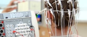
Holter monitoring (Holter ECG)
A complex, in-depth study that provides continuous recording of cardiac muscle parameters for 24-72 hours or up to 10 days (if necessary). Recording is carried out with a special device that is carried with you on a belt or belt. Electrodes are attached to the surface of the skin in certain places. After which the person leads a normal life (works, walks). At the same time, he notes in a notebook when he experiences pain in the heart area, takes medications, and begins active physical activity. The data obtained is reviewed and corrected by the doctor, after which he draws conclusions and prescribes treatment.
Interesting! There are portable devices that are implanted under the skin near the heart to record information throughout the year.
Rules for applying electrodes for ECG
I would like to immediately note that the electrodes for the ECG are located in different places. What is this connected with? This is the only way to catch all the heart impulses. There may be disturbances in heart rate and rhythm in different places.
The electrode application scheme includes the following:
- degreasing the skin with alcohol;
- abundant hair is treated with a soap solution;
- if the electrodes are disposable, then for high-quality recording of heart impulses it is necessary to remove the hair;
- Although some people use gauze, it is better to use conductive paste. As a last resort, you need to take saline solution.
ECG electrodes can be disposable or reusable
I would like to make a special mention regarding disposable electrodes. They are not included in all kits; most likely, they can be found in the latest brands of the device. According to experts, such electrodes are simple and easy to use, and also help reduce the preparation period for the patient before the procedure. They are usually used by private clinics. They are made of foil, which reduces the likelihood of disease transmission. In addition, disposable electrodes improve contact due to better conductivity. They are often used in cases where a cardiogram needs to be taken quickly, as well as for hyperhidrosis.
If we are talking about reusable electrodes, then we cannot fail to note their durability; of course, they are stronger. They are produced in the form of suction cups and clothespins. The signal passage is a little difficult, unlike the above mentioned electrodes for reusable use of a rougher design.
Basic rules for electrode placement
To record an ECG, you need to install electrodes on special points on the body. This is the only way to correctly record the electrical impulses produced by the heart. It is the correct location of the terminal that ensures a clear recording of the cardiogram. Be sure to lubricate the ECG electrode with alcohol.
If the patient has a lot of body hair, then you need to treat the skin with a soap solution or shave the hair. For better electrical conductivity, you need to lubricate each electrode with gel. If you don’t have the gel at hand, you can take an isotonic solution or gauze pads. But if the study is long, then gauze pads cannot be used.
If stationary ECG devices are used, they must be grounded.
Electrodes are either single-use or reusable. The choice of electrodes depends on the preferences of the healthcare professional performing the ECG.
ecg how to do it correctly
Electrode locations
Devices must be applied to the following areas of the body:
- lower limbs: right and left leg (black and green electrode, respectively);
- upper limbs. Red on the right hand, yellow on the left;
- rib cage.
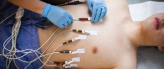
Chest electrodes are installed according to the following scheme:
- 1 lead. On the right side at the level of the fourth intercostal space;
- 2 lead. On the left side – the fourth intercostal space;
- 3 lead. Between the first and second;
- 4 lead. Fifth intercostal space on the left side;
- 5 lead. The same line goes, only along the anterior premuscular region;
- 6 lead. Corresponds to the left premuscular zone.
Despite the fact that an electrocardiogram seems to be a simple manipulation, to obtain correct results it is important to adhere to medical recommendations
In order to obtain reliable information about the functioning of the heart, you must adhere to the following recommendations:
- the day before you should have a good rest both physically and emotionally;
- Before the procedure, a light breakfast is allowed a maximum of two hours;
- strong coffee and tea, alcohol, smoking, energy drinks - all this is prohibited on the day of the study;
- if possible, it is necessary to reduce the amount of hygiene products, because they impair conductivity due to the formation of a fatty film;
- the room in which the study is carried out must be warm, the patient must be comfortable, but if physiological tremors are present, this will certainly affect the results.
Characteristic
The technique for taking an ECG at the Profmedpomoshch clinic in Moscow is not complicated. Its price is affordable (indicated in the price list). You do not undergo any punctures or radiation. You are simply connected to a special device that will provide information about the work of your heart. The data will indicate the frequency of contractions of the heart muscle, and will also identify possible diseases of the organ. In addition, using this device, diseases such as pulmonary embolism can be detected.
The device for taking an ECG was created at the beginning of the 20th century. Cardiologists all over the world still use it. The device consists of a galvanometer, an amplification mechanism and a device for recording electrical currents arising in the heart.
The monitoring procedure does not pose any danger to the body, even if used frequently. Doctors recommend performing an ECG as soon as pain and discomfort in the heart area occurs. The reason for frequent visits to the doctor may also be shortness of breath, extra pounds, stress, high or low blood pressure, rapid rhythm, etc.
People aged 40 years and above are recommended to undergo annual heart screening, and in some cases up to 4 times a year. In this way, the patient can be correctly diagnosed on time and receive timely medical care. Don't forget that preventive measures help prevent diseases.
Carrying out an ECG according to Slopak
The methodology is not very different from standard research. In this case, not six, but nine chest leads are used. The first six leads are also attached, and the next ones are presented below:
- 7 lead. Fifth hypochondrium, axillary region;
- 8 lead. Scapular line at the same level;
- 9 lead. Paravertebral line.
Electrocardiogram according to Slopak allows you to get a more detailed picture of the heart’s work
ECG by Sky
This type of research also differs from the standard technique in the placement of electrodes. The bottom line is that electrodes are placed on the chest area in three places, namely:
- 1 red lead is placed on the right side at the level of the second intercostal space;
- 2 yellow lead is installed on the left at the same level only along the posterior axillary line;
- Green lead 3 is the midclavicular line on the left side of the sternum.
As a result, a kind of triangle is created, the corners of which will serve as projection sites for the leads of the electrical activity of the heart. As for the indications, this technique is carried out in case of suspected heart attack, as well as for athletes during sports.
To increase the information content of the method, medication and stress tests are carried out. To identify areas of hidden ischemia, the test may be done after physical activity, such as squats, or after taking certain medications.
Sky leads will help identify coronary heart disease
Long registration
Data on the electrical work of the heart can be recorded over several hours or even days. Why is this necessary? Thus, doctors can identify transient disturbances in heart rhythm, and also compare the identified deviations with the patient’s activity during the day and his complaints of pain.
Let's consider the indications for long-term monitoring:
- complaints from patients about interruptions in the functioning of the heart, as well as a sensation of short-term palpitations, the interpretation of which is difficult on a conventional electrocardiograph;
- complaints in which angina pectoris can neither be excluded nor confirmed;
- the occurrence of attacks of weakness, fainting, dizziness, the cause of which has not been established;
- monitoring the functioning of an artificial heart pacemaker;
- IHD, as a control and identification of a type of arrhythmia that is asymptomatic;
- as a monitoring of the effectiveness of drugs and to identify undesirable effects on cardiac activity.
Preparing for an ECG
An ECG is an electrocardiogram of the heart. This is a very informative examination to assess the condition of the heart.
Electrodes are attached to certain areas of the body, the ECG is recorded on a special tape, on which electrical impulses are recorded in the form of curves, straight and jagged lines. A cardiologist, functionalist, therapist, or emergency physician is required to see any abnormalities on the electrocardiogram and evaluate acute cardiac pathology.
Often the patient asks the doctor questions: isn’t an ECG and an ultrasound of the heart (EchoCS) the same thing, when to do an ECG and when to do an EchoCS, what does an ECG show and what does an EchoCS? ECG, unlike EchoCS, does not show morphological and structural changes.
WHAT DOES THE ECG SHOW?
- rhythm and heart rate;
- hypertrophy of the walls of the left ventricle;
- enlargement of the cavities of the heart;
- electrolyte changes in the myocardium;
- circulatory disorders in the heart;
- regularity of the pacemaker (electrocardiostimulator).
Thus, many heart diseases, such as arrhythmias, tachycardias, blockades, ischemia, myocardial infarction, myocarditis, hypertension and many other cardiac pathologies, can be recognized by ECG recordings.
HOW TO PREPARE FOR ECG?
Although an ECG is a simple procedure, preparation for the study is necessary so that the doctor does not receive a false result and send the patient for additional examinations. To obtain the most reliable result, preparation is necessary the day before and on the day of the study.
PREPARATION FOR ECG BEFORE THE EXAMINATION:
MEDICINES. If the patient does not take medications on a regular basis, they must be discontinued 2 days before the procedure. If it is not possible to discontinue the drugs, you must inform the ECG specialist.
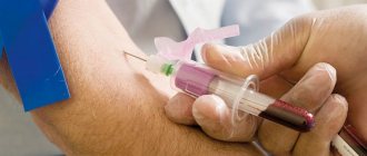
ALCOHOL. The day before, it is recommended to stop drinking alcohol, since alcohol affects the composition of the blood and the functioning of the heart, and the ECG readings may be distorted and the film may reveal arrhythmia, tachycardia, electrolyte disturbances in the myocardium, and, often, ischemia.
DREAM. If the body is tired, various abnormalities may be detected on the electrocardiogram. Therefore, 5-7 days before the examination, it is recommended to get enough sleep - at least 7-8 hours per night.
NUTRITION. It is undesirable to eat fatty, spicy foods on the eve of an ECG, and it is advisable to make breakfast as light as possible.
SPORT. It is advisable to refrain from physical exercise 2 days before the examination.
EMOTIONAL CALM. 2-3 days before the ECG is taken, it is advisable to find peace of mind - not to be nervous. Stress, psycho-emotional instability, depression, and anxiety can distort the ECG results.
THERMAL PROCEDURES. It is advisable to avoid visiting a solarium, bathhouse, or sauna for several days, as this can also affect the distortion of ECG results.
PREPARATION FOR ECG ON THE DAY OF EXAMINATION:
LIGHT BREAKFAST
ENERGY. It is necessary to exclude tea and coffee.
CLOTH. To avoid problems with connecting sensors, it is recommended to wear light clothing that is easy to remove, roll up the sleeves or the bottom of the trousers.
SMOKING. It is advisable not to smoke 3 hours before the examination, since nicotine leads to vasospasm, and this, in turn, affects the functioning of the heart, activating the work of the myocardium muscle.
CREAMS AND LOTIONS. On the day of the examination, you should not apply greasy products, because creams and lotions interfere with the attachment of sensors to the body for recording ECG.
IN WHAT CASES IS ECG PRESCRIBED?
- fainting;
- dyspnea;
- pain in the chest, neck, back;
- swelling of the legs;
- pregnancy;
- preparation for surgery;
- body check;
- trip to a sanatorium;
- sport competitions;
- as a preventive measure once a year.
SYMPTOMS FOR MANDATORY CONSULTATION TO A CARDIOLOGIST:
Proper functioning of the heart is the key to health!
Any disturbance in the functioning of the heart causes failure in all systems of our body. Nausea, tachycardia, weakness, dizziness, and neuroses appear. Preventing heart disease requires a more careful attitude to your health.
The heart is a very reliable organ; it takes a long time to warn a person about problems. The sooner we see a cardiologist due to heart problems, the better.
- A sharp decrease or increase in blood pressure.
Suddenly there is a headache, malaise, nausea, weakness, lightheadedness, and lightheadedness.
- Lip cyanosis (bluish color).
This is a sign of improper functioning of the lungs, there is not enough oxygen saturation of the blood, or improper functioning of the heart. It pumps blood weakly. Oxygenated blood, which has a bright color, does not enter the capillaries of the lips, and dark blood, saturated with carbon dioxide, does not drain well.
- Swelling of the legs. There are many reasons for swelling of the legs. One of the main reasons is heart problems. Edema often appears with cardiomyopathy, heart failure, aortic aneurysm, various heart defects, and after a myocardial infarction.
- Incorrect pulse. Normally, a person’s pulse is considered no higher than 90 and no lower than 60 beats per minute. With a low pulse, there can be fainting, even cardiac arrest. Due to insufficient oxygen supply to the brain, oxygen starvation occurs. Due to an excessively rapid pulse, there may be cerebral hemorrhage, arrhythmic shock, and much more.
- Interruptions in the heart. If the heart rhythm is abnormal, weakness, ripples in the eyes, and dizziness appear. This indicates direct heart problems.
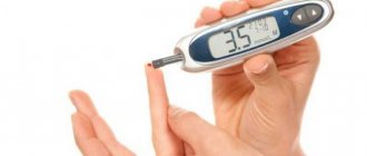
- Fainting. Presyncope. They can be a consequence of a stuffy room, diabetes mellitus, or accompany a stroke, myocardial infarction, cardiac arrhythmia until it stops completely.
- Chest pain. This is a signal about the destruction of the body. Malaise after physical activity, aching chest pain may indicate a pre-infarction condition. The pain radiates to the back, neck, and left arm.
For all of the above symptoms and complaints, it is necessary to do an ECG and, if possible, seek qualified help from a specialist, as well as perform all procedures for treatment.
Try to get rid of bad habits, adjust your diet, reduce your excess weight, increase your physical activity, try to avoid stressful situations in life, manage your attitude towards these situations.
Be healthy!
Sign up for functional diagnostics
Functional diagnostics doctor - Andrienko Olga Leonidovna
You can make an appointment by calling (391) 205−00−48 or through your personal account
Where to put children's electrodes?
An ECG of the heart helps to identify such pathological processes in children: defects, myocardial infarction, angina pectoris, etc. The study is easy to conduct and accessible. The manipulation can be carried out before planned surgical interventions, when heart murmurs are detected, before entering a sports section or school.
During the procedure, the child must behave calmly, so it is necessary to prepare him and explain that installation of the sensors is absolutely painless
The specialist installs nine to twelve electrodes on the child’s body: in the ankles, wrists and on the chest. Sensors of smaller sizes and special shapes are used, since children have more delicate skin. It is best to use electrodes for one-time use.
So, an electrocardiogram is an informative diagnostic method used for both adults and children. The procedure is simple, accessible, and does not cause any discomfort. An ECG will help identify serious cardiac disorders and determine the correct treatment. To obtain accurate results, it is important to adhere to the recommendations regarding preparation and the manipulation itself. Early diagnosis is the key to a speedy recovery!
How to prepare for the procedure, how it is carried out, who does the decoding
Electrocardiography does not require special preparation. The patient can undergo examination at any time. Before visiting the functional diagnostics office, you need to take care of your appearance, since you will have to expose your chest, forearms and shins, to which the electrodes are attached.
Important! Before the examination, it is not recommended to overeat, smoke, worry, go to the gym or run, drink alcohol, coffee, or energy drinks.
Taking an ECG looks like this: the patient removes the necessary areas of the body from clothing and lies down on the couch. The doctor or nurse degreases the skin with a special product (or water) so that the “suction cups” have better contact with the body, applies electrodes, turns on the device and makes a recording. With the results obtained and a preliminary transcript, the patient is sent to the attending physician.