Xanthelasma (translated from Greek as “golden-yellow plate”) is a slowly growing benign neoplasm localized on the skin of the eyelids. This tumor consists of plaques that rise above the healthy surface. The disease most often develops against the background of lipid metabolism disorders and increased blood cholesterol levels. In most cases, single or multiple growths with a smooth or wrinkled surface are found in middle-aged and elderly women.
Despite the fact that xanthelasma of the eyelids does not cause subjective symptoms and practically does not cause concern, it has a tendency to grow, often forming a whole chain of cholesterol plaques that are located around the eyes. Experts recommend getting rid of this aesthetic problem through removal.
| Removal of eyelid xanthelasma up to 5 mm | 3300 |
What are atherosclerotic plaques?
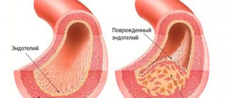
Atherosclerosis begins when cholesterol deposits appear on the walls of the arteries. Normal blood flow is ensured by the elasticity and smoothness of the inner surface of the vessel - the endothelium. When a lipid layer is formed, the lumen of the arteries narrows, the endothelium is damaged due to the introduction of harmful cholesterol into it, and the vessel walls become rigid. Gradually, the growths increase and atherosclerotic plaques form in the form of tubercles, which can partially or completely block the lumen. In addition to cholesterol, they contain calcium and foreign substances. As a rule, the process of plaque formation in blood vessels affects the entire body.
How is the course of treatment at the Juno clinic?
If during a diagnostic examination a connection was established between the occurrence of xanthelasmas and lipid metabolism disorders, external removal of the tumor will not be enough. In this situation, a specialist from the Juno clinic may recommend therapy for the primary disease, adjusting lifestyle and diet. If necessary, medications that regulate lipid metabolism disorders, anti-ischemic drugs, extracorporeal therapy methods, etc. will be prescribed.
Some of the main recommendations that must be followed for effective treatment of xanthelasma are: regular exercise, quitting smoking, maintaining optimal body weight and following a diet aimed at reducing blood cholesterol levels.
Causes
The exact causes of deposits in the arteries are unknown. It is believed that the main thing is a high level of bad cholesterol (LDL) in the blood. In addition, doctors identify a number of factors that contribute to the progression of the disease and more rapid formation of atherosclerotic plaques:
- smoking;
- high blood pressure;
- a large amount of animal fats in food;
- a small amount of vegetables, herbs, fruits;
- excess weight and abdominal fat deposition;
- constant overeating;
- increased blood glucose levels;
- physical inactivity;
- alcohol abuse;
- natural processes of aging of the body.
Why do xanthelasmas appear on the skin?
Xanthelasma of the eyelids is a flat, yellowish neoplasm of irregular shape, soft to the touch. It can appear on the skin of one or both eyelids, often having a symmetrical location.
The main cause of a benign tumor is a violation of fat metabolism. There are several factors that provoke its development:
- hypercholesterolemia (increased blood cholesterol levels by 6–10 times);
- primary and secondary hyperlipoproteinemia (lipid metabolism disorder);
- hereditary predisposition;
- congenital deficiency of the enzyme lipoprotein lipase;
- thyroid dysfunction;
- diabetes mellitus (provokes so-called diabetic xanthelasmas of the eyelids);
- diseases of the liver and biliary tract;
- hereditary diseases.
In some cases, the causes of xanthelasma remain unknown.
Plaque formation and composition
In healthy vessels, the formation of a growth is prevented by wall enzymes that dissolve fats. In order for the process of formation of a complex compound of fats, proteins and calcium to begin, certain conditions are necessary: disruption of the protective mechanisms, damage to the vascular wall, which becomes loose.
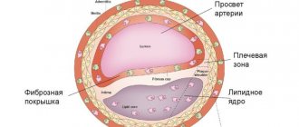
Plaque formation occurs gradually. First, cholesterol is deposited, over time it becomes overgrown with connective tissue and a pronounced tubercle appears
The plaque, consisting of lipids and connective tissue fibers, is a core with an outer shell. The core contains cholesterol and esters. Its cells are surrounded by macrophages with a foamy structure, including fats, which destroy the macrophages and enter the nucleus. The outer part of the plaque, located in the lumen of the artery, is a fibrous membrane, including elastin and collagen, the content of which determines the likelihood of its rupture.
At the beginning of their formation, lipid growths have a semi-liquid structure, so their parts can come off at any time, begin to move around the vessel and close its lumen. Also during this period, plaques can still be dissolved, so treatment is best started at an early stage of the disease.
Gradually, calcium is deposited in the growth shell, and it becomes more and more dense. This creates a calcified plaque that grows very slowly. It obstructs blood flow and leads to poor blood supply.
So, plaque formation occurs as follows:
- Accumulation of fat in the artery wall.
- Inclusion in the process of leukocytes that form inflammatory reactions (monocytes, T-lymphocytes).
- The transition of monocytes into the walls of blood vessels, the formation of macrophages with a foamy structure, pathological changes in the inner surface of the artery.
- Adhesion of platelets to the damaged part of the vascular wall.
- Immune response in the form of release of protective mediators and cell growth factors.
- Production and accumulation of elastin and collagen and the appearance of their areas in the endothelium.
- An increase in the size of the growth and its compaction.
What methods exist for removing xanthelasma?
In modern clinical practice, several methods are used to remove eyelid xanthelasma.
- Using a laser.
Laser removal of xanthelasma is the most progressive method of treatment, allowing to avoid the development of complications and recurrence of plaques. A non-contact laser beam burns out the tumor layer by layer without damaging nearby healthy tissue. This eliminates deformation of the eyelids and avoids the appearance of scars on the skin. The procedure does not require special preparation and does not require long-term rehabilitation. - Using liquid nitrogen.
Cryodestruction (freezing with liquid nitrogen) involves exposing xanthelasma to a temperature of about -200 °C. This is a fairly effective technique, but it requires a longer time for the skin to fully recover. - Using radio wave surgery.
Removal of xanthelasma using radio waves is a non-contact, non-traumatic method of tissue incision and coagulation. It is distinguished by the rapid healing of postoperative wounds and the minimal risk of scar formation. - Using electrocoagulation.
The electrocoagulation method requires the use of local anesthesia. After cauterization of xanthelasma with electric current for several days, the wound is treated with an antiseptic solution to avoid the development of inflammation. - Using surgical excision.
Surgical removal of xanthelasma is used in the presence of large tumors (more than 1 cm in diameter). The operation takes place under local anesthesia. After excision of the plaque with a scalpel and treatment of the surgical field, sutures are applied to the wound. Often, spots and scars remain on the skin, which can be removed with laser resurfacing.
The price of a procedure to get rid of xanthelasma depends both on the treatment method and on the number of plaques. In most cases, the final price for xanthelasma removal is determined after consultation with a doctor.
Types of plaques
Depending on the size, structure and structure, cholesterol plaques are divided into unstable and stable. Complicated forms include heterogeneous.
Unstable ones consist mainly of fats. They are more loose and prone to rupture with the formation of a blood clot and blocking the lumen in the vessel.
Stable ones contain a lot of collagen fibers, which means they are more elastic, which prevents rupture. Such plaques are permanent and progress slowly. Calcified growths are stable, less dangerous than semi-liquid ones, practically do not ulcerate, but do not resolve.
Heterogeneous has depressions and growths, has a loose structure, and is prone to hemorrhage and ulceration.
Atherosclerotic plaques can manifest themselves in different ways:
- They remain in the artery wall, grow slowly, then stop growing, do not block the path to blood flow, and do not manifest themselves in any way.
- They grow slowly inside the arterial lumen and can completely or partially block it.
- They can rupture and blood will clot inside the vessel. If this happens in the heart, a heart attack will occur, if in the brain, a stroke will occur.
Why are they dangerous?
Cholesterol plaques contribute to the development of serious diseases that can result in human death:
- IHD (cardiac ischemia). Plaques in the blood vessels of the heart lead to angina. When they rupture and blood clots form, myocardial tissue dies, that is, a heart attack (infarction).
- Rupture of plaques in the blood vessels of the brain leads to stroke and death of brain cells. TIA (transient ischemic attack) develops when there is temporary blockage of blood vessels without brain damage. This condition is a precursor to a stroke.
- When peripheral arteries, for example, in the legs, are narrowed, blood circulation in the lower extremities worsens, pain appears, wounds do not heal well, and gangrene may develop, leading to amputation of the legs.
How to find out that there are plaques in the vessels
The formation of plaques and stenosis (narrowing of the lumen) of the arteries is a long process, and at an early stage there are no signs. If the growths are stable, do not collapse, have stopped growing and do not close the lumen of the vessel, then there are no symptoms.
The manifestations of plaques may vary depending on their size, location, etc. But they all boil down to symptoms of circulatory disorders and malnutrition of organs and tissues.
If plaques on blood vessels grow and increasingly close the lumen, preventing blood flow, then the main symptom is pain in the area of the affected vessel, especially after physical activity.
Symptoms appear when the plaque disintegrates and its particles migrate through the bloodstream. In this case, there is a high probability of blood clots and the development of stroke and heart attack.
In general, the clinical picture can unfold as follows:
- From time to time there comes a sharp weakness.
- A crawling sensation in the right or left side of the body or in one arm or leg.
- Sudden numbness.
- Numbness in one arm or one leg.
- Deterioration of vision in one of the eyes.
- Confused speech.
Symptoms may vary depending on the location of the pathological process.
In the thoracic aorta
Quite strong pains appear in the heart area, behind the sternum, radiating to the neck, shoulder, arm, shoulder blade, and they do not go away with nitroglycerin. Blood pressure may increase, shortness of breath and signs of ischemia may appear:
- headache;
- pale facial skin;
- fast fatiguability;
- memory loss;
- clouding of consciousness;
- convulsions.
Headache may be one of the symptoms of the formation of atherosclerotic plaques in the vessels of the brain
In my head
With stenosis or blockage of the paravertebral, common carotid (CCA) and subclavian arteries that supply blood to the brain, mental disorders are usually observed:
- decreased performance;
- fast fatiguability;
- memory loss;
- depressive moods;
- delusional-anxious state;
- speech and hearing disorders;
- dementia;
- stroke.
Signs are divided depending on the stage of development:
- Fatigue, decreased performance, headache, decreased attention, memory loss, moodiness.
- Depression, anxiety.
- Speech and hearing disorders, disorientation in space, paresis, strokes, development of dementia.
Fatigue and decreased performance are early signs of cerebral atherosclerosis
In the lower extremities
At the beginning of the disease there may be no symptoms. Pain in the leg muscles when walking, lameness, and later trophic manifestations gradually appear:
- pale skin;
- hair loss;
- trophic ulcers;
- muscle tissue atrophy.
Stages of obliterating atherosclerosis of the lower extremities
In the abdominal region
When plaques appear in the abdominal aorta, the following symptoms appear:
- poor appetite, weight loss;
- pain in the navel area;
- difficulty defecating, flatulence;
- cold extremities;
- numbness of the limbs;
- intermittent claudication;
- swelling of the legs.
What to do if xanthelasma appears?
If a neoplasm appears in the eyelid area, with a description similar to xanthelasma, consult a dermatologist. As a rule, making a diagnosis does not cause difficulties and is carried out during a visual examination. However, one should take into account the fact that a neoplasm that appears on the skin is an external sign of a malfunction of the internal organs. Therefore, in this situation, a dermatologist may recommend additional consultation with a therapist, endocrinologist, cardiologist, nutritionist and undergo a comprehensive examination.
A set of diagnostic tests may include:
- diascopy (pressure on the affected area of the skin to determine the nature of the tumor);
- subcutaneous node biopsy;
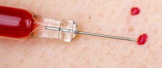
- blood test for lipid profile;
- calculation of body mass index;
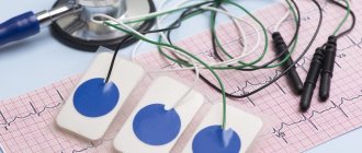
- ECG;
- EchoCG;
- MSCT of the coronary arteries;
- Ultrasound of the liver, thyroid gland, etc.
Diagnostics
The diagnostic scheme is as follows:
- Examination of the patient.
- Anamnesis collection.
- Laboratory blood tests.
- Instrumental methods: Vascular ultrasound (duplex scanning and triplex scanning) gives an idea of the blood flow and structure of blood vessels; X-ray of the aorta allows you to determine calcification, expansion of the aortic window, and aneurysm; angiography is an x-ray examination of blood vessels with the introduction of a contrast agent.
When diagnosing cholesterol plaques, it is important to distinguish them from other pathologies:
- with atherosclerosis of cerebral vessels - from head injuries, neurasthenia, cerebral syphilis and others;
- with damage to the aorta - from diseases of the digestive system and abdominal organs;
- for blockage of blood vessels in the legs - from varicose veins, conditions after injuries and others.
Angiography allows you to identify atherosclerotic plaques, determine their size and location
Treatment
Atherosclerosis is better treated in the early stages. Particularly good results can be expected with an integrated approach. It is important to know that it is impossible to completely remove the blockage, but there is a chance to stop the growth of plaques or at least slow it down.
The main points of treatment are diet and drug therapy. In addition, traditional methods, homeopathy and surgical removal of plaques are used. Lifestyle and habits are of great importance.
Lifestyle
First of all, you need to eliminate risk factors associated with bad habits and diet. You need to quit smoking, try to drink alcohol as little as possible, establish proper nutrition, and exercise. As a result, the likelihood of stroke and heart attack will be reduced, although the blockage will remain.
Nutrition
Without following a special diet, the fight against atherosclerosis will not be successful. First of all, you will have to give up foods high in cholesterol or limit the following in your diet:
- animal fats;
- offal;
- meat;
- canned food (fish and meat);
- fatty dairy foods;
- eggs;
- cocoa and chocolate.
In addition, you need to significantly reduce your intake of salt, sugar and sweets.
Risk factors:
- Age over 40 years;
- Living in hot countries;
- Frequent tanning and sunburn;
- Light skin type, blond or red hair, blue or green eyes;
- Previous actinic keratosis or skin cancer;
- Weak immune system due to chemotherapy, leukemia, AIDS, or previous organ transplantation.
Rice. 3a. Actinic keratosis capitis
Rice. 3b. Actinic keratosis on the nose
Rice. 3c. Actinic keratosis on the face
Rice. 3g. Actinic keratosis on the forehead
Rice. 3d. Actinic keratosis at the corner of the eye