- Home >
- Symptoms >
- Heaviness in the head
Each of us at least once in our lives experienced an unpleasant feeling of weakness throughout the body. Most people do not attach any importance to this symptom, because almost all diseases in the body occur with weakness, malaise, and loss of performance. However, in some cases, it is weakness in the body that is the first alarming symptom, when it is not too late to do something, get examined and treated. Let's deal with this insidious symptom.—>
At the MART clinic on Vasilyevsky Island
- Evidence-based medicine
- Experienced specialists
- Monitoring of patients for 6 months.
- Diagnostics (MRI, ultrasound, tests)
- Daily 8:00 – 22:00
Make an appointment
Heaviness in the head is an extremely unpleasant feeling, which, unfortunately, is familiar to almost everyone. In this state, a person often suffers from a bad mood, irritability, lack of restraint, mental fog and confusion in the head. There are difficulties with the ability to think, concentrate and even do ordinary things, and the only desire is to fall asleep as quickly as possible and wake up as a healthy person.
But despite all the difficulties it brings, heaviness in the head is usually not perceived as a serious problem, especially if it overtakes a generally healthy person only periodically and disappears without a trace after, for example, a walk in the fresh air. In this case, a temporary deterioration in well-being is usually associated with overwork, lack of sleep, stress, psycho-emotional overload or changes in weather. At the same time, this kind of ailment can be a sign of many serious diseases. Let's try to figure out what heaviness in the head can signal.
Inflammation
Inflammatory causes of ear congestion:
- external otitis of any etiology (fungal, bacterial, viral);
- eustacheitis (tubootitis);
- catarrhal otitis media, bullous otitis (with influenza);
- purulent otitis media (including chronic);
- exudative otitis;
- adhesive otitis media
With inflammation in the nasal cavity, paranasal sinuses or pharynx, swelling of the nasopharyngeal mouth of the auditory tube occurs, due to which the pressure in the tympanic cavity and the environment ceases to equalize, and the drainage function of the auditory tube is disrupted. The infectious agent, together with the discharged contents from the nasal cavity and paranasal (paranasal) sinuses, enters the auditory tube and tympanic cavity, causing inflammation of the auditory tube - eustacheitis (tubo-otitis) and otitis media.
Due to inflammation, swelling of the mucous membrane of the middle ear and eardrum occurs, sound-conducting mechanisms stop working normally, and ear congestion occurs, and hearing decreases. A common cause of dysfunction of the auditory tube and the development of otitis in children is hypertrophy of the nasopharyngeal tonsil and adenoiditis. In adults, vasomotor and allergic rhinitis and deviated septum are common causes of swelling at the mouth of the auditory tube. The cause may also be a violation of blood circulation and innervation in the inner ear.
With inflammation in the external auditory canal, swelling of its walls appears, fungal or purulent masses clog the ear canal, and the surface layers of the eardrum become inflamed (myrinigitis). Because of this, the mobility of the eardrum, necessary for proper sound transmission, is reduced.
Most of the described forms of otitis often occur as complications of acute respiratory diseases, sinusitis, rhinitis, especially in the case of improper cleansing of the nasal cavity. In addition to the feeling of congestion during otitis media, shooting, sharp or aching pains, hearing problems, bloody, fungal, purulent discharge, itching, a feeling of pulsation or fluid transfusion in the ear can be added.
A feeling of ear fullness also occurs when:
- water getting into the ears (usually when swimming), the water usually evacuates on its own and does not require intervention;
- when a cerumen or epithelial plug forms (can also occur after water gets into the ear due to swelling of the cerumen plug);
- foreign bodies getting into the ears: insects, earbuds, cotton wool from cosmetic sticks, etc.
After eliminating the provoking factor, the discomfort disappears and hearing is restored.
Dizziness and tinnitus
S.Ya.Kosyakov, G.Z.Piskunov Dizziness
Introduction
Dizziness is a general term that characterizes a large number of symptoms.
In general, it means a pathological sensation of movement, it can also mean imbalance, lightheadedness, darkening of the eyes, disorientation, weakness and other sensations. Symptoms can vary in intensity from mild and short in duration to severe attacks of rotation accompanied by nausea and vomiting. To more accurately define symptoms, the following definitions are used: Dizziness:
a general concept describing symptoms of imbalance and stability.
Balance problems:
Difficulty maintaining balance, especially while standing and walking.
Presyncope:
A feeling of being “switched off”, similar to the sensation that occurs when holding your breath for a long time.
Systemic dizziness:
sensation of rotation, spinning, twisting of surrounding objects.
The ability to maintain balance is the result of a complex interaction of various organs and systems. The brain is the main center for processing all information about balance coming from the senses to the muscles that maintain balance. Information in the form of nerve impulses comes from the main systems: visual, vestibular, proprioceptive and tactile (joints and feet). Visual information is the most important for the brain and signals movement in relation to surrounding objects. Anatomy
There are two components of hearing: mechanical and electrical (neural).
The mechanical component ensures the delivery of sound waves through the external auditory canal, the movement of the eardrum and the three auditory ossicles in the middle ear. The inner ear is represented by a cochlea, consisting of two halves connected to each other and filled with liquid. The cochlea is responsible for the electrical component of hearing and converts a mechanical signal into an electrical one, which in turn goes to the brain. The other part of the inner ear is responsible for balance and the vestibular system. The three semicircular canals are located in mutually perpendicular planes. Depending on the direction of movement of the head, the fluid moves in the channels, the resulting electrical impulse is transmitted to the brain through the vestibular nerve, transmitting information about the direction of movement. The fluid in the inner ear is renewed daily. Its source of origin is cerebrospinal fluid, absorption occurs in the endolymphatic sac. In Meniere's disease, the absorption capacity of the endolymphatic sac deteriorates. Increased pressure in the inner ear leads to dizziness and decreased hearing. The facial nerve exists in close relationship with the ear. The facial nerve carries out movements of the facial muscles and allows the tip of the tongue to distinguish taste. When it is affected, the eye closes poorly, fluid pours out of the corner of the mouth, and facial movements on the affected side are impossible. Balance function
The balance function is ensured by the interaction in the brain of nerve impulses coming from the inner ear, neck muscles, muscles and joints of the lower extremities.
Disturbances in any of these systems can lead to a subjective feeling of dizziness and instability. General problems with body functions (such as low or high blood pressure, nearsightedness, and many others) can lead to dizziness by affecting the coordination of impulses in the brain. The brain's response to distorted or inconsistent impulses can lead to false sensations of movement (dizziness), which in turn leads to unsteady gait and falls. Dizziness is often accompanied by cold sweats, nausea and vomiting. Visual and muscle and joint signals (tactile and proprioceptive) to the brain warn us that we are moving on the right path or that our head is tilted. The brain interprets this information along with information from the vestibular system and gives the appropriate command to the muscles to maintain balance. Dizziness occurs when sensory information is distorted. Some people experience dizziness when they are in a high place, for example. This is partly due to the inability to focus on nearby objects. While standing on the ground, a person may sway slightly. A person maintains balance, identifies his body position relative to something. When standing in a high place, it is difficult for a person to correlate the position of his body relative to objects in the distance and, accordingly, it is more difficult to maintain balance. As a result, anxiety, fear, and dizziness may occur, which sometimes forces the person to sit down. There is an opinion that motion sickness, a disorder that occurs during sea motion, in a car, or in flight, occurs when the brain receives conflicting sensory information about the movement and position of the body. For example, when reading while driving in a car, the inner ear perceives the movement of the vehicle, but the gaze is fixed on a stationary book that does not move. As a result, sensory conflict can lead to typical symptoms of motion sickness, dizziness, nausea, and vomiting. Another form of dizziness occurs when you spin repeatedly and suddenly stop. Rotation causes movement of the endolymph. The movement of the endolymph causes impulses, which in turn tells the brain that we are moving, but other sensory systems report that we have stopped, so the patient feels dizzy. Causes of Dizziness
Dizziness can be classified into types depending on the part of the vestibular system that is not working properly.
Disturbances can occur at the level of the inner ear, brain, eyes and limbs (muscles of the back, neck, legs and joints that react to maintain our position). Dizziness due to the inner ear
Part of the inner ear (cochlea) is used for hearing, the other part is used for balance (labyrinth).
If there are disturbances in the labyrinth or in the nerve connecting it to the brain, this leads to dizziness. Various types of disorders in the inner ear can lead to vertigo, including Meniere's disease, labyrinthitis, positional vertigo, vestibular neuronitis, and nerve tumors. These disorders typically cause problems with balance, a sensation of spinning objects, and nausea. Also, these phenomena may be accompanied by tinnitus and hearing loss on the corresponding side. Dizziness of a central nature
The cause of dizziness of a central nature is usually a disturbance in the area of the brain responsible for balance.
Symptoms may include lightheadedness, confusion, unsteadiness, and sometimes loss of consciousness. Causes of central vertigo include low blood sugar, low blood pressure, stroke, multiple sclerosis, migraines, head injuries, tumors and age-related changes. Treatment of this type of dizziness is usually associated with the elimination of problems leading to disruption of brain function. Muscular-articular dizziness
This type of dizziness is rare.
If there are diseases of the muscles, joints, or the sensitivity of the lower extremities is impaired, then difficulties arise in the body’s reaction to movement and in maintaining an upright position. Musculo-articular dizziness can be caused by: atrophic changes in muscles (muscular dystrophy), severe forms of diabetes, arthritis, joint implantation and trauma. Symptoms: Typically unsteadiness and poor balance. Visual dizziness
Unsteadiness of the eye muscles and poor vision can impair balance function.
The brain relies on visual information to maintain balance. Motion sickness in a car or at sea are examples of visual vertigo because the eyes are constantly fixated on a moving object and “confuse” the vestibular part of the brain. This leads to dizziness, nausea and vomiting. Dizziness is not a fatal condition and may improve with treatment, but balance problems may remain. Diagnosis of dizziness
Dizziness can be caused by various disorders in the body.
Based on the medical history and examination data, the doctor selects the required scope of examination to obtain a more complete picture of the disease. The usual set of examinations includes testing of hearing and vestibular function, computed tomography and nuclear magnetic resonance, blood tests, and ultrasound examination. The most commonly used test for dizziness is electronystagmography (ENG). This test measures inner ear endurance and eye coordination. The method involves observing eye movements while cold and warm air is blown into the external auditory canal. This usually causes a brief feeling of dizziness. It is important not to take medications before the test that could affect the test results (for example, Valium, alcohol, etc.). When ordering such an examination, it is necessary to find out from the doctor the effect of the medications taken on the test results. Transcranial Doppler sonography is another test specific for the examination of dizziness of vascular origin. It is a safe, quick way to see disturbances in blood flow in the parts of the brain responsible for balance. In addition, a computed tomography (CT) scan of the temporal bones and, in some cases, magnetic resonance imaging (MRI) may be performed. The purpose of these examinations is to achieve confidence in the absence of a life-threatening pathology and to determine the exact location of the disorder. This is the basis for effective treatment. The scope of the examination is determined by the doctor in each specific case. Several tests are necessary to diagnose the cause. Perseverance and understanding are necessary for both the doctor and the patient, which is also the basis for effective treatment. The most common types of vertigo Benign paroxysmal positional vertigo (BPPV)
BPPV is the most common type of vertigo.
With this disease, dizziness occurs only when the head position changes (usually when turning in bed, tilting the head backwards or forwards). This type of dizziness is caused by microcrystals that float in the fluid of the inner ear and cause a spinning sensation. The most common cause of BPPV is head trauma or viral infections, but sometimes it begins without any obvious cause. Treatment for BPPV consists of certain exercises to return the crystals to a place where they will not cause dizziness. When left at rest in a certain position for 48 hours, they often lock into place. Exercise may reduce symptoms. If these actions are ineffective, then surgical treatment (for example, occlusion of the posterior semicircular canal) may be necessary. Vestibular neuronitis
Neuronitis (nerve inflammation) usually occurs due to a viral infection and can affect the balance centers or the vestibular nerve.
When this happens, the balance centers in the brain are overstimulated, resulting in significant imbalance and systemic dizziness. Fortunately, vestibular neuronitis usually subsides over time and does not recur. Drugs such as betaserc help in the initial stage and reduce the severity of the main symptoms; later, vestibular rehabilitation exercises can speed up the recovery process. In some cases of persistent disease, surgical treatment is recommended. Meniere's disease (Endolymphatic hydrops)
Meniere's disease is a consequence of disorders in the inner ear due to increased pressure in the endolymphatic space.
This is usually due to increased sodium concentration in the fluids of the inner ear. In addition to imbalance that lasts for hours, patients may experience hearing fluctuations, tinnitus, and a feeling of fullness in the affected ear. Sometimes the lesion affects both ears. The full cause of this disorder is not fully known. Sometimes attacks can be caused by excessive salt intake, anxiety, changes in weather, and other reasons. Treatment usually includes limiting salt intake and using diuretics, fluid restriction, sedatives and some other vestibular suppressants. Betaserc is the only drug created for long-term treatment of dizziness. Treatment helps reduce the severity of attacks, but complete cure of the disease cannot be achieved. Vestibular rehabilitation exercises can speed up the recovery process and increase the patient's resilience to vestibular disorders. All prescriptions of drugs should be carried out only by a doctor. For severe cases of Meniere's disease, surgical treatments are available. The list of these methods is long and more often they are destructive in nature for the structures responsible for balance. Vascular vertigo
The correct functioning of the balance system requires not only the flow of information into the inner ear, but also the appropriate transmission of impulses along the nerves to the brain.
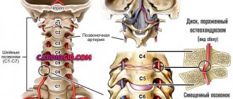
If there is not enough blood flow to the areas of the brain responsible for balance, even for a short time, dizziness can occur. The causes of vascular dizziness are different. The phenomena of osteochondrosis in the cervical spine can lead to compression of the arteries leading to the brain; atherosclerotic plaques can narrow the arteries, also causing a decrease in blood flow. Often, blood pressure in the vessels leading to the brain may temporarily decrease when standing up suddenly, especially in older patients receiving blood pressure-lowering medications. Special examinations such as MRI or Doppler sonography help in diagnosing such diseases. Another rather rare cause of dizziness is Perilymphatic Fistula
. The inner ear is a space filled with fluid located in the temporal bone.
If fluid leaks from the structures of the inner ear, hearing loss may occur, which may be greater or less, and dizziness. Most often, fluid leaks through the membranes of the windows of the inner ear, which can occur after physical activity or injury. In some cases, there are congenital disorders that characterize an enlarged connection between the inner ear and the brain ("enlarged vestibular aqueduct"). Sometimes this can be seen with a special x-ray examination - computed tomography. Sometimes the site of the membrane rupture heals on its own, sometimes minor surgery is required. A perilymphatic fistula, or as it is also called, a labyrinthine fistula, can occur as a result of chronic inflammation of the middle ear, especially with cholesteatoma. Cholesteatoma is compacted skin scales. If there is a hole in the eardrum, the skin grows into the cavity of the middle ear, and its metabolic products, like the formation of a pearl, form a lump of cholesteatoma, which presses on the walls of the middle ear cavities and destroys the bone, in particular, the semicircular canal. Therefore, the treatment of chronic otitis media is very important, and when the hole is localized in the upper part of the eardrum (epitympanitis), it must be surgical, because Most often, cholesteatoma is found in these cases. Tumors
Rarely, tumors may be the cause of dizziness.
Most tumors are benign. Acoustic neuroma is a benign tumor of the vestibular nerve. The presence of a neuroma can lead to instability, hearing loss and noise. The most effective treatment method is surgery. Treatment of dizziness All questions regarding the treatment of dizziness and, in particular, taking medications should be discussed with your doctor.
Treatment in each specific case is selected individually and depends on age, severity of dizziness, concomitant diseases and many other factors.
Ear noise
Ear noise is a very common symptom.
The noise can be constant or periodic, of varying severity and frequency. The noise can be subjective (audible only to the patient) or objective (audible to others), and may or may not be associated with hearing loss. Noise is a symptom and not a disease and can occur in various diseases, such as pain in the arm or leg are symptoms of various diseases. Noise appears when the auditory nerve is irritated for various reasons. The noise may or may not be accompanied by hearing loss. Hearing is measured in decibels (dB). A hearing level of 0 to 25 dB is considered normal for the perception of spoken language. Hearing mechanisms
to understand the possible causes of noise in the ear, it is necessary to have some idea of hearing mechanisms.
The mechanism of auditory perception is provided by the five main components: the outer ear, the middle ear, the inner ear, the paths and the brain. The outer ear
of the outer ear consists of an auricle and an external auditory pass.
These structures collect sound waves and transmit them to the eardrum. middle ear
is located between the eardrum and the inner ear.
This space contains three auditory bones: a hammer, an anvil and a stirrup. The fluctuations of the eardrum are transmitted through the auditory bones on the liquid of the inner ear. The middle ear lined with the mucous membrane is identical to the nose and contains mucous glands and blood vessels. The drum cavity is connected to the rear parts of the nose using the Eustachian pipe. Eustakhiev’s pipe is served to maintain equal pressure between the middle ear and the outer atmosphere. The feeling of clicking or congestion when height changes is a demonstration of the ventilation function of the Eustachian pipe. The inner ear
in the inner ear is in a dense bone capsule and contains liquids and auditory cells.
The cells are covered with delicate membrane with microscopic blood vessels. In the inner ear, fluctuations in the fluid as a result of the movements of the stirrup, are converted into electrical impulses in the nerve. Electric impulses arising in the inner ear are transmitted to the brain along the auditory nerve. The auditory nerve going into the brain is located in a small bone channel along with the vestibular and facial nerve. The brain
of the auditory nerve by reaching the brain is divided into many internal connections.
In the brain, nerve impulses are recognized as recognizable sounds. Ear noise,
most ear noise are heard only by patients - this is a subjective noise.
The noise that the patient himself hears, and someone is also called objective. Objective noise can be as a result of muscle cramps in the middle ear or auditory pipe, or due to anomalies of blood vessels of the surrounding the ear. Ear noise of muscle nature,
noise can be the result of muscle spasm attached to one of the auditory bones or the result of a spasm of muscles attached to the auditory pipe.
In the middle ear, there are two muscles: a stirmer that is attached to the stirrup and muscle pulling the drum overlain attached to the hammer. Usually these muscles quickly decline in response to loud noise or at fear. Sometimes one or two of these muscles begin to rhythmically contract for no apparent reason. These abbreviations can cause repeated noise in the ear. An annoying clicking usually passes on its own. The ear noise of muscle nature as a result of a spasm of various muscles of the pharynx is quite rare, but sometimes it can be if muscle spasm is long, then drug treatment (muscle relaxants) or surgical treatment (intersection of spasmodic muscles) is used. The ear noise of vascular nature
has two large blood vessels closely related to the middle and outer ear: jugular vein and sleepy artery.
These are large blood vessels supplying the brain with blood and carrying out its outflow. To hear your own heartbeat or noise of blood passing through these large vessels is not a normal phenomenon. Sometimes this phenomenon can occur at high temperature, infection of the middle ear, after intense physical activity. The noise of circulation in these situations is temporary and is not heard by others. Sometimes the noise of blood circulation becomes heard by another. This can happen due to the thickening of the wall of the blood vessel, the presence of bending or narrowing in the vessel. Further examination is necessary to identify the cause and choose the treatment of this pathology. Ear noise due to the outer ear
is the closure of the external auditory passage with a gray, foreign body, edema is leading to a decrease in hearing and pressure on the eardrum.
Often this leads to a pulsating type. Ear noise due to the middle ear,
impaired function of the middle ear may be the result of an allergic reaction, infection, injury, cicatricial changes and limitations of the mobility of the auditory bones.
These disorders often lead to hearing impairment and ear noise. However, there is no direct dependence between the degree of hearing loss and noise intensity. Ear noise due to the inner ear
any condition that violates the balance of fluid pressure in the inner ear can lead to an ear noise.
This may be the result of an allergic reaction, infection, circulatory disorders, which lead not only to changes in the liquids of the labyrinth, but also in the membrane structures of the inner ear. Ear noise due to damage to the tracking paths,
the most delicate structures of the mechanism responsible for the hearing.
Hair cells convert fluctuations in the fluid into nerve impulses. The most insignificant edema and violation of interference in the hair cells, regardless of the cause, lead to function and irritation. This can happen for various reasons: an allergic reaction, infection, edema, systemic diseases, both acute and chronic, toxic effects, sudden loud sounds and sensitive subjects, injuries, the effects of medications, minute changes in blood supply and diet changes. Pressure changes can cause swelling both outside and inside the nerve when passing in the bone tunnel to the brain. In these cases, ear noise occurs on one side. Because The bone tunnel cannot stretch, then due to compression, not only the auditory and vestibular functions suffer, but also the facial nerve. The gap or spasm of a small vessel that occurred somewhere in the auditory path causes compression and violation of circulation. Accordingly, under such conditions, a sudden noise may occur with complete or partial loss of auditory function. If the blood clot, then it can resolve with minimal consequences. Auro noise of brain nature
as a result of edema, pressure or circulation disorders for hypertension, atherosclerosis, as a result of the consequences of injuries, one or more complexes of the passing paths at the entrance and end of their brain can be involved.
In such situations, symptoms are usually localized on the one hand, in addition, the development of symptoms and signs can tell the doctor the place and prevalence of the lesion. The ear noise accompanying the decrease in
ear noise can be associated or not with hearing impairment.
With the coexistence of ear noise and hearing loss, the intensity of ear noise is not an indicator of the further development of hearing loss. Many patients with the advent of ear noise are afraid of the progression of hearing loss. However, these are often not interconnected things. All issues of ear noise treatment should be discussed with your doctor.
Thus, the treatment of dizziness and ear noise is a difficult task, which is possible only by the joint efforts of the doctor and the patient. High -quality hearing diagnosis plays a very important role in such a treatment. The sequence in identifying the causes of these conditions, in treatment and rehabilitation is an integral condition for achieving success.
Sensorineural hearing loss
The cause of hearing loss and a feeling of fullness in the ears may be sensorineural hearing loss, which occurs as a result of a violation of the blood supply or innervation in the inner ear.
Sensorineural hearing loss can result from:
- changes in blood pressure due to hypertension;
- acute viral diseases;
- barotrauma;
- fracture of the base of the skull;
- ischemia of the central areas of the auditory analyzer;
- neuroma of the vestibulocochlear nerve;
- compression from the outside by neoplasms and vascular aneurysms.
When is it due to nerves?
At the time of panic attacks or as they approach, patients often experience increased blood pressure, which can cause severe discomfort in the ears, temples and head. It is known that hypertension provokes the occurrence of severe spasms.
We recommend that you read: What is depersonalization?
Often, floaters appear before the eyes - various spots, stripes, dots.
Other accompanying symptoms include:
- tachycardia, interruptions, body tremors, fever or chills;
- respiratory disorders (shortness of breath, suffocation);
- frequent urination;
- pain, tension, tightness in the chest area;
- nausea, diarrhea;
- the appearance of red spots on the skin, “nervous” allergies;
- numbness of the limbs.
Symptoms of ear congestion
Associated symptoms for various ear diseases:
| Cause | Symptoms | |||||||
| Pain | Ear discharge | Itching | Hearing loss | Autophony | Noise | Sensation of fluid transfusion | Manifestations of general intoxication | |
| Sulfur plug | No | No | Not really | Yes | Not really | Not really | No | No |
| Otitis externa | Yes | Not really | Yes | Yes | Not really | No | No | Not really |
| Tubootitis | Not really | No | No | Yes | Yes | Not really | No | Not really |
| Otitis media | Yes | No | No | Yes | Yes | Not really | No | Yes |
| Suppurative otitis media | Yes | Yes | Not really | Yes | Not really | Not really | No | Yes |
| Exudative otitis media | No | No | No | Yes | Yes | Not really | Yes | No |
| Sensorineural hearing loss | No | No | No | Yes | Not really | Yes | No | No |
Diagnosis of ear congestion
Standard examinations for any ear pathology:
- video endoscopy or microscopy of the ear;
- video endoscopy of the nose and nasopharynx;
- audiometry;
- tympanometry;
If necessary, additional studies are prescribed:
- computed tomography or magnetic resonance imaging of the temporal bones and brain;
- culture of nasal discharge and external auditory canal for flora and sensitivity to antibiotics;
- general blood test, blood glucose (other additional laboratory tests are possible after consulting a doctor);
- Doppler ultrasound of neck vessels;
- consultations with other specialists.
Treatment
Treatment for ear congestion and otitis media involves treating the underlying cause of the disease:
- in case of an inflammatory process in the nasal cavity and paranasal sinuses, antibacterial therapy is prescribed, in addition, lavage of the nasal cavity is prescribed, which eliminates the discharge of discharge into the auditory tube during self-washing, treatment with a YAMIK catheter, puncture of the maxillary sinuses, physiotherapy;
- if a chronic pathology of the nasal cavity or nasopharynx is detected, leading to the closure of the mouth of the auditory tube, surgical treatment is performed (adenotomy, vasotomy, septoplasty, etc.);
- To remove wax plugs or foreign bodies, the doctor performs lavage (under visual control) or instrumental removal of the foreign body;
- for external otitis, the outer ear is washed and its walls are treated with antifungal and antibacterial drugs;
- in case of dysfunction of the auditory tube or exudate in the tympanic cavity, medical procedures are necessary: pneumomassage of the tympal membrane, blowing of the ears according to Politzer, catheterization of the eustachian tube, paracentesis or shunting of the eardrum;
- for sensorineural (sensorineural) hearing loss, a course of vascular and vitamin therapy is prescribed to improve the nutrition of the inner ear and brain.
Methods for treating heaviness in the head
As already mentioned, heaviness in the head in itself is not a disease. This is only a symptom, and it is the root cause of this condition that needs to be treated.
Treatment of the underlying disease is specific, selected taking into account the individual characteristics of the patient’s body and entirely depends on the identified disorders.
However, symptomatic treatment of heaviness in the head and similar accompanying symptoms is also possible. Most often, non-steroidal anti-inflammatory drugs, analgesics, antispasmodics and other drugs are used in therapy to relieve discomfort. In some cases, they resort to blockades.
However, do not forget that long-term drug treatment is addictive, and the drugs lose their effectiveness over time. So this approach to treatment can be truly justified only in a situation where heaviness in the head is caused by serious organic lesions. In other cases, it is recommended to solve the problem, if possible, through safer therapeutic methods without the use of “chemistry”.
Thus, heaviness in the head can be easily eliminated with the help of self-massage of the biologically active zones of the back of the neck, the back of the head, the temporal region, and the crown. Impact on these areas not only relieves tension from the neck muscles, but also stimulates the flow of fresh blood, which helps improve well-being and restore performance.
In addition, you should pay attention to a number of treatment methods that have proven effective in improving blood flow in the cervical spine and normalizing blood supply to the brain. First of all, these include manual therapy and all kinds of physiotherapeutic procedures.
Physical therapy exercises can also give good results in the fight against heaviness in the head, drowsiness, fatigue and other associated symptoms. Physical activity helps saturate the blood and tissues with oxygen. In addition, regular performance of even simple exercises allows you to strengthen the muscle corset and, accordingly, reduce the load on the spinal column itself and protect it from the development of destructive processes.
Basic treatment regimens for ear pathology*
| Pathology | Basic treatment methods |
| Sulfur plug | Removal by rinsing or instrumental removal. |
| Otitis externa | Toilet of the external auditory canal, treatment with medications, ear drops, vasoconstrictor nasal sprays, systemic antibacterial or antifungal therapy, FTL**. |
| Tubootitis | Vasoconstrictor nasal sprays, blowing of the eustachian tubes according to Politzer, pneumomassage of the eardrum, FTL**, catheterization of the auditory tubes if necessary. |
| Otitis media | Vasoconstrictor nasal sprays, systemic antibacterial therapy, ear drops, Politzer ear blowing, pneumomassage of the eardrum, FTL**, if necessary, catheterization of the auditory tubes, lavage of the nasal cavity using the moving method. If necessary, surgical treatment - eardrum bypass or paracentesis. Complemented by the treatment of diseases of the ENT organs. |
| Suppurative otitis media | Vasoconstrictor nasal sprays, systemic antibacterial therapy, ear drops, Politzer blowing, if necessary, transtympanic injection of drugs, catheterization of auditory tubes, nasal lavage using the displacement method. If necessary, surgical treatment - eardrum bypass or paracentesis and other operations, FTL**. It is supplemented by treatment of pathologies of the nasal cavity, nasopharynx and paranasal sinuses, if indicated. |
| Exudative otitis media | Vasoconstrictor nasal sprays, mucolytics, Politzer blowing, catheterization of auditory tubes. If necessary, surgical treatment - eardrum bypass or paracentesis, FTL**. It is complemented by the treatment of pathologies of the nasal cavity, nasopharynx and paranasal sinuses. |
| Sensorineural hearing loss | Catheterization of auditory tubes, systemic vascular and vitamin therapy. |
*These treatment regimens are not a recommendation for self-treatment and serve to familiarize patients with basic treatment methods. The final decision on treatment and examination methods is made by the doctor after the examination!
** FTL – physiotherapeutic treatment.
general characteristics
With ear congestion, patients report a feeling of discomfort, fullness in the external auditory canal, and decreased hearing, accompanied by a constant hum or ringing.
Some people compare the sensation to the ear canal filling with water. With short-term physiological congestion, bilateral symptoms are more often detected; inflammation and other pathological causes can cause unilateral manifestations. Congestion can last from several minutes to several weeks. Patients often notice a connection between discomfort in the ear and acute bacterial or viral infections, injuries to the auricle or head in the temporal region. If congestion bothers you constantly or occurs periodically over several days, this is an indication for a visit to a specialist. It is also necessary to visit a doctor when the symptom is combined with other manifestations - headaches or pain in the ear canal, dizziness, increased body temperature.