Telangiectasia (spider veins) is a vascular pathology that is characterized by persistent dilation of small blood vessels of the dermis (capillaries) due to hemodynamic disturbances and stagnant processes in the blood vessels.
As a result of persistent vasodilation, hyperemia appears, against which dilated capillaries can be seen. The color and size of the spots depends on the type of affected vessels: arterial capillaries form a pattern of thin red “networks”, venous capillaries form blue and purple spots.
If appropriate treatment and correction measures are not taken, under the influence of provoking factors, the visibility of capillaries through the skin increases, their shade becomes brighter, the affected area increases - the number and size of spider veins becomes larger.
Causes of telangiectasia
There are many reasons for the appearance of a pronounced vascular pattern. Basic —
this is a genetic predisposition (congenital low elasticity of the walls of blood vessels), endocrine diseases and hormonal changes (pregnancy, childbirth, hormone therapy), cardiovascular diseases. In addition, telangiectasias can be a symptom of dermatological diseases (rosacea) and systemic connective tissue diseases (scleroderma, lupus erythematosus).
With reduced vascular elasticity, the following provoking factors lead to the appearance of spider veins:
- bad habits (smoking and alcohol disrupt the functioning of the circulatory system, nicotine contributes to the narrowing of blood vessels, and alcohol causes their persistent dilation);
- frequent traumatic cosmetic procedures and the use of aggressive facial skin care products;
- regular insolation (ultraviolet rays increase the fragility of capillaries, since tanning causes superficial dilation of the vessels of the dermis. 50% of UVA penetrates to the papillary and reticular layers, damaging blood vessels and the basement membrane of lymphatic vessels);
- diseases of the gastrointestinal tract lead to impaired absorption of certain vitamins that play an important role in maintaining vascular health (PP, K, C) and unhealthy diet (excessive sugar consumption, spicy, hot foods, marinades, coffee have a strong stimulating effect on circulatory processes). Helycobacter pylori bacteria are capable of disrupting neurochemical regulatory processes, which leads to vasodilation.
- Strength physical activity worsens the condition of blood vessels and provokes the appearance of new spider veins.
- Excess weight is also harmful to blood vessels, especially those of the legs.
- sudden changes in temperature are especially harmful to the face (they provoke the development of rosacea).
Diagnostics
In order to accurately determine the disease and the degree of its development, you need to make an appointment with a phlebologist. The specialist will conduct an external examination of telangiectasia and palpation of the limbs. As a result, the doctor can draw conclusions about the location of the stars and the nature of the disease.
After this, the specialist will prescribe the necessary studies. They usually include duplex ultrasound scanning, which allows you to assess the condition of blood vessels, determine pathologies and their locations, and assess the direction and speed of blood flow. After this, the phlebologist can draw conclusions about the patient’s condition. For greater accuracy, the doctor may also prescribe tests and studies of other organs, including the heart, thyroid gland, liver, and reproductive system. This will allow us to identify concomitant diseases and establish the cause of the appearance and progression of varicose veins.
Conclusion 3:
You should contact a vascular surgeon immediately after you discover spider veins. Using a series of studies, a specialist will determine the cause of their appearance and prescribe therapy.
Classification of telangiectasia
Spider veins most often appear on the face, ankles and thighs, and less commonly on the body and arms. The presence of stars is often not accompanied by pain or discomfort.
Depending on what type of blood vessels is affected, they are divided into the following types:
- arterial (large, have a clearly defined contour, star-shaped and reddish tint, usually located on the face).
- capillary (thin linear lines of vascular pathology located on the wings of the nose, cheeks, chin).
- venous (prevail on the body, mainly localized on the lower extremities, have a blue or purple tint).
The shape of telangiectasias depends on the pattern of branching of the vessels (tree-like, punctate, linear, arachnid). They can take the form of dots, lines, branched formations ( asterisks) and sometimes cover large areas.
Spider veins on the face (rosacea) most often appear on the wings of the nose or cheeks.
WHAT PROCEDURES DO YOU NEED?
Indicate what you would like to improve in your appearance and we will offer you an individual rejuvenation and care program
Morphofunctional features of the structure of skin vessels
The skin has two networks of blood vessels - deep and superficial.
The deep network of vessels (DNV) delivers blood to the sweat glands and hair follicles. The GSS is formed by arteries and comes from subcutaneous fat. In the dermis, branching into small blood vessels occurs; from this vascular network, smaller blood vessels extend upward, which form the superficial vascular network (SVN), located parallel to the surface of the skin in the papillary layer of the dermis.
The PSS delivers blood to the sebaceous glands, sweat ducts and the top of the hair follicles.
From the GSS, arterial capillaries extend deeper into the skin veins. There are four types of venous plexuses.
One of the main features of the blood vessels of the skin is the ability to reflexively narrow or expand from external influences on nerve endings.
Cuperosis occurs due to impaired blood circulation in the upper layers of the skin - in the PSS. If blood stagnates, it constantly creates stress on the walls of the capillaries, which causes them to become fragile and brittle.
The color and size of the spots depends on the type of affected vessels:
- expansion of capillary and arterial vessels leads to the formation of thin red “networks” that do not protrude above the surface of the skin and turn pale when pressed;
- spots of dark blue and purple colors are formed from the venous vessels, which stand out against the background of a homogeneous skin.
About 50% of women are susceptible to this disease. The incidence of telangiectasias is associated with age. In women under the age of 30 they are found in 8-9%, by the age of 50 this figure increases to 40%, by the age of 70 – to 75%. Men also often suffer from this disease. The prevalence of vascular anomalies among the male population reaches 25-30%.
Removal of spider veins at the ROSH Medical Center
Methods for removing telangiectasia include: radio wave removal, electrocoagulation, a combined technique combining the effects of electric current and light radiation (Elos), laser photocoagulation.
Doctors at the ROSH Medical Center always work as a team to achieve maximum aesthetic results. Therefore, when a patient presents with vascular changes on the face at the initial appointment, the dermatologist determines whether this is a cosmetic defect or a sign of some general disease that causes a decrease in the elasticity of the capillary walls, local dilation of small vessels and disruption of microcirculation processes.
If a patient consults a doctor for laser correction of blood vessels on the face with a progressive stage of the disease (dry sensitive skin with multiple telangiectasias, pastosity and locally pale areas caused by vascular spasm), and dilated vessels are also present on the lower extremities (venous insufficiency, varicose veins), then a phlebologist must be involved to diagnose and prescribe comprehensive medical treatment.
A comprehensive body correction program also requires the elimination of venous congestion, the normalization of microcirculation processes, which is provided by the arteriolar and venular vascular plexuses, and the restoration of lymphatic outflow.
Removal of spider veins using the Sciton Joule laser
Laser photocoagulation of blood vessels
Modern minimally invasive laser techniques are used to remove telangiectasia on the face and, if the “star” is small in diameter, on the body. Due to the fact that the dilated capillaries are located in the surface layer, the laser beam penetrates the skin, under the influence of its radiation the hemoglobin and oxyhemoglobin in the dilated vessels heats up and coagulates, forming a clot of destroyed blood cells that covers the damaged area. The walls of the vessel stick together and lose their rich color. After some time, in the previously affected area, the vessel atrophies and disappears (replaced by connective tissue after the completion of the acute phase of the inflammatory reaction). Sometimes several sessions of laser coagulation are required (in the first session, the doctor removes larger vessels, and in the subsequent session, the remaining small capillaries).
To remove vascular pathologies, medical specialists chose a non-invasive and effective method using the Sciton Joule platform together with the BBL modules (broadband light, removes medium and small vessels, port-wine stains, rosacea, post-traumatic pigmentation) and Glear ScanYAG (1064 nm neodymium laser with adjustable pulse duration ). Let's take a closer look at the advantages of the Sciton Joule platform:
- The ability to select a program with the required wavelength and power allows you to most effectively influence only pathologically altered (dilated) vessels, producing the effect of selective photothermolysis. Areas of adjacent tissue are not injured, which ensures a minimal recovery period and increases the effectiveness of the procedure.
- High speed and maximum efficiency reduce the duration of the procedure (vessel removal will take from 5 to 15 minutes).
- A unique deep contact cooling system reduces the temperature of the epidermal layer at the treatment site, allowing the procedure to be carried out as painlessly as possible, preventing overheating of the skin and eliminating the risk of burns. In this case, cooling is supplied continuously (not only during the pulse, but also after).
- Ability to work with any skin phototype.
- The ability to work with vascular pathologies in children from one year of age, taking into account the individual characteristics of the youngest patients (thin skin and fragile capillaries, low pain threshold).
For patients with vascular changes, the Sciton BBL Forever Yong procedure is recommended as a complex therapy - a method of effective and efficient phototherapy, which is based on 6 light filters. Scientists at Stanford University conducted clinical studies of the capabilities of the BBL laser module to influence the genome of aging skin cells. Histological research results have shown that BBL rejuvenates damaged cells, changing their functional properties, which are characteristic of young cells. Activation of genes occurs without damaging tissues and triggering repair mechanisms. Thus, by carrying out the Forever Yong procedure once every 3 months, we achieve a comprehensive result - chronic rosacea does not progress, cellular rejuvenation processes are launched and the aging process slows down. Moreover, the procedure is painless and well tolerated, which is especially important for sensitive skin.
A high level of safety, painlessness, maximum efficiency and the ability to combine modules to solve various problems are both the characteristics and main competitive advantages of the Sciton Joule laser platform.
Preparation for the procedure:
- The innovative Sciton Joule laser allows correction at any time of the year, but three weeks before the procedure it is recommended to avoid exposing the skin to direct sunlight and use medications with a high SPF factor. A protective cream with a combination of effective filters must be used daily and after the procedure to avoid pigmentation.
- In a few weeks, you need to stop using cosmetics containing hydroxy acids, retinol, and some types of alcohols. After laser correction of blood vessels, we recommend a restorative cream for sensitive skin with a vascular strengthening effect.
- Be sure to warn your doctor about taking antibiotics, antispasmodics, glucocorticosteroids and retinoids.
Contraindications to the procedure:
- Diabetes.
- Oncological diseases.
- Epilepsy.
- Pregnancy and lactation period.
- Presence of a pacemaker.
- Tendency to form keloid scars.
- Dermatological diseases (acute inflammatory conditions, infectious diseases, damaged skin surface). Laser coagulation is carried out only after undergoing treatment for skin diseases prescribed by a doctor and in a state of remission.
Side effects
:
- redness and swelling of the epithelial layer (this is normal and goes away quickly enough if you follow the doctor’s recommendations),
- pigmentation - can appear when the skin is exposed to direct sunlight. Highly qualified medical doctors make it possible to eliminate complications after the procedure and achieve excellent aesthetic results.
Prevention
There is no specific prevention of capillary dilatation, but you can reduce the risk of certain forms of telangiectasia:
- balanced diet;
- quitting smoking and drinking alcohol;
- using UV protective creams;
- excluding sudden temperature changes;
- careful use of hormonal medications.
If the defect does appear and begins to develop, then it is better to consult a doctor; the pathology may be a symptom of dangerous diseases.
Author of the article: Yulia Dmitrieva (Sych) - In 2014, she graduated with honors from Saratov State Medical University named after V. I. Razumovsky. Currently working as a cardiologist at the 8th City Clinical Hospital in the 1st clinic.
Removal of telangiectasia using a high-frequency device ONETEC (Germany)
Coagulation of blood vessels in case of rosacea, telangiectasia, hemangiomas can be carried out using a non-contact high-frequency device ONETEC (Germany).
The method is based on the use of high-frequency modulated radiation (a combination of radio waves and electric current). At the same time, when choosing the necessary program, the doctor can use a different frequency range (from 500 kHz to 1.5 MHz). The use of low-frequency radio waves for the removal of rosacea, hemangiomas, and spidernevi allows even sensitive areas to be treated painlessly and without a rehabilitation period. German quality standards guarantee safety for patients and pronounced results after the first procedure.
Forecast
In general, telangiectasia, photos of the manifestations of which you can see in the article, has a favorable prognosis. In most cases, the disease does not require special treatment, as it goes away on its own after some time. Sometimes adjustments are needed, and timely seeking help contributes to a complete recovery in the future. There are only isolated cases where bleeding occurs in the stomach or intestines. Emergency help is needed here.
Only ataxia (Louis-Bar syndrome) has a poor prognosis, since there are currently no effective treatments. Such diseases become the cause of death already in adolescence or young adulthood, as a lung infection and malignant damage to the lymphatic system develops. Therefore, the appearance of spider veins on the skin should prompt a person to visit a medical facility. Do not forget that the disease is not only a cosmetic defect, but can also indicate the presence of serious pathologies in the body.
Radio wave apparatus "Surgitron"
Elimination of vascular defects using the Surgitron apparatus is another method used by our specialists, non-contactly, using radio waves, to block blood flow in an enlarged vessel and allow the removal of medium and large venous spider veins on the face and body, eliminating the risk of scar tissue formation.
A wide selection of the most modern equipment allows medical doctors to choose a method for removing telangiectasias in accordance with the individual characteristics of the patient, taking into account contraindications. Many years of experience of specialists allows us to eliminate telangiectasia, rosacea, port-wine stains, hemangiomas, and rosacea in more than 90% of cases without complications and restrictions in the usual lifestyle.
It must be remembered that rosacea and other vascular changes can be signs of a systemic disease of the body and progress with age. If you limit treatment to only removing a cosmetic defect, the vascular pattern may appear in another area. The sooner the patient turns to a specialist for comprehensive treatment of vascular pathology, the greater the chance of complete elimination of telangiectasia. Skin with fragile capillaries requires regular and careful care, selection of special cosmetic preparations (must contain vitamins, peptides, flavonoids and venotonics that restore connective tissue structures and improve microcirculation processes), supportive procedures, regular examination and correction of body conditions. Medical doctors provide all the necessary information at training seminars for our patients, as well as conduct facial skin diagnostics and share invaluable advice.
Clinical experience
Patient, 52 years old. Skin is dry, sensitive; skin type II; pronounced redness mainly in the area of the cheeks, wings of the nose and chin, hyperpigmentation in the area of the cheeks; superficial static and dynamic wrinkles; Skin turgor is slightly reduced.
History: The patient came to the clinic with complaints of redness on her face, which has been bothering her since a young age: she has to constantly use decorative cosmetics before going out. Previous methods of combating blood vessels only temporarily reduced the pronounced manifestations of redness on the face.
From the medical history, the patient has a burdened hereditary factor (her mother has the same manifestations on her face). Taking hormone replacement therapy for about a year (femoston). Likes to visit the sauna.
On examination: the skin is smooth with a matte surface, not shiny, with a grayish tint; has small, almost imperceptible pores, the level of skin moisture is very low. In the area of the cheeks, nose and chin, the small vascular network is bright red, diffuse, stagnant, and does not disappear with pressure against the background of unchanged skin. Also among the network of capillary vessels there are single venous vessels of a larger diameter of a dark blue, red-purple color. Homogeneous, clearly defined, 1-4 cm in size, single spots of hyperpigmentation in the cheek area.
Superficial facial wrinkles in the area between the eyebrows and forehead; periorbital and perioral. Moderate nasolabial and labiomental folds.
There is slight fine-plate peeling in the forehead and cheeks.
According to the patient, discomfort and tightness are felt after washing. The skin does not tolerate temperature changes and climate changes, so areas of irritation and peeling most often appear in the cheeks and forehead. To reduce the severity of peeling, she uses fatty creams, periodic injections of biorevitalizants and plasma lifting, which does not completely solve the problem.
Diagnosis: Cuperosis stage II, photoaging.
Before treatment
Additional procedures after hardware removal of telangiectasias
In our clinic, the vascular treatment program includes additional procedures that prolong the effect after hardware removal of telangiectasias:
- Photodynamic monochrome LED therapy - affects the skin using a light spectrum that does not contain ultraviolet waves and does not have a thermal effect (contraindications for vascular pathologies), is well tolerated by sensitive skin with signs of rosacea. The medical device has several programs (“Erythema”, “Rosacea”) that suppress pathological processes caused by persistent vasodilation, restore blood and lymph flow, normalize cell nutrition, and stimulate the formation of a new healthy network of capillaries. After coagulation of blood vessels, LED therapy optimizes healing processes and regeneration of new cells and tissues.
- A hardware procedure using microcurrents to restore cell functions.
- Therapeutic mesotherapy for the purpose of additional strengthening of capillary walls, tissue drainage and restoration of normal skin tone.
Traditional methods
Before we begin discussing traditional methods, we would like to note that self-medication can lead to deterioration in health. Therefore, if you intend to use any of the methods listed below, consult a specialist in advance.
To treat telangiectasia with folk remedies, the following substances are used:
- apple cider vinegar and almond oil for rubbing;
- kombucha or green tomato - for compresses;
- baking soda or herbs that have an anti-inflammatory effect (calendula, chamomile and others) - for the bath.
Attention ! Folk remedies do not treat, but act directly on the symptom of telangiectasia. To make them more effective and bring the desired effect, use them in conjunction with the prescriptions of specialists.
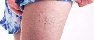
Telangiectasia (spider veins) of the lower extremities.
Telangiectasias of the lower extremities are dilated intradermal veins with a diameter of no more than 1 mm. Their formation, as a rule, indicates a chronic venous disease, the development of which is based on inflammatory processes and changes in venous outflow. The process of blood movement through the veins is ensured by cardiac and muscle contractions, and respiratory processes. At the same time, circulation to the lower extremities is easier than to the heart, so the help of valves is required. Venous valves open during heart contractions, blood flow moves to the heart area, and then close until the next contraction. This prevents backflow of blood in the veins. With chronic venous disease, the valves cannot cope with the load, cannot close tightly, and the blood flow is blocked, forming stagnation. Together with an increase in pressure inside the vessel, these processes lead to stretching of the thin walls of the venous vessels and deformation of the valve leaflets. Dilated vessels form a visible network, can be purple or blue, usually have a tree-like shape.
The main causes of the formation of venous telangiectasia are damage to the venous wall of the capillaries and valvular insufficiency. Additional factors leading to impaired venous outflow: excess weight, heart failure, professional activities associated with the body being in one position for a long time. For example, in people who, due to their profession, are forced to stand, the pressure in the veins of the lower extremities increases, the vessels are stretched and the risk of disease progression is inevitable. Provoking factors include a sedentary lifestyle and high-instep shoes that compress the foot. Muscle contractions during physical activity accelerate blood flow, the volume of blood in the veins decreases and pressure decreases, the outflow of lymph through the lymphatic system increases - these are necessary processes for the prevention and treatment of all disorders of the venous system.
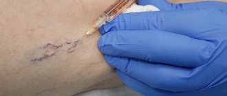
A consultation with a phlebologist includes a clinical examination (detailed history taking, analysis of patient complaints, examination) and, if necessary, instrumental diagnostics. At the same time, the clinical class of the disease and the exact diagnosis are determined, methods of treatment of a certain type of chronic venous disease are discussed. To accurately assess the degree of pathology of the veins and arteries of the lower extremities, accurate diagnosis is necessary. Most often in his practice, a phlebologist uses duplex scanning (visualization of blood vessels, determination of blood flow) and Dopplerography as an additional method (determines the direction of blood flow in the vessels, its presence or absence in the main veins, assesses the condition of peripheral veins). More complex diagnostics are indicated for patients with trophic tissue changes and before surgery. Only after consultation and diagnosis does the doctor choose a method for removing venous telangiectasias. To achieve a high aesthetic result, several techniques are sometimes combined: laser technologies and sclerotherapy. The treatment regimen is supplemented by the prescription of pharmacotherapy and the mandatory selection of compression stockings.
Laser photocoagulation is indicated for the treatment of small-caliber vessels (up to 0.5 mm), and for the elimination of venous telangiectasias with a diameter of 1 mm, sclerostherapy shows a good cosmetic effect and elimination of the symptoms of the disease - the introduction under local anesthesia into the lumen of the vessel of a sclerosant in liquid or foam form, which leads to to gluing of its walls (vein fibrosis).
An arsenal of modern medications, sclerotherapy, laser and radio wave coagulation allow the phlebologist to treat vascular pathologies of the first and second clinical classes without resorting to radical methods.
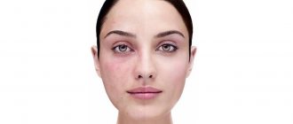
What it is?
Telangiectasia is a type of vascular disease that affects small vessels of the skin . It is expressed in their significant expansion, and appears in the form of capillary networks and stars. The expansion size reaches 0.5 - 1 mm in diameter.
They are divided into two types:
- congenital;
- acquired.
The photo below will allow you to understand in more detail what telangiectasia is.
Telangiectasis and reticular veins - cosmetics or a serious problem?
Many women paid attention to spider veins and stars on their legs, which clearly did not add to their beauty. Often these formations significantly worsen the appearance of the lower extremities. Quite often, despite the pronounced disfiguring veins in the legs, patients have no signs of main varicose veins. In this case, the problem becomes primarily cosmetic. But despite the fact that the likelihood of thrombophlebitis and thrombosis is quite low (due to the small diameter of the affected veins), blood stagnation occurs in telangiectasis and reticular veins. Of course, the symptoms of CVI will be much more modestly expressed than with the main form of varicose veins, but they will manifest themselves to one degree or another. Therefore, reticular varicose veins and telangioctases cannot be attributed to a purely cosmetic problem. In most public hospitals, the treatment of this pathology is not regulated and patients must go to private clinics.