Compression therapy - why and what it is needed for
If you notice dark, bulging, unsightly veins on your legs and/or thighs, you likely have varicose veins. You may have heard that compression stockings can prevent them or relieve symptoms. You may already have varicose veins, but you haven't worn compression garments before and don't want to start using them. Therefore, the benefits of using compression therapy for varicose veins need to be well explained.
Modern compression jersey looks very nice
To do this, it is necessary to once again explain what varicose veins are, as well as the causes and risk factors for its development. In addition, you need to figure out when it is better to wear compression hosiery for varicose veins, how to buy it, put it on and use it correctly.
Team of Doctors
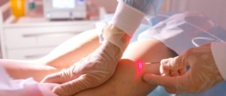
Miniphlebectomy is performed with special phlebological microinstruments. The “sick” veins are removed through small punctures under local anesthesia. An elastic bandage is also applied for a week. Surgical treatment methods have minimal postoperative pain, which is easily relieved with painkillers, are performed on an outpatient basis, last several minutes and do not require inpatient monitoring of the patient. After these operations there are no scars or scars.
The effect after laser surgery and miniphlebectomy is obvious within a week. After sclerotherapy - a few weeks.
In conclusion, we recommend contacting a phlebologist at the first manifestations of varicose veins of the hands. Treatment in the initial stages is much more effective than in its final stages. Man is so perfect that it is possible to return health from illness from almost any condition; but with the progression of the disease and with age, this requires more and more effort and certain conditions.
What are varicose veins?
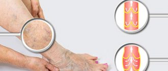
If you have blue, twisted veins that are located near the surface of the skin, bulging and uncomfortable, you probably have varicose veins. Although theoretically these veins can occur in any part of the body, in the vast majority of cases they are located in the lower extremities.
Spider veins are much smaller than varicose veins and are usually located near the surface of the skin. Varicose veins can be located both close to the surface of the skin and deep in the subcutaneous tissue. In the latter case, they are difficult to determine without instrumental research methods. Spider veins can be blue, red, or purple in color and often look like spider webs or tree twigs. Spider veins often appear not only on the legs, but also on the face. They are caused by the same mechanisms as varicose veins, but do not usually lead to the complications caused by varicose veins.
In addition to clearly visible signs of bulging and curving blood vessels, symptoms of varicose veins include:
- Swelling of ankles and feet.
- Feelings of pain, heaviness, or aching in the legs.
- Pulsation or spasms in the lower leg muscles.
- Itching in the legs, especially in the lower leg and ankle, manifests itself as dry skin.
- Darkening and change in skin texture.
- Venous ulcer.
Although varicose veins are not aesthetically pleasing, they do not always lead to serious serious complications if the disease is treated correctly. Varicose veins can also lead to a condition called varicothrombophlebitis, which occurs when blood clots form in a vein near the surface of the skin. This condition is characterized by swelling, pain and redness. The development of varicothrombophlebitis signals a significant circulatory disorder, which also causes other complications of varicose veins. If you have varicose veins or just spider veins, don't ignore them. Instead, you need to visit a phlebologist, undergo a consultation and duplex ultrasound scan of the veins of the lower extremities. Next, you need to discuss with your doctor the tactics of treatment and prevention, the necessary changes in lifestyle to prevent complications and progression of varicose veins.
Arm pain
Gout
Arthritis
Rheumatism
Ulcer
32638 March 26
IMPORTANT!
The information in this section cannot be used for self-diagnosis and self-treatment.
In case of pain or other exacerbation of the disease, diagnostic tests should be prescribed only by the attending physician. To make a diagnosis and properly prescribe treatment, you should contact your doctor. Pain in the hand: causes of occurrence, what diseases it occurs with, diagnosis and treatment methods.
Definition
Pain can be accompanied by various pathological processes of the upper extremities - injuries, degenerative and inflammatory lesions, neurological syndromes. Pain in the arm can be one of the symptoms of diseases of the cardiovascular, respiratory and digestive systems of the body.
Types of hand pain
The variety of pain syndromes is determined not only by the complexity of the structure of the upper limbs, but also by the variety of functional loads. By its nature, the pain can be pulling, shooting, aching. It may bother you when there is strain on the arm, or it may occur at night and disturb sleep. In addition, irradiation (return) of pain to the arm is possible due to myocardial infarction, angina pectoris, colic, cholecystitis, and stomach ulcers.
Possible causes of arm pain
Arm pain can be physiological in nature and be caused by muscle fatigue
after heavy or unusual stress.
It occurs due to the accumulation of anaerobic metabolic products (lactate) in muscle tissue and resolves within two to three days.
After significant physical activity exceeding the muscle endurance threshold, delayed pain may occur (1–2 days after physical activity). Experts believe that they are caused by damage to muscle cells, their membranes, and connections between microfibrils. Such pain is usually long-lasting.
Injuries
– bruises, fractures, tendon sprains, muscle tears – are characterized by a sharp onset of pain and its intensity. Bruises and fractures are accompanied by severe pain, swelling, and hemorrhage from small or large vessels.
A sprain occurs when there is a sudden movement in the joint, lifting significant weights, or a fall with emphasis on the arm and is characterized by varying degrees of damage to the connective tissue fibers. This injury is accompanied by severe pain, swelling and limited mobility in the joint. Often, sprains occur in the area of the wrist and elbow joints after heavy loads or monotonous movements repeated many times.
Muscle ruptures and tears occur when overused. In addition to severe pain, there is hemorrhage in the area of injury and the inability to tense the muscle.
One of the largest groups of diseases that causes severe pain in the arm is tunnel syndromes
caused by compression and inflammation of the nerve in a narrow space (tunnel) formed by the muscles, ligaments and bones of the arm.
Additional factors that increase the risk of such inflammation are endocrine diseases (diabetes mellitus, hypothyroidism), impaired joint mobility due to arthritis or rheumatism, tumor formations in the nerve area (neurofibroma, schwannoma) or beyond it (hemangioma, lipoma). As a rule, tunnel syndromes develop during prolonged similar movements (when working at a computer, playing tennis) or injuries. This is facilitated by incorrect posture, scoliosis, and osteochondrosis.
Inflammation of tendons and joint ligaments
also develops with significant physical stress and injury, especially in cases where the tendons are attached to powerful muscles. Stretching and microtrauma at the attachment site are accompanied by aching, aching pain, which can become unbearable during physical activity. Inflammation leads to swelling and limited mobility in the joint. A common pathology of the hand is epicondylitis, in which inflammation affects the junction of the muscle and ligament of the elbow joint. In this case, the pain is localized in the elbow area and accompanies movement in the elbow, hand and fingers.
Joint damage due to arthritis
causes not only pain, but also stiffness of movement. The causes of such damage may be osteoarthritis, gout, etc.
In any case, all signs of inflammation are present: pain, swelling, local increase in temperature, redness of the skin in the projection of the joint, impaired movement in the joint.
Rheumatoid
arthritis
is characterized by damage to first the small and then large joints of the hand, which is accompanied by stiffness of movement in the morning. In addition to increased pain, joint deformation and limited mobility, the disease is also characterized by general manifestations of the disease - fatigue, sweating of the palms and feet, weight loss.
Deposition of uric acid salts (urates) in the joints is the main manifestation of gout
– leads to severe pain. Its intensity is so great that even simply touching the sore joints causes discomfort. The basis of the disease is a metabolic disorder.
With inflammation of the ulnar periarticular synovial bursa - bursitis
– pain occurs in the elbow and is accompanied by swelling of the joint. In addition to severe pain in the arm, which limits movement, local redness of the skin and increased temperature over the joint area may be observed.
Arm pain may be due to anterior scalene syndrome
and be accompanied by muscle spasm and compression of the nerves and vessels of the shoulder girdle. The disease may be based on degenerative changes in the vertebrae (osteochondrosis) of the cervical spine or trauma.
The patient is bothered by pain in the neck and shoulders, which prevents him from raising his arms up and to the sides, taking a deep breath, or tilting his head.
Osteochondrosis of the cervical spine and its complications
(disc protrusion, intervertebral hernia) cause pain in the arm due to pinching of the nerve that innervates it. In this case, not only sharp pain may occur, but also numbness of the hand and impaired sensitivity.
There is another reason that should always be kept in mind when pain in the arm occurs - myocardial infarction
. The patient feels severe pain behind the sternum, which can radiate to the neck, back and left arm. Such pain is not the only sign of this dangerous condition. As a rule, shortness of breath, cold sweat, and difficulty breathing are present.
Diagnosis and examinations for arm pain
To diagnose a condition that may be causing arm pain, it is necessary to consider how acute the pain is and the events that precede its onset. With severe injuries, making a diagnosis is usually not difficult, but to distinguish a bruise from a fracture, radiography is necessary.
Chronic venous insufficiency and varicose veins
Varicose veins often manifest as symptoms of chronic venous insufficiency, which occurs when venous outflow in the lower extremities is impaired. The valves in our veins control blood flow; when they work properly, the blood in the veins flows in only one direction, from the distal portions to the heart. When the valves in the veins stop functioning normally, blood flow slows, contributing to the development of symptoms of chronic venous insufficiency.
Chronic venous insufficiency and varicose veins do not occur spontaneously on their own. The following factors often contribute to the development of varicose veins and chronic venous insufficiency:
- Sitting or standing for long periods of time.
- Deep vein thrombosis.
- Trauma to the lower limb as a result of surgery.
- Family history of venous insufficiency and varicose veins.
- Gender (women are more likely to develop varicose veins than men).
- Pregnancy. The growing fetus puts pressure on the veins of the abdominal cavity, changing hemodynamics in the lower extremities.
- Age (aging processes weaken the valves of blood vessels).
- Overweight or obesity (excess weight puts extra strain on your veins).
- Weak motor activity.
- Smoking.
If you have a combination of these risk factors, you need to visit a phlebologist and undergo an appropriate examination. You should also pay close attention to your doctor's recommendations on how to keep your veins healthy and prevent problems such as varicose veins from developing in the future.
Thrombophlebitis of the superficial veins: diagnosis and treatment
D
This type of pathology is a very common disease of the venous system, which is encountered by doctors of any specialty.
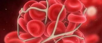
Currently, terms such as phlebothrombosis and varicothrombophlebitis are also often used in medical practice. All of them are legal to use, but the following points should be taken into account. Phlebothrombosis is considered as an acute obstruction of a vein as a result of hypercoagulation, which is the leading mechanism. But after 5–10 days, the resulting thrombus causes reactive inflammation of the tissues surrounding the vein with the development of phlebitis, that is, phlebothrombrosis transforms into thrombophlebitis
.
The term “varicothrombophlebitis” clearly indicates, in fact, the initial cause of thrombosis that occurs against the background of varicose veins that the patient already had.
The pathology of the venous system listed above in the overwhelming majority of clinical cases occurs in the large saphenous vein system and much less often in the small saphenous vein system.
Thrombophlebitis of the veins in the upper extremities is extremely rare, and the main provoking factors for their occurrence are multiple punctures for administering medications or prolonged placement of a catheter in a superficial vein.
Particular attention should be paid to patients with spontaneously occurring thrombi on the upper and lower extremities, not associated with iatrogenic effects. In such cases, the phenomenon of thrombophlebitis can be suspected as a manifestation of a paraneoplastic reaction caused by the presence of an oncological pathology in the patient, requiring an in-depth multifaceted examination.
Thrombosis in the superficial venous system is provoked by the same factors that cause thrombosis of the deep venous system of the lower extremities. These include: age over 40 years, the presence of varicose veins, cancer, severe disorders of the cardiovascular system (cardiac decompensation, occlusion of the main arteries), physical inactivity after major operations, hemiparesis, hemiplegia, obesity, dehydration, common infections and sepsis, pregnancy and childbirth, taking oral contraceptives, limb trauma and surgical interventions in the area of venous trunks.
Thrombophlebitis can develop in any part of the superficial venous system
, with the most common localization on the lower leg in the upper or middle third, as well as the lower third of the thigh. The overwhelming number of cases of thrombophlebitis (up to 95–97%) were noted in the basin of the great saphenous vein (Kabirov A.V. et al., Kletskin A.E. et al., 2003).
The further development of thrombophlebitis can actually occur in two ways:
1. Relatively favorable course of the disease
, against the background of the treatment, the process stabilizes, thrombus formation stops, inflammation subsides and the process of organizing a blood clot begins, followed by recanalization of the corresponding part of the venous system. But this cannot be considered a cure, because... Damage to the initially altered valve apparatus always occurs, which further aggravates the clinical picture of chronic venous insufficiency.
Clinical cases are also possible when a fibrotic thrombus tightly obliterates the vein and its recanalization becomes impossible.
2. The most unfavorable and dangerous option
in terms of the development of local complications - ascending thrombosis along the great saphenous vein to the fossa ovale or the transition of the thrombotic process through the communicating veins to the deep venous system of the leg and thigh.
The main danger of the course of the disease according to the second option is the threat of developing a complication such as pulmonary embolism (PE), the source of which can be a floating thrombus from the system of the small or great saphenous vein, as well as secondary thrombosis of the deep veins of the lower extremities.
It is quite difficult to judge the frequency of thrombophlebitis among the population, but if we take as a basis the fact that among patients hospitalized in surgical departments with this pathology, more than 50% had varicose veins, then taking into account the millions of patients with this pathology in the country, this figure looks very impressive and The problem is acquiring great medical and social significance.
The age of patients ranges from 17 to 86 years and even older, and the average age is 40–46 years, that is, the working population.
Considering the fact that with thrombophlebitis of the superficial veins, the patient’s general condition and well-being, as a rule, do not suffer and remain quite satisfactory, this creates for the patient and his relatives the illusion of relative well-being and the possibility of various methods of self-medication.
As a result, such behavior of the patient leads to late referral for qualified medical care, and often the surgeon is faced with complicated forms of this “simple” pathology, when high ascending thrombophlebitis or deep vein thrombosis of the limb occurs.
Clinical picture
The clinical picture of the disease is quite typical in the form of local pain in the projection of the saphenous veins at the level of the leg and thigh
with the involvement of the tissues surrounding the vein in the process, up to the development of sharp hyperemia of this zone, the presence of compactions not only of the vein, but also of the subcutaneous tissue. The longer the zone of thrombosis, the more pronounced the pain in the limb, which forces the patient to limit its movement. Hyperthermic reactions in the form of chills and an increase in temperature to 38–39°C are possible.
Quite often, even a banal acute respiratory disease becomes a provoking moment for the occurrence of thrombophlebitis, especially in patients with varicose veins of the lower extremities.
The examination is always carried out from both sides - from the foot to the groin area. Attention is drawn to the presence or absence of pathology of the venous system, the nature of changes in skin color, local hyperemia and hyperthermia, and swelling of the limb. Severe hyperemia is typical for the first days of the disease; it gradually decreases by the end of the first week.
When thrombophlebitis is localized in the small saphenous vein, local manifestations are less pronounced than when the trunk of the great saphenous vein is affected, which is due to the peculiarities of the anatomy. The superficial layer of the shin's own fascia, covering the vein, prevents the transition of the inflammatory process to the surrounding tissues. The most important point is to find out the timing of the onset of the first symptoms of the disease, the speed of their increase, and whether the patient has attempted to influence the process with medication.
So, according to A.S. Kotelnikova et al. (2003), the growth of a blood clot in the great saphenous vein system is up to 15 cm per day. It is important to remember that in almost a third of patients with ascending thrombosis of the great saphenous vein, its true upper limit is located 15–20 cm above the level determined by clinical signs (V.S. Savelyev, 2001), that is, this fact should every surgeon should take into account when advising a patient with thrombophlebitis of a vein at the hip level, so that there is no undue delay in the operation aimed at preventing pulmonary embolism.
It should also be considered inappropriate to administer local anesthetics and anti-inflammatory drugs to the area of the thrombosed vein on the thigh, since, while relieving pain, this does not prevent the growth of the thrombus in the proximal direction. Clinically, this situation becomes difficult to control, and duplex scanning can really only be used in very large medical institutions.
Differential diagnosis
should be carried out with erysipelas, lymphangitis, dermatitis of various etiologies, erythema nodosum.
Instrumental and laboratory diagnostics
For a very long time, the diagnosis of thrombophlebitis of the superficial veins was made by a doctor based only on the clinical symptoms of the disease, since there were virtually no non-invasive methods for characterizing venous blood flow. The introduction of ultrasound diagnostic methods into practice has opened a new stage in the study of this common pathology. But the clinician must know that among ultrasound methods for diagnosing venous thrombosis, the decisive role is given to duplex scanning, since only with its help can one determine a clear boundary of thrombosis, the degree of organization of the thrombus, the patency of the deep veins, the condition of the communicants and the valve apparatus of the venous system. Unfortunately, the high cost of this equipment still sharply limits its practical use in outpatient and inpatient settings.
This study is indicated primarily for patients with suspected embologenic thrombosis, that is, when there is a transition of a thrombus from the superficial to the deep venous system through the sapheno-femoral or sapheno-popliteal anastomosis.
The study can be carried out in several projections, which significantly increases its diagnostic value.
Phlebographic examination
The indications for it have been sharply narrowed. The need to perform it arises only in the event of a thrombus spreading from the great saphenous vein to the common femoral and iliac vein. Moreover, this study is carried out only in cases where the results of duplex scanning are doubtful and their interpretation is difficult.
Laboratory diagnostic methods
In a routine clinical blood test, attention is paid to the level of leukocytosis and the level of ESR.
It is advisable to study C-reactive protein, coagulogram, thrombelastogram, prothrombin index level and other indicators characterizing the state of the coagulation system. But the scope of these studies is sometimes limited by the capabilities of the laboratory service of a medical institution.
Treatment
One of the important points that determine the outcome of the disease and even the fate of the patient is the choice of tactics for the optimal treatment option for the patient.
If thrombophlebitis is localized at the level of the lower leg, the patient can undergo treatment on an outpatient basis, under the constant supervision of a surgeon. In these conditions, it is necessary to explain to the patient and his relatives that if signs of thrombosis spreading to the hip level appear, the patient may need to be hospitalized in a surgical hospital. Delay in hospitalization is fraught with the development of complications, including the occurrence of pulmonary embolism.
In cases where thrombophlebitis at the level of the leg, treated for 10–14 days, does not respond to regression, the question of hospitalization and more intensive treatment of the disease should also be raised.
One of the main issues in the treatment of patients with thrombophlebitis of the superficial veins is the discussion of the need for the patient to comply with strict bed rest
.
It is now a recognized fact that strict bed rest is indicated only for patients who already had clinical signs of pulmonary embolism or there are clear clinical data and the results of instrumental studies indicate the embologenic nature of thrombosis.
The patient's physical activity should be limited only to severe physical activity (running, lifting weights, performing any work that requires significant muscle tension in the limbs and abdominals).
General principles of treatment of superficial vein thrombophlebitis
These principles are truly general for both conservative and surgical treatment of this pathology. The main objectives of treatment
these patients are:
- Act as quickly as possible on the source of thrombosis and inflammation to prevent its further spread.
- Try to prevent the transition of the thrombotic process to the deep venous system, which significantly increases the risk of developing pulmonary embolism.
- Treatment should be a reliable method of preventing recurrent thrombosis of the venous system.
- The treatment method should not be strictly fixed, since it is determined primarily by the nature of the changes occurring in the limb in one direction or another. That is, it is quite logical to switch or complement one treatment method with another.
Of course, conservative treatment
indicated for the absolute majority of patients with “low” superficial thrombophlebitis of the saphenous veins.
Once again, it should be emphasized that reasonable physical activity of the patient improves the function of the muscle pump, which is the main determinant in ensuring venous outflow in the inferior vena cava system.
The use of external compression (elastic bandage, stockings, tights) in the acute phase of inflammation can cause some discomfort, so this issue should be resolved strictly individually.
The issue of using antibiotics in this category of patients is quite controversial. The doctor must remember the possible complications of this therapy (allergic reactions, intolerance, provocation of blood hypercoagulation). Also, the question of the advisability of using anticoagulants (especially direct action) in this group of patients is far from clear.
The doctor must remember that the use of heparin after 3-5 days can cause thrombocytopenia in the patient, and a decrease in the platelet count by more than 30% requires discontinuation of heparin therapy. That is, difficulties arise in controlling hemostasis, especially in an outpatient setting. Therefore, it is more appropriate to use low molecular weight heparins (dalteparin, nadroparin, enoxaparin), since they extremely rarely cause the development of thrombocytopenia and do not require such careful monitoring of the coagulation system. The positive thing is that these drugs can be administered to the patient once a day. A course of treatment requires 10 injections, and then the patient is transferred to indirect anticoagulants.
In recent years, ointment forms of heparin (Lioton-gel, Hepatrombin) have appeared for the treatment of these patients. Their main advantage is fairly high doses of heparin, which are delivered directly to the site of thrombosis and inflammation.
Of particular note is the targeted effect on the zone of thrombophlebitic changes of the drug Gepatrombin
(“Hemofarm” – Yugoslavia), produced in the form of ointment and gel.
Unlike Lyoton, it contains 2 times less heparin, but additional components - allantoin and dexpanthenol, which are part of the ointment and gel "Gepatrombin", as well as pine essential oils, which are part of the gel, have a pronounced anti-inflammatory effect, reduce skin itching and local pain in the area of thrombophlebitis. That is, they help relieve the main symptoms of thrombophlebitis. The drug Gepatrombin has a strong antithrombotic effect.
It is used topically by applying a layer of ointment to the affected areas 1-3 times a day. If there is an ulcerative surface, the ointment is applied in the form of a ring up to 4 cm wide around the perimeter of the ulcer. The good tolerability of the drug and the versatility of its effect on the pathological focus puts this medicine at the forefront in the treatment of patients with thrombophlebitis both on an outpatient basis and during treatment in hospitals. Hepatrombin can be used in a complex of conservative treatment or as a means aimed at relieving inflammation of the venous nodes after the Troyanov-Trendelenburg operation, as a method of preparation for the second stage of the operation.
The complex of conservative treatment of patients should include non-steroidal anti-inflammatory drugs
, which also have an analgesic effect. But the clinician must remember to exercise extreme caution when prescribing these drugs to patients with diseases of the gastrointestinal tract (gastritis, peptic ulcer) and kidneys.
Phlebotonics, already well known to doctors and patients, have proven themselves well in the treatment of this pathology.
(rutoside, troxerutin, diosmin, gingko biloba and others) and
disaggregants
(acetylsalicylic acid, pentoxifylline). In severe cases with extensive phlebitis, intravenous transfusions of rheopolyglucin 400–800 ml IV for 3 to 7 days are indicated, taking into account the cardiac status of the patient due to the danger of hypervolemia and the threat of developing pulmonary edema.
Systemic enzyme therapy has limited use in practice due to the high cost of the drug and a very long course of treatment (from 3 to 6 months).
Surgery
The main indication for surgical treatment of thrombophlebitis, as previously indicated, is the growth of a blood clot along the great saphenous vein above the middle third of the thigh or the presence of a blood clot in the lumen of the common femoral or external iliac vein, which is confirmed by phlebography or duplex scanning. Fortunately, the latter complication does not occur so often, in only 5% of patients with ascending thrombophlebitis (I.I. Zatevakhin et al., 2003). Although individual reports indicate a significant frequency of this complication, reaching even 17% in this group of patients (N.G. Khorev et al., 2003).
Methods of anesthesia - different options are possible: local, conduction, epidural anesthesia, intravenous, intubation anesthesia.
The position of the patient on the operating table is of certain importance - the foot end of the table should be lowered.
The generally accepted operation for ascending thrombophlebitis of the great saphenous vein is the Troyanov–Trendelenburg operation.
.
The surgical approach used by most surgeons is quite typical - an oblique incision below the inguinal fold according to Chervyakov or the inguinal fold itself. But at the same time, it is important to take into account the main clinical point: if there are instrumental data or clinical signs of a thrombus moving into the lumen of the common femoral vein, then it is more advisable to use a vertical incision that provides control over the thrombosed great saphenous vein and the trunk of the common femoral vein, when it is sometimes necessary to clamp it moment of thrombectomy.
Some technical features of the operation:
1. Mandatory isolation, intersection and ligation of the trunk of the great saphenous vein in the area of its mouth.
2. When opening the lumen of the great saphenous vein and detecting a thrombus in it that extends beyond the level of the ostial valve, the patient must hold his breath at the height of inspiration during surgery under local anesthesia (or this is done by an anesthesiologist for other types of anesthesia).
3. If the thrombus “does not form on its own,” then a balloon catheter is carefully inserted through the saphenofemoral anastomosis at the height of inspiration and thrombectomy is performed. Retrograde blood flow from the iliac vein and antegrade blood flow from the superficial femoral vein are checked.
4. The stump of the great saphenous vein must be sutured and ligated; it must be short, since a stump that is too long is an “incubator” for the occurrence of thrombosis, which creates a threat of the development of pulmonary embolism.
In order to discuss options for this routine operation, it should be noted that during the Troyanov-Trendelenburg operation, some surgeons suggest performing thrombectomy from the great saphenous vein and then injecting a sclerosant into it. The expediency of such manipulation is questionable.
The second stage of the operation - removal of thrombosed varicose nodes and trunks is carried out according to individual indications in a period of 5-6 days to 2-3 months as local inflammation is relieved, in order to avoid suppuration of wounds in the postoperative period, especially with trophic skin disorders.
When performing the second stage of the operation, the surgeon must ligate the perforating veins after preliminary thrombectomy, which improves the healing process.
All conglomerates of varicose nodes must be removed to avoid the further development of severe trophic disorders.
A very wide range of general surgeons and angiosurgeons are involved in the surgical treatment of this group of patients.
The apparent simplicity of treatment sometimes leads to tactical and technical errors. Therefore, this topic is almost constantly present at scientific conferences. Literature:
1. Zatevakhin I.I. with co-authors. “Angiology and Vascular Surgery” No. 3 (supplement) 2003, pp. 111–113.
2. Kabirov A.V. with co-authors. “Angiology and Vascular Surgery” No. 3 supplement 2003, pp. 127–128.
3. Kletskin A.E. with co-authors. “Angiology and Vascular Surgery” No. 3 (supplement) 2003, pp. 161–162.
4. Kotelnikov A.S. with co-authors. “Angiology and Vascular Surgery” No. 3 (supplement) 2003, pp. 168–169.
5. Revskoy A.K. “Acute thrombophlebitis of the lower extremities” M. Medicine 1976
6. Savelyev V.S. "Phlebology" 2001
7. Khorev N.G. “Angiology and Vascular Surgery” No. 3 (supplement) 2003, pp. 332–334.
Compression therapy for varicose veins (mechanism of action)
Compression therapy for varicose veins helps to apply a certain pressure on the blood vessels, preventing the accumulation of blood and promoting proper one-way blood flow. Compression stockings can also help manage swelling, especially if you wear compression stockings in the morning before you begin physical activity, which is when leg swelling occurs. The dense tissue prevents the veins from expanding as usual, which helps blood flow faster. Thanks to the combination of these effects, compression stockings for varicose veins can help relieve soreness, aching sensations, as well as muscle throbbing and spasms. However, you should discuss the use of compression stockings with your doctor to avoid possible side effects and complications.
The mechanism of action of compression hosiery for varicose veins
If you already have varicose veins, compression stockings can help relieve symptoms and improve the appearance of your lower legs. In modern practice, compression products of varying degrees of compression, and therefore clinical effect, are used.
- Compression stockings class 1, 15-20 mm Hg. They provide moderate compression and are recommended for prophylaxis in healthy people, as well as in case the patient refuses a higher level of compression.
- Level 2 compression, 20-30 mm Hg, is more effective and relevant for patients with varicose veins.
- 3rd compression level, 30-40 mm Hg. Art., provides significant pressure. In some clinical situations it will be the most appropriate.
- 4th compression level, 40-50 mm Hg. Art. This knitwear is used in selected clinical cases under close medical supervision.
How to choose compression hosiery for varicose veins
Once you have determined the required level of compression with your healthcare provider, you will need to purchase a specific product.
The best compression stocking available today
Leading criteria for choosing compression hosiery:
- Length of compression product. Varicose veins can occur both only on the legs and throughout the entire lower limb. The location of your varicose veins will influence which type of compression garment you should choose. If you have varicose veins only within the shins, then you should pay attention to knee-length compression socks. If varicose veins are on the legs and thighs, then groin-length stockings will be the best choice. Some patients in this case prefer compression tights.
- Choosing the size of compression hosiery. This aspect can be entrusted to specialists in a specialized salon or you can measure the parameters of the lower limb yourself. In the latter case, you may need to measure the circumference of your ankle, calf and/or thigh. On your own or with the help of a specialist, you will need to compare the measurement data with the size table to find your personal size. Choosing the right compression garment size is very important: if it is too small or large, the compression stockings will not provide the right level of pressure. This will lead to the ineffectiveness of compression therapy or the impossibility of using hosiery.
- Choosing a compression product style. The modern compression products industry offers a wide selection of fabrics, styles and product shapes. You need to make a choice between opaque or transparent fabric, the presence or absence of a pattern, closed or open toe. You will also need to choose a color, which varies from product to product. Basic neutral colors such as black, beige and white are almost always available. Certain types of compression products are presented in bright solid colors, and there are options with various patterns.
Using elastic bandages for varicose veins
Currently, there are not many situations where modern experts recommend compression bandages, but still in some clinical cases this is not without meaning.
Compression bands can be adjusted to achieve different levels of pressure, which is both an advantage and a significant disadvantage. Even medical workers do not always apply compression bandages correctly, which leads to undesirable effects. What can we say about ordinary patients? In situations with unstable edema, compression bandages may have a better effect than compression stockings. The difficulty lies in applying compression bandages, which is best left to professionals. Otherwise, the target effect of compression therapy may not be achieved. And in the worst case scenario, complications arise from the improper use of compression bandages. Therefore, in the vast majority of cases, modern specialists choose compression hosiery as a more controlled and universal means of helping patients with varicose veins.
The use of compression therapy in the treatment of varicose veins
Compression therapy today is an integral part of the process of radical treatment of varicose veins. It is difficult to imagine any intervention on the veins of the lower extremities today without an important component of compression. In the version that has become classic, compression stockings or knee socks are used for three to four weeks after intervention on the veins of the lower extremities. The goals of compression therapy in this case are: prevention of complications and side effects of the intervention, relief of symptoms of chronic venous insufficiency. In some cases, the period of use of compression hosiery may be increased or decreased.
Most people can wear compression stockings for varicose veins without any side effects. However, you should discuss the situation with your healthcare provider if you experience redness, skin imperfections, pain, numbness, or swelling while using compression products. If you want to stop using compression stockings due to discomfort, talk to your doctor first. Perhaps the reason is an unsuccessfully chosen size, type, or insufficiently high quality of the compression product.
Quality, comfort and convenience of compression hosiery
Compression therapy cannot cure varicose veins on its own, but it can help manage symptoms and improve circulation and overall quality of life. If you are already at risk of developing varicose veins based on the criteria discussed above, you can consult a phlebologist and use products with a certain degree of compression. In combination with lifestyle changes, compression stockings or knee socks will also be the best prevention of the development or recurrence of varicose veins.
Patient questions about compression therapy
Can varicose veins (varicose veins) be treated with compression stockings?
Compression stockings or knee socks will help cope with some of the symptoms of varicose veins (swelling, heaviness), and compression is also a good prevention of complications of varicose veins. However, only removal of varicose veins can completely rid you of such a disease as varicose veins.
Is it useful to wear compression hosiery for varicose veins? Will it impair blood circulation?
The use of compression hosiery improves pathologically altered hemodynamics in varicose veins and has an extremely positive effect.
Can compression stockings be used for trophic venous ulcers?
The use of compression therapy in the presence of a trophic venous ulcer is a necessary component of treatment, without which it is almost impossible to achieve healing of the ulcer.
How long should I wear compression after surgery?
Most patients are advised to wear compression stockings for about three to four weeks after varicose vein surgery. In some cases, the period of wearing compression can be changed upward or downward.
Symptoms of phlebitis
A typical inflammatory disease is characterized by a symptomatic “quartet”:
- pain;
- redness;
- edema;
- dysfunction.
Their intensity, duration and localization are determined by the location and nature of the vessel damage, and related factors.
If the superficial veins become inflamed, the disease manifests itself locally:
- Tension, density and swelling of tissues.
- Severe pain along the vessel.
- General hyperemia and increased skin temperature, bright stripes along the inflamed vein.
If the inflammation affects the deep veins, the symptoms change. Pain, tension and swelling remain, but instead of redness, the skin becomes milky white. Severe weakness appears in the affected limb (most often the deep veins of the lower limb are affected). Its cooling and weakening of pulsation may be observed (especially in complicated cases - thrombophlebitis).
The general condition of phlebitis of superficial and deep localization is characterized by:
- weakness and dizziness;
- high temperature;
- enlargement of nearby lymph nodes.
With phlebitis of the deep veins, these signs are more pronounced, and the malaise itself is longer lasting.
Similar symptoms are characteristic of both the acute form of phlebitis and during the period of exacerbation of a chronic disease.
Prolonged, intense and/or frequent inflammation of the veins (especially deep ones) is fraught with the involvement of neighboring tissues (including lymph nodes) in the inflammatory process with its subsequent generalization. And the development of such serious illnesses as:
- chronic venous stagnation and venous insufficiency;
- abscesses and phlegmons;
- gangrene;
- formation of ulcers;
- thrombosis;
- elephantiasis;
- pulmonary embolism.