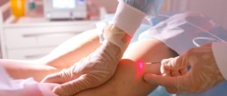
What are varicose veins?
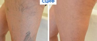
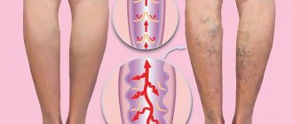
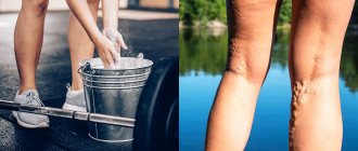
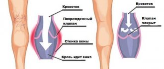
Varicose veins (in common parlance - varicose veins ) are overstretched, irregularly shaped, tortuous blood vessels that have lost their elasticity. They are increased in length and width and look like thick, convoluted blue strands that are visible under the skin. Veins become this way when the venous valves are missing or for some reason cannot perform their functions. If the valves do not work properly, blood flows through the veins in the opposite direction, downwards, accumulating in the lower sections of the veins and bursting their walls. As a result, the veins lose their natural shape, and a pathological chain of various complications begins.
Differences
How are arteries different from veins? These blood vessels differ significantly in many ways.
Arteries and veins, first of all, differ in the structure of the wall
According to the structure of the wall
Arteries have thick walls, they have a lot of elastic fibers, smooth muscles are well developed, they do not fall off unless they are filled with blood. Due to the contractility of the tissues that make up their walls, oxygenated blood is quickly delivered to all organs. The cells that make up the layers of the walls ensure the smooth passage of blood through the arteries. Their inner surface is corrugated. The arteries must withstand the high pressure that is created by powerful surges of blood.
The pressure in the veins is low, so the walls are thinner. They fall off when there is no blood in them. Their muscle layer is not able to contract like arteries. The surface inside the vessel is smooth. Blood moves through them slowly.
In veins, the thickest membrane is considered to be the outer one, in arteries it is the middle one. Veins do not have elastic membranes, arteries have an internal and an external one.
By shape
The arteries have a fairly regular cylindrical shape, they are round in cross section.
Due to the pressure of other organs, the veins are flattened, their shape is tortuous, they either narrow or expand, which is due to the location of the valves.
In count
In the human body there are more veins and fewer arteries. Most middle arteries are accompanied by a pair of veins.
According to the presence of valves
Most veins have valves that prevent blood from flowing backwards. They are located in pairs opposite each other throughout the entire length of the vessel. They are not found in the portal cava, brachiocephalic, iliac veins, as well as in the veins of the heart, brain and red bone marrow.
In arteries, valves are located as vessels exit the heart.
By blood volume
Veins circulate approximately twice as much blood as arteries.
By location
The arteries lie deep in the tissues and approach the skin only in a few places, where the pulse is heard: on the temples, neck, wrist, and instep of the feet. Their location is approximately the same for all people.
Veins are mostly located close to the surface of the skin
The location of the veins may vary from person to person.
To ensure blood movement
In the arteries, blood flows under the pressure of the force of the heart, which pushes it out. At first the speed is about 40 m/s, then gradually decreases.
Blood flow in the veins occurs due to several factors:
- pressure forces depending on the push of blood from the heart muscle and arteries;
- the suction force of the heart during relaxation between contractions, that is, the creation of negative pressure in the veins due to the expansion of the atria;
- suction effect on the chest veins of respiratory movements;
- contractions of the muscles of the legs and arms.
In addition, approximately a third of the blood is in the venous depots (in the portal vein, spleen, skin, walls of the stomach and intestines). It is pushed out from there if it is necessary to increase the volume of circulating blood, for example, during massive bleeding or during high physical exertion.
How common are varicose veins?
Varicose veins are one of the most common diseases of the vascular system. According to some statistical estimates, from varicose veins . The number of people who have varicose veins increases with age, and women are affected much more often than men. According to statistics, in the age group under 25 years only 8% of women suffer from varicose veins, and in the older age group - 55 years and older - 64% of women are affected by varicose veins.
How can you recognize varicose veins in yourself?
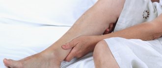
The most common sign of varicose veins is fatigue, dull pain, a feeling of heaviness and fullness in the legs after sitting or standing for a long time. Often these symptoms appear or worsen in the evening. However, it is usually impossible to determine exactly where it hurts. And if these unpleasant symptoms - fatigue, heaviness, pain - go away after resting with your legs elevated, then they are really caused by varicose veins (unless some other cause is reliably identified).
However, do not rush to blame everything on varicose veins, especially if there are no external signs in the form of dilated veins. Some other painful conditions may also exhibit the same symptoms.
Leg cramps
With varicose veins, painful nighttime cramps in the leg muscles can actually occur (in other words, “leg cramps”). Most often, cramps appear in the calf muscles and can sometimes be so painful that the patient wakes up. Moreover, night cramps usually occur after a hard day, when the patient had to stand or sit a lot.
Main functions of the vein
The human venous system, the functions of which are practically invisible in everyday life unless you think about it, ensures the life of the body.
Blood, dispersed to all corners of the body, is quickly saturated with the products of all systems and carbon dioxide.
In order to remove all this and make room for blood rich in useful substances, the veins work.
In addition, hormones that are synthesized in the endocrine glands, as well as nutrients from the digestive system, are also distributed throughout the body through the veins.
And, of course, a vein is a blood vessel, so it is directly involved in regulating the process of blood circulation throughout the human body.
Thanks to it, there is a supply of blood to every part of the body, during paired work with the arteries.
Is varicose veins inherited?
It is now known that varicose veins are hereditary. Scientists even believe that they were able to isolate a separate gene responsible for the development of varicose veins. It is not yet clear whether this gene causes malformations of the venous valves or malformations of the vein walls themselves. But there is no doubt that these studies will help develop a gene therapy technique - perhaps the most promising way to prevent and treat varicose veins. Unfortunately, this is still a matter of the rather distant future, and gene therapy is not yet available to patients with varicose veins.
Varicose veins during pregnancy
Pregnancy does not cause varicose veins, but it is often a trigger for the appearance of varicose veins in those women who are predisposed to it. For example, in people with congenital insufficiency or even absence of venous valves. This fact has already been established quite definitely, because many pregnant women do not develop any varicose veins. Sometimes varicose veins appear only during the fourth, fifth or tenth pregnancy.
And in some women, they appear during pregnancy and disappear immediately after the birth of the child. Pregnancy acts as a triggering factor for varicose veins due to the fact that during pregnancy the content of sex hormones - estrogen and progesterone - in a woman’s blood increases sharply. These hormones, in high concentrations, help soften the venous walls, the veins stretch, and the valves cannot close normally because of this.
Other causes of varicose veins
Such a widespread prevalence of varicose veins in highly developed Western countries is probably associated with the lifestyle of the population. For example, we spend a lot of time sitting on chairs. From kindergarten until graduation, a person sits for at least 40 hours a week (counting approximately 5 hours during the day in class, 3 hours in the evening doing homework, watching TV, and so on 5 days a week). Now let's multiply these hours by 10 months a year, and so on - up to 17 years. Then - work in some institution where you have to sit even longer. When a person sits in a chair, the veins running along the back of the thighs are compressed, and the calf muscles (the rhythmic contractions of which help move venous blood to the heart) do not work.
Another important factor is nutrition. In Western countries, people prefer a low fiber diet. With such a diet, fecal matter becomes denser, and constipation often occurs. When straining to move hard stool, the abdominal muscles tense and the pressure in the abdominal cavity increases significantly. High pressure spreads to the veins running along the back of the abdominal cavity and to the veins of the legs, which dilate, causing the venous valves to leak.
Angioplasty and stenting
The chronic form of NVC syndrome is more difficult to treat. When the venous outflow is decompensated, it becomes necessary to restore the patency of the vessel. Open operations involving the isolation of the IVC and its replacement with a vascular prosthesis are feasible, but very traumatic and ineffective. The artificial vena cava prosthesis often thromboses again and the complex operation becomes completely useless. With the advent of new composite materials for large-diameter stents, our clinic began to perform endovascular methods for restoring the patency of the vena cava. Angioplasty and IVC stenting are performed by experienced endovascular surgeons at the Innovative Vascular Center. The point of the intervention is to restore the patency of the closed segment of the inferior vena cava with a special conductor, a high-pressure balloon and the installation of a metal frame - a stent.
Varicose veins in older people
Why varicose veins more common in older people, and especially common in women?
1. To answer briefly - because their vascular system wears out with age and, sooner or later, fails. However, there are still many objective reasons why older women suffer from varicose veins more often than younger men and women. Firstly, since women generally live somewhat longer than men, there are correspondingly more elderly women than elderly men, and their veins have been working harder for a longer period of time.
2. Men don't get pregnant. Even if varicose veins that appeared in a woman during pregnancy disappear soon after the birth of the child, these veins were still abnormally enlarged within a few months. And with age, all the muscles of the human body, including the smooth muscles of the vascular walls, become less elastic than in youth. And the veins, which had already expanded once, during pregnancy, in old age again become a little wider than normal.
3. Nowadays, many women over the age of 30 are resorting to hormone replacement therapy, which was originally intended to relieve the unpleasant symptoms of menopause. There is no doubt that hormone replacement therapy helps women look younger, feel better, and generally cope with the menopause years more easily. Doctors' observations also confirm that hormone replacement therapy to some extent reduces the frequency of angina attacks and prevents a decrease in bone strength due to osteoporosis.
However, hormonal supplements at the same time soften the vein walls in the same way as increased levels of estrogen and progesterone during pregnancy. This side effect of hormonal pills is all the more dangerous because the walls of the veins are already becoming weaker - due to natural age-related changes in the muscle layer. So, additional clinical studies are needed to definitively clarify this issue.
Veins of the lower extremities
Vienna
- these are vessels that ensure the outflow of blood from organs and tissues to the heart. The wall of the veins consists of three layers: internal (endothelium), middle (muscular) and external (adventitia). Unlike arteries, the walls of veins are thinner and contain few elastic fibers. Therefore, the veins are less elastic and collapse easily. In this case, the diameter of the veins is larger than that of the arteries. The peculiarity of the veins is that their diameter depends on many factors: body position, blood pressure, blood flow speed, condition of the valves and breathing phase.
The peculiarity of the veins in the legs is that they have valves. Venous valves are folds of the inner lining; they allow blood to flow towards the heart and prevent it from flowing back.
The flow of blood is carried out due to respiratory movements, the existence of constant muscle tone of the venous wall, constant support of blood from the arterial end of the capillary bed, and the suction action of the right parts of the heart. The main role in moving blood is played by the so-called “muscular-venous pump”. The deep veins that run in the legs are surrounded on all sides by muscles. When walking and physical activity on the legs, the muscles contract and squeeze blood upward.
If the valves are malfunctioning, during the operation of the muscle pump, there is no drop in pressure in the deep veins during muscle contraction. Venous blood is retained in the sinuses and venules, which leads to changes in capillary exchange parameters and the development of edema, pigmentation, itching and other symptoms of venous insufficiency.
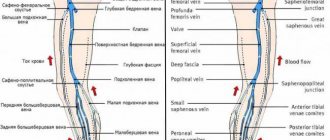
The outflow of blood from the lower extremities is provided by three interconnected and clearly interacting systems: superficial veins, deep veins and communicating veins (perforators) connecting them.
Superficial veins
and their tributaries form venous networks under the skin. They can be felt and are quite visible. This network is especially clearly visible on the dorsum of the foot. From the superficial veins of the leg, it is customary to distinguish the large and small saphenous veins of the leg. Both saphenous veins receive other superficial veins along their path. Varicose veins on the legs concern the superficial veins.
Deep veins
. These veins are located between the muscles and connective tissue. The main outflow of blood (85-90%) is through the deep veins. These veins have valves that prevent blood from flowing back.
Superficial and deep veins are connected to each other by communicating veins
. The reason for the existence of these veins is to equalize the pressure between them. Damage to the valves of the communicating (perforating) veins leads to blood flowing from the deep veins to the superficial ones. Normally, the valves of these veins allow blood to flow in only one direction - from superficial to deep.
Types of varicose veins
Varicose veins are divided into two main groups:
- The first group includes primary varicose veins, caused by a hereditary predisposition to varicose veins.
- The second group includes varicose veins that appear after damage to the venous walls as a result of injury with the formation of blood clots in the veins or thrombosis.
When a clot or thrombus passes through a vein, the integrity of the venous valves is disrupted and secondary varicose veins are formed.
Features of blood movement through the veins
At some stages of movement, for example, from the lower extremities, the blood in the venous canals is forced to overcome gravity, rising almost one and a half meters on average.
This occurs due to the phases of breathing when negative pressure occurs in the chest during inhalation.
Initially, the pressure in the veins located near the chest is close to atmospheric.
In addition, blood is pushed through contracting muscles, indirectly participating in the blood circulation process, raising the blood upward.
Varicose veins
Varicose veins are bundles of thin, purple or red veins that appear around the knees or ankles. (Sometimes such vascular “webs” can appear on the face, near the nose.) These vessels cannot be called varicose veins, since, by definition, varicose veins are veins that are increased in length and in diameter. In fact, these are slightly dilated venules (vessels connecting capillaries to the veins themselves), which are located close to the surface of the skin.
Such dilated venules appear due to increased levels of female sex hormones in the blood and are often found in women taking oral contraceptives. But venules can expand even in the presence of varicose veins of larger veins that do not appear externally. However, women with varicose veins often experience symptoms very similar to those of varicose veins.
Structure and characteristics
The circulatory system has two circles, small and large, which have their own tasks and characteristics. The diagram of the human venous system is based precisely on this division.
Pulmonary circulation
The lesser circle is also called the pulmonary circle. Its task is to carry blood from the lungs to the left atrium.
The capillaries of the lungs have a transition to venules, which then unite into large vessels.
These veins go to the bronchi and parts of the lungs, and already at the entrances to the lungs (gates), they unite into large channels, of which two come out of each lung.
They do not have valves, but go, respectively, from the right lung to the right atrium, and from the left to the left.
Systemic circulation
The large circle is responsible for supplying blood to every organ and tissue area in a living organism.
The upper part of the body is attached to the superior vena cava, which at the level of the third rib flows into the right atrium.
Veins such as the jugular, subclavian, brachiocephalic and other adjacent veins supply blood here.
From the lower part of the body, blood flows into the iliac veins. Here the blood converges through the external and internal veins, which converge into the inferior vena cava at the level of the fourth lumbar vertebra.
For all organs that do not have a pair (except for the liver), blood flows through the portal vein first to the liver, and from here to the inferior vena cava.
Treatment of varicose veins
Treatment depends on the severity of the disease. If the disease does not manifest itself too strongly, then conservative treatment is best:
- regular rest with your feet up,
- elastic bandaging (or special elastic stockings),
- physical exercises for leg muscles.
If these measures are not enough, the veins affected by varicose veins must be surgically removed at the Phlebology Center. Or using new, experimental methods - elastic strengthening of the venous walls is carried out surgically. That is, a special elastic plastic cover is placed on the outer surface of the affected veins in places of varicose veins, where incompetent venous valves are located. And finally, to treat dilated venules or varicose veins of small veins remaining after surgery, sclerotherapy - that is, the introduction of sclerosing substances into the areas of dilation, which causes clogging of the pathological vein. Blood returns to the heart through normal venous vessels.
Introduction:
The circulatory system is responsible for moving blood throughout the body. The main components of this system are the heart and blood vessels. The heart is the central organ of the circulatory system. With each heartbeat, blood is pumped through the arteries and delivers oxygen and nutrients to various organs and tissues, after which the blood returns back to the heart through other blood vessels - veins.
There are three types of blood vessels that play different roles. The two main types are arteries and veins. Arteries carry oxygenated and nutrient-rich blood away from the heart, and veins return waste blood back to the heart. Lymphatic vessels are the third component; they filter and “purify” the liquid part of the blood - plasma, before returning it to the general bloodstream.
Complications during treatment
The main danger with conservative treatment (elastic stockings, exercise and resting with legs elevated) is its possible ineffectiveness.
Surgical treatment of varicose veins of the lower extremities should currently be performed by experienced vascular surgeons and phlebologists . Often complications and relapses after surgical treatment are caused by the fact that the operation was not performed by a specialist from the phlebology center.
With sclerotherapy, the main nuisance is small dark spots that can remain at the injection sites for several months, or in some cases forever.
Dilated veins after surgical treatment
If varicose veins have been removed, varicose veins will no longer appear in their place. However, sometimes varicose veins are found after surgery - in veins that were not previously affected, or in small veins that were not identified during the preoperative examination. Varicose veins after surgery appear because the blood is forced to find new outflow paths. At the same time, a larger volume of blood is redistributed to the remaining veins than before, and if there were any defects in the valves or walls, then new problems arise. New varicose veins, as a rule, bring cosmetic inconvenience and can be easily eliminated by a phlebologist using modern sclerotherapy techniques.
Puncture of the right subclavian vein
Place the patient on his back, arms along the body, turn his head to the left. To move your shoulders back and down, place a bolster between your shoulder blades. To increase the filling of the central veins and reduce the risk of air embolism, place the Trendelenburg position (the head end of the table is lowered 15° downwards), if the bed design does not allow this - horizontal.
Feel the jugular notch of the sternum, sternoclavicular and acromioclavicular joints. Next, treat the skin with an antiseptic solution and limit the puncture site with sterile wipes. The puncture point is located 2-3 cm below the clavicle, on the border of the middle and medial thirds of it. Infiltrate the skin and subcutaneous tissue around the puncture site with 5-10 ml of 1% lidocaine solution.
Insert the needle through the indicated point until it touches the collarbone. Gradually push the end of the needle down until it is just below your collarbone. Then rotate and point the needle at the jugular notch. Slowly advance the needle forward, maintaining a vacuum in the syringe, until blood is drawn. The cut end of the needle should be turned towards the heart - this increases the likelihood of correct installation of the catheter. Try to keep the needle parallel to the plane of the bed (to avoid puncture of the subclavian artery or pleura);
If you miss a vein, slowly withdraw the needle under the skin while maintaining vacuum in the syringe. Rinse the needle and make sure it is clear. Try again, taking the injection direction a little more cranial.
