- Gallery
- Reviews
- Articles
- Licenses
- Vacancies
- Insurance partners
- Partners
- Controlling organizations
- Schedule for receiving citizens for personal requests
- Online consultation with a doctor
- Documentation
Increased intracranial pressure occurs with an increase in the volume of brain tissue, cerebrospinal fluid, or a voluminous pathological process in a relatively closed cranial cavity.
You need to imagine that the head is, first of all, the tightly connected bones of the skull, inside which the brain is located. And naturally, an increase in internal volume increases pressure on the bones of the skull and, accordingly, on the tissues and vessels of the brain.
The accumulation of cerebrospinal fluid (CSF) in the cranial cavity is called hydrocephalus. It can be either external, i.e. accumulation of cerebrospinal fluid around the brain and internally (enlarged cerebral ventricles). This pathology can occur at any age - from newborns to the elderly. Each age interval has its own causes, there are age-related characteristics of manifestations and age-related characteristics of treatment.
False alarm
My three month old son regurgitates food frequently. Could this be a sign of increased intracranial pressure?
Olga, Kaliningrad
Article on the topic
Burped again! How to understand that a child has a symptom of a congenital disease - In 90% of cases, regurgitation in children under one year of age, especially under the age of 4 months, is caused by immaturity or a disorder of the digestive, less often, nervous system.
And in very rare cases, children with the symptom you mentioned have a syndrome of increased intracranial pressure, in which regurgitation is not associated with food intake and can be observed 1.5–2 hours after eating (although this can also occur with other disorders).
Ask your friends: did their children spit up before they were 1 year old? Many people will answer that they vomited and with age it went away without treatment.
What are the indications for epidural anesthesia?
So, let's touch on the main reasons that may be indications for the use of epidural anesthesia:
- Various kidney diseases;
- Increased blood pressure, while the use of epidural anesthesia will reduce the pressure of the woman in labor;
- Diseases of the cardiovascular system, in which any stress on the heart is contraindicated;
- Low pain threshold, which was already mentioned above, and, accordingly, unbearable pain during childbirth, as a result of which it happens that women even lose consciousness and then doctors have to save the life of not only the expectant mother, but also the child;
- Slow dilatation of the cervix;
- The need for a caesarean section.
In some cases, epidural anesthesia may also be necessary for postpartum procedures, such as tubal ligation.
Epidural anesthesia blocks pain in the lower part of the mother's body
Trust but check!
My six-month-old grandson was prescribed neurosonography. A neurologist from a children's clinic suspects he has increased intracranial pressure. How informative is this method and what does it consist of?
Tamara, Moscow
— Neurosonography is an ultrasound of the brain. This method is exclusive to young children. It can be performed in the presence of open fontanels (a non-ossified area of the cranial vault in an infant. - Ed.) and thin cranial sutures. After one year of age, when the fontanelles close, it is almost impossible to carry out this research method, except in rare cases.
Typically, neurosonography allows doctors to detect increased intracranial pressure. However, it is necessary to evaluate not only the results of this, but also of Doppler examination of cerebral vessels, since in this case too, overdiagnosis of the above-mentioned syndrome often occurs. This is why the correct interpretation of the data obtained by a neurologist is so important. If you doubt his conclusions, consult another specialist.
Article on the topic
Pain, what pain! What are the causes of migraines in a child?
Mechanism of action of epidural anesthesia
Epidural anesthesia during childbirth is considered the most modern method of pain relief and is safe for both the expectant mother and her baby, and therefore many women in labor agree to use it due to fear of labor pain. As for the mechanism of action of this type of anesthesia, during the process of epidural anesthesia, a special anesthetic drug is introduced into the places located around the dura mater of the spinal cord.
As a result, pain signals that travel to the brain from the muscles of the lower extremities are blocked. However, the process of administering an anesthetic is repeated every 30 minutes, during which a soft catheter is inserted at the level of the 3-4 lumbar vertebrae of the woman in labor. And if in the case of anesthesia there was complete anesthesia of the lower part of the body, then when using epidural analgesia the nerves still remain partially in working order, which means the woman can move.
Often, women independently insist that doctors use epidural anesthesia, but the need for such anesthesia can only be determined by an anesthesiologist, and it can only be used when the dilation of the mother’s cervix is already 3-4 cm.
Low pain threshold is one of the reasons for using epidural anesthesia
A special case
Tell me, can cerebral hypoxia during pregnancy or childbirth lead to increased intracranial pressure in the child?
Galina, Perm
— Hypoxia (oxygen starvation. — Ed.) of the brain suffered by a child will not necessarily lead to disorders of the child’s nervous system. At least not as often as this diagnosis is given by pediatricians and neurologists to children under 1 year of age in Russia.
As a rule, increased intracranial pressure is observed with traumatic brain injury, brain tumors, meningitis, and occlusive hydrocephalus.
Benefits of labor analgesia
Only the woman in labor and the doctor can finally decide whether to perform epidural anesthesia or not, but let’s take a look at the main advantages of this procedure, which at first glance does not seem so harmless.
— Firstly, the expectant mother remains fully conscious when the anesthetic drug is administered, which means she can monitor the birth process;
— Secondly, the introduction of epidural anesthesia does not suppress the natural process of labor;
— Thirdly, the use of this type of anesthesia can normalize uteroplacental circulation, and also gives an effective and long-lasting result.
Carefully! The use of epidural anesthesia can be dangerous not only for the mother, but also for the baby
Take note
A sign of increased intracranial pressure is not:
- increased muscle tone;
- pulsation of the parietal fontanel;
- the child supports or walks on the forefoot (toes, toes);
- tongue sticking out;
- restless sleep, frequent waking up and screaming in sleep;
- trembling (tremor) of the chin.
Article on the topic
Pediatrician secrets. What to do if a child complains of pain The following symptoms may indicate increased intracranial pressure:
- an increase or decrease in the size of the head by more than 3 cm from the norm (see table), however, it is important to look at the ratio of the head circumference to the chest;
- large size and tension of the parietal fontanel (when you touch it, it is tight);
- divergence of cranial sutures;
- significant changes in the fundus.
News
Author: neurologist S.V. Zaitsev
This article is over 18 years old, all this time it has been actively multiplying and spreading across websites, social networks and blogs.
The main goal was achieved, many parents (and doctors) gained some clarity in perinatal neurology, many babies were “saved” from hospitalization, unnecessary examinations and pills. For many doctors and massage therapists, the “volume of work” has decreased significantly.
It seems that everything has been clear for a long time, unfortunately, the article has not lost its relevance at all, so I am writing again and again...
Despite free access to any scientific information, and so far more than 90%! Children in their first year of life come for consultation to specialized neurological centers about a non-existent diagnosis - perinatal encephalopathy (PEP). Child neurology is a relatively new field, but is already going through difficult times. At the moment, many doctors practicing in the field of infant neurology, as well as parents of infants with any changes in the nervous system and mental sphere, find themselves “between two fires.” On the one hand, the position of the school of “Soviet child neurology” is still strong - excessive diagnosis and incorrect assessment of functional and physiological changes in the nervous system of a child in the first year of life, combined with long-outdated recommendations for intensive treatment with a variety of medications. On the other hand, there is often an obvious underestimation of existing psychoneurological symptoms, inability to strategically plan, ignorance of the possibilities of modern neurocorrection (orthopedics, ophthalmology, neuropsychology, speech therapy, defectology, etc.), therapeutic nihilism and fear of the practical application of modern methods of neurorehabilitation and drug therapy; and, as a result, lost time, unused internal reserves and the development of neuropsychic disorders in preschool, school and adolescence. At the same time, unfortunately, a certain “formality-automaticity” and “cost-effectiveness” of modern medical technologies lead, at a minimum, to the development of psychological problems in the child and his family members. The concept of “norm” in neurology at the end of the 20th century was sharply narrowed, but is now intensively and, not always justifiably, expanding. The truth is somewhere in the middle...
According to perinatal neurologists of the country's leading medical centers, so far, no less 80-90%!
Children in their first year of life are referred by a pediatrician or neurologist from a district clinic for a consultation regarding
a non-existent
diagnosis - perinatal encephalopathy (PEP):
The diagnosis of “perinatal encephalopathy” (PEP or perinatal lesion of the central nervous system (PP CNS), in the old days was very common in pediatric neurology and extremely convenient: it described almost any, real or imaginary, dysfunction (and even structure) of the brain in the perinatal period of a child's life (from approximately 7 months of intrauterine development of the child until 1 month of life after birth), arising as a result of pathology of cerebral blood flow and oxygen deficiency.Later, bringing to life the “continuity of neurological diagnostics,” perinatal encephalopathy (PEP) necessarily smoothly transformed into two other favorite neurological diagnoses: MMD (minimal cerebral dysfunction) and VSD (vegetative-vascular dystonia).
The diagnosis of “perinatal encephalopathy” (PEP) was usually based on one or more sets of any signs (syndromes) of a probable nervous system disorder, for example, hypertensive-hydrocephalic syndrome (HHS), muscular dystonia syndrome (MDS), hyperexcitability syndrome.
After a thorough clinical examination, sometimes in combination with additional studies, the percentage of reliable diagnoses of perinatal brain damage (hypoxic, traumatic, toxic-metabolic, infectious, etc.) quickly decreases to 3-4% - this is more than 20 times! The most bleak thing about these figures is not only the certain reluctance of individual doctors to use the knowledge of modern neurology and conscientious delusion, but also the clearly visible psychological (and not only) comfort of such overdiagnosis.
Hypertension-hydrocephalic syndrome (HHS): increased intracranial pressure (ICP) and hydrocephalus
As before, the diagnosis of hypertension
“
hydrocephalic syndrome” (HHS)
or “intracranial hypertension” (increased intracranial pressure (
ICP)
), one of the most common and “favorite” medical terms among pediatric neurologists and pediatricians, which can explain almost everything! and at any age, complaints from parents. This is extremely comfortable for a doctor!
For example, a child often cries and shudders, sleeps poorly, spits up a lot, eats poorly and gains little weight, eyes widen, walks on tiptoes, his arms and chin tremble, there are convulsions and there is a lag in psycho-speech and motor development: “only he is to blame — hypertensive-hydrocephalic syndrome (HHS)
or increased intracranial pressure." Isn’t it a very useful and convenient diagnosis?
Quite often, as the main argument for parents, “heavy artillery” is used - data from instrumental research methods, completely outdated and uninformative echoencephalography ( ECHO-EG
) and rheoencephalography (
REG
), or examinations “from the wrong opera” (
EEG
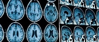
), or incorrect, in isolation from clinical manifestations, subjective interpretation of normal variants during neurosonography or tomography.
In fact, intracranial hypertension is a very serious and quite rare neurological and neurosurgical pathology. It accompanies severe neuroinfections and brain injuries, hydrocephalus, cerebrovascular accidents, brain tumors, etc.
Hospitalization is mandatory and urgent!
In addition, there is no direct and reliable connection between intracranial hypertension and translucent vessels on the face and scalp, walking on tiptoes, trembling hands and chin, hyperexcitability, developmental disorders, poor academic performance, nosebleeds, tics, stuttering, bad behavior, etc. d. and so on.
That’s why, if your baby has been diagnosed with “perinatal encephalopathy (PEP) or perinatal damage to the central nervous system (PP CNS), intracranial hypertension or hypertensive-hydrocephalic syndrome (HHS)”, based on the “bulging” eyes (not to be confused with the true Graefe symptom , a symptom of the “setting sun”!) and walking on tiptoe, then you shouldn’t go crazy in advance. In fact, these reactions may be characteristic of easily excitable young children. They react very emotionally to everything that surrounds them and what happens. Sensitive parents will easily be able to notice such a relationship.
It is absolutely unreasonable to begin treatment for this unspecified “serious” pathology on the recommendations of one doctor based on the above “arguments”; in addition, such unfounded treatment may not be safe at all.
But! There is another, no less important aspect of the problem that must be taken into account in this situation. Sometimes medications are really necessary, and wrongful refusal of them, based only on mom’s (and more often dad’s) own conviction that medications are harmful, can lead to serious troubles.
Now a few words about the equally “adored” hydrocephalus
and
hydrocephalic syndrome
.
Unfortunately, in ordinary life such an erroneous “diagnosis” occurs in almost every fourth or fifth baby. It turns out that some doctors often incorrectly call a stable (usually slight) increase in the ventricles and other cerebrospinal fluid spaces of the brain hydrocephalus (hydrocephalic syndrome). This does not manifest itself in any way through external signs or complaints and does not require treatment. Sometimes, when performing neurosonography, an ultrasound doctor finds pseudocysts
- but this is not a reason to panic at all! Pseudocysts are single round tiny formations (cavities) containing cerebrospinal fluid and located in typical areas of the brain. The reasons for their appearance, as a rule, are not reliably known; they usually disappear by 8-12 months. life. It is important to know that the existence of such cysts in most children is not a risk factor for further neuropsychic development and does not require treatment. However, although quite rare, cysts form at the site of subependymal hemorrhages, or are associated with perinatal cerebral ischemia or intrauterine infection. The number, size, structure and location of cysts provide specialists with very important information, taking into account which, based on a clinical examination, final conclusions are formed.
Description of NSG is not a diagnosis and not a reason for treatment!
Most often, NSG data provide indirect and uncertain results, and are taken into account only in conjunction with the results of a clinical examination.
Once again, I must remind you of the other extreme: in difficult cases, sometimes there is a clear underestimation on the part of parents (less often, doctors) of the problems the child has, which leads to a complete refusal of the necessary dynamic observation and examination, as a result of which the correct diagnosis is made late, and the treatment does not lead to the desired result. Therefore, if increased intracranial pressure and hydrocephalus are suspected, diagnosis should be carried out at the highest professional level.
What is muscle tone and why do doctors and parents “love” it so much?
Look at your child’s medical record: is there no such diagnosis as “muscular dystonia”, “hypertension” and “hypotension”? — you probably just didn’t go with your baby to the neurologist’s clinic until he was a year old. This is, of course, a joke. However, the diagnosis of “muscular dystonia” is no less common (and perhaps more common) than hydrocephalic syndrome and increased intracranial pressure.
Changes in muscle tone can be, depending on the severity, either a variant of the norm (most often) or a serious neurological problem (this is much less common).
It is sometimes quite difficult for even a competent doctor to notice the difference between physiological changes and pathological symptoms in one consultation. The fact is that changes in muscle tone are not only associated with neurological disorders, but also strongly depend on the specific age period and other characteristics of the child’s condition (excited, crying, hungry, drowsy, cold, etc.). Thus, the presence of individual deviations in the characteristics of muscle tone does not always cause concern and require any treatment.
But even if functional disorders of muscle tone are confirmed, there is nothing to worry about. A good neurologist will most likely prescribe massage and physical therapy (exercises on large balls are very effective). Medicines are prescribed extremely rarely, usually for severe muscle hypertension of a spastic nature.
Hyperexcitability syndrome (syndrome of increased neuro-reflex excitability)
Frequent crying and whims with or without cause, emotional instability and increased sensitivity to external stimuli, sleep and appetite disturbances, excessive frequent regurgitation, motor restlessness and shuddering, trembling of the chin and arms (etc.), often combined with poor growth weight and bowel dysfunction - do you recognize such a child?
All motor, sensitive and emotional reactions to external stimuli in a hyperexcitable child arise intensely and abruptly, and can fade away just as quickly. Having mastered certain motor skills, children constantly move, change positions, constantly reach for and grab objects. Children usually show a keen interest in their surroundings, but increased emotional lability often makes it difficult for them to communicate with others. They have a subtle mental organization, they are very impressionable, emotional and easily vulnerable! They fall asleep extremely poorly, only with their mother, they constantly wake up and cry in their sleep. Many of them have a long-term reaction of fear when communicating with unfamiliar adults with active reactions of protest. Typically, hyperexcitability syndrome is combined with increased mental exhaustion and fatigue.
The presence of such manifestations in a child is just a reason to contact a neurologist, but in no case is it a reason for parental panic, much less drug treatment.
Constant hyperexcitability is not causally specific and can most often be observed in children with temperamental characteristics (for example, the so-called choleric type of reaction).
Much less frequently, hyperexcitability can be associated and explained by perinatal pathology of the central nervous system. In addition, if a child’s behavior is suddenly disrupted unexpectedly and for a long time for virtually no apparent reason, and he or she develops hyperexcitability, the possibility of developing an adaptation disorder reaction (adaptation to external environmental conditions) due to stress cannot be ruled out. And the sooner the child is examined by specialists, the easier and faster it is possible to cope with the problem.
And, finally, most often, transient hyperexcitability is associated with pediatric problems (rickets, digestive disorders and intestinal colic, hernia, teething, etc.).
At an early age, the child’s condition is extremely changeable, so minimal developmental deviations and other disorders of the nervous system can sometimes be detected only during long-term dynamic monitoring of the baby, with repeated consultations. For this purpose, specific dates for planned consultations with a pediatric neurologist in the first year of life have been determined: usually at 1, 3, 6 and 12 months. It is during these periods that most serious diseases of the nervous system of children in the first year of life can be detected (hydrocephalus, epilepsy, cerebral palsy, metabolic disorders, etc.). Thus, identifying a specific neurological pathology in the early stages of development makes it possible to begin complex therapy on time and achieve the maximum possible result.
And in conclusion, I would like to remind parents: be sympathetic and attentive to your kids! First of all, it is your active and meaningful interest in the lives of children that is the basis for their future well-being. Do not try to cure them of “supposed illnesses,” but if you have any concerns or concerns, seek independent advice from a qualified professional.
Author: neurologist S.V. Zaitsev
September 27, 2017
Source
Memo to parents
Head and chest circumference standards
| Age | Head circumference | Chest circumference |
| 1 month | 38.0 cm | 37.5 cm |
| 2 months | 39.0 cm | 38.5 cm |
| 3 months | 40.0 cm | 39.5 cm |
| 4 months | 41.5 cm | 41.0 cm |
| 5 months | 42.5 cm | 42.0 cm |
| 6 months | 43.5 cm | 43.5 cm |
| 7 months | 44.0 cm | 44.5 cm |
| 8 months | 44.5 cm | 45.0 cm |
| 9 months | 45.0 cm | 45.5 cm |
| 10 months | 45.5 cm | 46.0 cm |
| 11 months | 46.0–46.5 cm | 46.5 cm |
| 12 months | 47–49.0 cm | 48–50 cm |


