- Symptoms of intracranial pressure in adults
- Headache
- Nausea and vomiting in the absence of gastrointestinal diseases
- Increased fatigue
- Irritability
- Changes in blood pressure, tachycardia, arrhythmia
- Decreased libido
- Meteosensitivity
- What to do if the above symptoms appear?
- Lumbar puncture
- Treatment of intracranial pressure in adults
- Treatment with medications
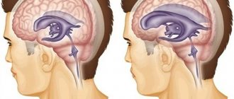
The condition of increased intracranial (intracranial) pressure is called liquor-hypertension syndrome, intracranial hypertension. This condition differs from the familiar arterial hypertension. The disease can occur for unknown reasons (idiopathic form), and it also appears with head injuries, tumors and inflammatory diseases of the brain.
Measurement methods
It is impossible to independently measure intracranial pressure at home.
We need the help of competent doctors and medical equipment. There are two main measurement methods - invasive and non-invasive. The first non-invasive method is fundus ophthalmoscopy. The doctor will perform an examination and estimate the eye pressure based on the diameter of the optic nerve head. With intracranial hypertension, the disc will be swollen, enlarged, with unclear contours.
Transcranial Doppler ultrasound shows pulsation and blood flow in the cerebral arteries. An increase in the index above accepted values means an increase in pressure in the cranial cavity.
scan of the brain
detect possible injuries and damage, swelling, hematomas and tissue displacement. In children, you can perform an ultrasound through an open fontanel and see the structures of the brain.
An invasive but reliable method is a spinal puncture. The procedure is performed strictly according to indications. The state of the cerebrospinal fluid is assessed: pressure and physical characteristics. Based on the rate of its flow, one can guess the nature of the liquor pressure.
Preventative measures against the disease
For the purpose of prevention, it is necessary to follow a number of rules:
- eliminate bad habits;
- avoid strong and prolonged stressful situations;
- minimize the risk of traumatic brain injuries when engaging in traumatic sports by using protective helmets;
- periodically undergo a course of massage of the cervical-collar area;
- prevent exacerbation of concomitant diseases such as hypertension, epilepsy, etc.;
- do not lift heavy objects;
- avoid excessive physical activity;
- provide a sufficient amount of potassium-rich foods in your daily diet;
- adhere to a rational daily routine;
- do gymnastics for the neck muscles;
- lead a healthy lifestyle.
These actions are aimed not only at preventing the development of the disease, but also at improving brain function in general. Preventive measures promote proper circulation of cerebrospinal fluid and blood, as a result of which the brain is more fully saturated with nutrients and oxygen.
It is very important to consult a doctor in a timely manner; only with the help of adequate treatment can you get rid of the disease, prevent relapse and the development of serious complications that can be caused by intracranial pressure.
Reasons for the increase
The normal intracranial pressure in adults is 7-15 mmHg. The main causes of intracranial hypertension:
- an increase in the volume of normal structural components due to edema,
- the appearance of additional space-occupying formations: cysts, tumors, abscesses,
- extra- and intracranial diseases: sinus thrombophlebitis, subdural and epidural hematomas (spontaneous or after injury),
- common inflammatory process: meningitis, meningoencephalitis, encephalitis,
- exo- and endogenous intoxications.
Increased intracranial pressure: how to recognize and reduce?
Increased intracranial pressure - what does this concept really mean? How does intracranial pressure relate to arterial pressure?
We looked for answers to these and other questions with Yulia Vladimirovna Roshchupkina, a neurologist at the Expert Tula Clinic.
— Yulia Vladimirovna, what is intracranial pressure? What should it be normally and when it comes to increasing it?
This is a parameter that reflects the strength of the influence of cerebrospinal fluid (CSF) on brain tissue. The cerebrospinal fluid is under a certain pressure, which is called intracranial. Today it is measured in millimeters of mercury (mmHg) and is normally between 10 and 15 mmHg.
INTRACRANIAL PRESSURE IS MEASURED IN MILLIMETERS OF MERCURY (MM.Hg) AND IS NORMALLY FROM 10 TO 15 MM. RT. ST.
The danger is represented by pressure above 25 mmHg.
When it approaches 35 mmHg. it is designated as critical, loss of consciousness and death of brain cells are possible.
— Is increased intracranial pressure a diagnosis or a symptom?
This is a syndrome. Its synonym is intracranial hypertension. It occurs with a number of pathologies, as well as tilting the head, sneezing, physical activity, etc.
— By what signs can you understand that a person has increased intracranial pressure?
Symptoms of intracranial hypertension include a pressing, bursting headache, mainly in the morning. May be in different parts of the head.
Read the material on the topic: What to do if the head is “cast iron”?
Nausea and vomiting are also noted; drowsiness; disorders of memory, attention, thinking; autonomic disorders (fluctuations in blood pressure, decreased heart rate, increased sweating); blurred vision and blindness.
— What happens with increased intracranial pressure - do the vessels narrow or, on the contrary, expand?
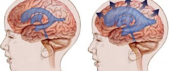
Its increase is caused by dilation of the arteries leading to the brain, which is accompanied by an increase in blood flow to it, as well as a decrease in outflow through the veins. As a result, blood vessels overflow, and the surrounding tissue is saturated with blood plasma.
SYMPTOMS OF INTRACRANIAL HYPERTENSION INCLUDE PRESSING, BURNING PAIN, MAINLY IN THE MORNING. MAY BE IN DIFFERENT PARTS OF THE HEAD
In addition, this is also possible when a space-occupying formation or process develops in the cranial cavity.
— Increased intracranial pressure occurs only with high blood pressure or can it also occur with hypotension?
It is also possible with hypotension (one example is brain injury with concomitant significant blood loss).
Read the material on the topic: When there is nowhere lower. What are the reasons for hypotension?
On the question of the relationship between increased arterial and intracranial pressure: if the regulatory mechanisms work normally, then arterial hypertension does not necessarily lead to intracranial hypertension.
— What is the danger of high intracranial pressure?
As it increases, daily morning vomiting is possible against the background of intense headache. Mental functions are depressed. Lethargy appears, there may be disturbances of consciousness, even coma.
Blood pressure rises, breathing becomes depressed and becomes less frequent, and the pulse slows down. Generalized seizures may occur. In advanced cases, displacement and infringement of brain structures is possible, disrupting the functioning of vital centers of respiration and circulation, which leads to death.
— Tell us what leads to the occurrence of intracranial hypertension.
These are infections of the brain and its membranes; traumatic brain injury; neoplasms of various nature in the cranial cavity; hematomas, brain abscesses; prolonged lack of oxygen; difficulty in outflow through the jugular veins; dropsy of the brain; overweight, obesity, metabolic pathology; chemical poisoning.
Read the material on the topic: Will an MRI show meningitis?
— Are the reasons why intracranial pressure increases in children and adults the same or different?
In some ways they are similar. At the same time, in children, in particular, prolonged intrauterine oxygen deficiency, neuroinfections, and other pathologies of pregnancy and childbirth can lead to its increase.
— How is the cause of increased intracranial pressure diagnosed?
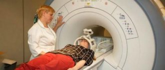
Mostly MRI and CT, ultrasound of the brain, echoencephaloscopy, and fundus examination are performed.
Read the material on the topic: What will the fundus tell you?
Intracranial pressure is measured using a pressure gauge. In this case, a special catheter is inserted into the spinal canal or into the ventricles of the brain.
You can sign up for a brain MRI in your city here
— Yulia Vladimirovna, does the condition of intracranial hypertension require mandatory treatment or can increased intracranial pressure go away on its own?
It depends on its cause. The solution to this issue is within the competence of the attending physician; self-medication is unacceptable. If any of the above complaints occur, contact your doctor for timely treatment.
— Are traditional methods used to reduce increased intracranial pressure? How effective are they?
And here everything depends on the cause and mechanism of development of this pathology. Folk remedies are not able to eliminate the cause; they are never used as primary means. However, theoretically, in some cases they can slightly alleviate the patient’s condition. In any case, the decision about the possibility of taking this or that drug is made by the doctor.
— What specialty should you consult with a doctor if you suspect intracranial hypertension?
To a neurologist. After clarifying the cause, consultation and, if necessary, treatment by a neurosurgeon may be necessary.
You can make an appointment with a neurologist in your city here
Attention: the service is not available in all cities
You may also find useful:
It's off scale! Looking for reasons for high blood pressure
What is hidden behind the diagnosis of migraine?
Vegetative-vascular dystonia: diagnosis or fiction?
For reference:
Roshchupkina Yulia Vladimirovna
In 2001 she graduated from the Faculty of Medicine of Kursk State Medical University.
From 2001 to 2002 she completed an internship in the specialty “Neurology”.
She specializes in acupuncture.
Currently he holds the position of neurologist at Clinic Expert Tula. Receives at the address: st. Boldina, 74.
Symptoms in adults
Intracranial hypertension manifests itself through a sequence of symptoms.
This is a progressive deterioration of neurological status not associated with other pathology. Men and women experience general cerebral symptoms - nausea, vomiting and headache. In severe cases, there may be impairment of consciousness from stupor to coma. There is a disturbance in the movement of the eyeballs in the form of strabismus, dilated pupils and a decreased reaction to light.
The presence of one of the listed symptoms does not indicate increased intracranial pressure. However, this is a reason for a more detailed examination. It is necessary to evaluate the clinical picture as a whole, and also take into account the results of additional diagnostic methods.
Non-invasive research methods
Non-invasive methods are used at the initial stage of diagnosis; measurements are carried out using external hardware systems. They make it possible to identify pathological changes in brain structures, as well as diagnose cranial pressure. Non-invasive techniques are safe and painless. However, the assessment of indicators is indirect, since the data obtained as a result of the examination may be mistakenly taken for signs of increased ICP, namely the expansion of the interventricular gap and the ventricles themselves. These methods are necessary to establish the reason that provoked the change in indicators.
Fundus examination is the most informative method of non-invasive monitoring of intracranial hypertension
To identify increased cranial pressure in a clinic, a specific algorithm is used, including the following methods:
- Fundus examination. Detection of swollen optic discs in the fundus and expansion of the vascular network are signs of pathology that require additional research. In the absence of pathological changes, additional techniques are not required, since ICP is within normal limits.
- Tomography, ultrasound of the brain. They allow you to determine the cause of the disorder, but do not determine the exact amount of cranial pressure.
- Electroencephalography. Determines changes in brain electrical activity, indicating an increase in ICP indicators (chaotic excitation of structural elements of the brain, formation of high-frequency rhythms, diffuse changes).
- Otoacoustic examination. The technique is carried out through the ears. With cerebral hypertension, blood pressure values in the inner ear increase.
- Transcranial Dopplerography. Blood pressure is measured by determining the decrease in blood flow velocity resulting from the development of cerebral hypertension.
The results obtained during the studies do not provide an accurate picture of the indicators of cerebral hypertension; they are used to establish the primary diagnosis of the disease.
Reduced intracranial pressure
The clinical picture is not as obvious as with increased blood pressure, but some nonspecific signs occur.
They interfere with human performance and daily activities. Possible symptoms:
- periodic headache of varying intensity,
- irritability, poor attention and weakness,
- dizziness and drop in blood pressure,
- nausea, chills,
- changes in the emotional background (emotional lability),
- blurred vision.
Causes of low blood pressure: performing medical interventions or operations that lead to hypotension. For example, diagnostic lumbar puncture.
Injuries leading to rupture of the dura mater also have an effect. Systemic changes: uremia or dehydration.
Examination and treatment
Intracranial hypertension is an increase in pressure exerted by cerebrospinal fluid moving along the brain pathways.
This pathology is included in the list of the most common brain diseases and is a dangerous disease that has a destructive effect on its structures. More often, hypertension is a secondary disease that develops against the background of various factors, including traumatic or oncological etiology. According to statistics from neurologists around the world, representatives of the stronger sex are more susceptible to intracranial hypertension, although in children it is diagnosed with equal frequency in both boys and girls.
Important: high blood pressure can be provoked not only by cerebrospinal fluid, but also by arterial blood or the substrate of a brain tumor.
Get diagnosed with intracranial hypertension at Clinic No. 1:
- Fundus examination
- Ultrasound of cerebral vessels
- X-ray of the head
- EEG
- Angiography
For one-time payment for services - 20% discount
Call
Ways to reduce pressure in intracranial hypertension
Since such a pathology is most often a symptom of a disease, it is necessary to eliminate the cause of its occurrence.
To ensure the flow of cerebrospinal fluid, in some cases, surgery is performed to remove a tumor, abscess or hematoma. If the cause is an infectious inflammation of the meninges, then massive antibiotic therapy is carried out. It is possible to introduce them into the subarachnoid space.
To eliminate symptoms, methods are used that include non-drug and drug therapy. First, you need to raise the victim's head and limit fluid intake.
Diuretics are used among medications to remove excess fluid from tissues. Acetazolamide and Furosemide have this effect. The therapy is selected by a neurologist. Diacarb effectively reduces the production of cerebrospinal fluid.
To improve microcirculation and blood supply to tissues, neuroprotectors and nootropics are used. In severe cases, hormonal therapy is required under monitoring of vital signs.
Carrying out a spinal puncture with mechanical extraction of cerebrospinal fluid has a hypotensive property. Surgery is often required. Shunt surgery is planned to artificially drain fluid from the brain.
Diagnostic methods for children
As a rule, cranial pressure in a child can be determined using neurosonography (ultrasound), which is performed through the fontanel. This examination is safe and painless. It is used to examine young children and allows you to accurately determine the functional state of the ventricles of the brain. An increase in their volume is a sign of increased intracranial pressure.
Neurosonography procedure in young children
In older children, when the fontanelle has already closed, computed tomography and magnetic resonance imaging are used to visualize brain structures. Studies make it possible to establish the blood supply of blood vessels, the state of liquorodynamics and the presence of neoplasms.
Today, an echoencephalogram is also widely used, with the help of which most indicators are assessed, including the pulsation of cerebral vessels. During the study, the amplitude of oscillations of the ultrasonic signal is taken as a basis, due to which the ICP is assessed. The disadvantage of the method is the inaccuracy and unreliability of the results obtained.
Reasons for the development of the disorder
The factor that provokes an increase in pressure is the impaired outflow of cerebrospinal fluid from the brain. Normally, it occupies about 10% of its total volume, performing various functions, which include:
- protection of the brain from trauma - in the event of an impact or fall, the cerebrospinal fluid will “soften” the contact of the cranial bones and delicate tissues;
- it is also with the movement of cerebrospinal fluid that toxins and decay products are removed from the brain;
- Finally, it ensures that the correct electrolyte-water balance is maintained.
The most common causes of ICP include metabolic disorders that provoke insufficient flow of fluid into the blood, as well as spasm of the vessels through which the cerebrospinal fluid moves. In addition, oxygen deprivation of the brain, excess fluid in the body, obesity, severe intoxication, and inflammation of the brain can also provoke ICP. The presence of tumors in the body - benign or malignant - can also cause disruption of the outflow of cerebrospinal fluid.
If the outflow of cerebrospinal fluid from the intracranial space is disrupted for any reason, this provokes the accumulation of its excess volume and an increase in pressure. At the same time, in both a child and an adult patient, an increase in intracranial pressure to more than 30 mm Hg can lead to the development of irreversible changes in tissues and, as a consequence, disability and even death.
standard treatment
To reduce intracranial pressure, drug therapy is prescribed: diuretics, vasodilators and sedatives; drugs to improve the tone of the vascular wall of veins and stimulate brain activity; vitamin complexes and homeopathic preparations.
For some types of pathology, physiotherapy, acupuncture, massage, and special gymnastic exercises are indicated.
If drug treatment is ineffective and ICP levels are high, surgical treatment is prescribed.
Diagnosis of high cerebral pressure
Patients with suspected hypertension are immediately prescribed an MRI. Also carried out:
- Fundus examination is a painless diagnostic method that reveals papilledema, a characteristic sign of hypertension.
- Doppler ultrasound examination of cerebral vessels will reveal if there are “obstacles” in the path of cerebrospinal fluid flow.
- Head X-ray is a simple and accurate method for diagnosing the condition of the brain; it can be performed with or without contrast.
- An electroencephalogram (EEG) is a non-invasive test aimed at measuring the electrical activity of brain cells.
- Angiography.


