The disease is congenital in nature. The anomaly is a capsule with a cavity filled with liquid. A coelomic pericardial cyst can be located on a small stalk (pedicle) or be fused to the structures of the outer pericardial sac.
The initial stages of minor dysplasia are latent; in more complex cases, shortness of breath, cough, discomfort in the left side of the chest and heartbeat disturbances develop. This benign formation is more often diagnosed in females aged 20 to 45 years. Men encounter these types of cysts three times less often. Treatment consists of surgical removal of the neoplasia.
Pericardial cyst (coelomic pericardial cyst)
What kind of education is this
Panatomically, the coelomic pericardial cyst is a benign anomaly of a round shape, the average size of which in diameter is about 3-8 cm. Closed neoplasms are characterized by large sizes, while diverticula are characterized by smaller values.
The capsule is represented by thin smooth layers of fabric, with a shiny surface on the inside. Their microscopic structure is identical to the cytohistological structure of the pericardium. The outer surface of the capsule consists of layers of connective tissue with many vessels. There are no muscle structures in the cyst.
The liquid contained inside has no color and is therefore transparent. It may contain a small amount of protein formations and dissolved salts. Pus indicates an infectious lesion, and the presence of blood in the exudate may indicate injury to the formation.
A little anatomy
Externally, a coelomic cyst looks like a thin-walled cavity neoplasm with a smooth gray-yellow surface, which is filled with clear or straw-yellow serous fluid. Sometimes fatty inclusions are found on the surface of the cyst. A thin vascular network is visible on its wall.
The basis of the walls of the coelomic cyst is a connective tissue permeated with elastic fibers. Its inner surface consists of cubic or flat mesothelium, and its outer surface consists of loose vascularized connective tissue with inclusions of adipose tissue.
The thickness of the walls of coelomic cysts usually does not exceed the thickness of writing paper, and their diameter can reach 3-10 cm (sometimes more). The shape of such formations can be pear-shaped, oval or round.
The liquid found in the formation cavity usually contains very little protein and a lot of salts. In more rare cases, it contains a large amount of protein.
In 60% of patients with such formations, the cyst is located in the right cardiophrenic sinus. In 30% of cases, the tumor is localized in the left cardiophrenic sinus and only in 10% of patients is it detected in other areas of the mediastinum.
Histological analysis of the tissues of coelomic cysts reveals fibrous connective tissue with inclusions of monocytic and lymphoid cells, lined with mesothelium. Muscle fibers are not found in such formations.
Causes
Doctors name two main factors for the formation of coelomic pericardial cysts:
- Embryonic development disorder . During the formation of the fetus, weak zones are formed in the pericardial sac, which causes protrusion of tissue structures in the form of diverticula (not completely closed cavities). If they become isolated and lose communication with the pericardium, then an isolated cyst is formed. The most likely cause is considered to be the uneven development of lacunae in the fetus, which, when fused, form the pericardial space.
- Another (less frequent) reason is the development of neoplasia, as a secondary phenomenon against the background of an infectious inflammatory process or after injury . The formation of coelomic cysts is possible as a result of hematomas, pericarditis, and destruction of heart tumors.
The note. The first description of the disease dates back to the mid-19th century. At first, the cysts were called pericardial diverticula, because the presumed genesis was a protrusion of the tissue of the pericardial sac. But in the forties of the 20th century, it was found that the main reason was the abnormal development of the coelom, therefore since then the name of the diagnosis has been revised and clarified.
Features of the pathology
A cyst on the heart is a benign cavity with thin walls and liquid contents. The capsule has an irregular round shape, the diameter depends on the intensity of the development of the pathology. A clear liquid remains inside, but if infected or injured, it can change color.
The capsule has an irregular round shape, the diameter depends on the intensity of the development of the pathology.
A pericardial cyst on the heart is found in 22% of cases of all types of organ pathologies. Often the capsule is located in the right diaphragmatic region. The cyst is rarely diagnosed in the lower part of the heart. According to statistics, in females, the development of cysts of the cardiac sac is observed 3 times more often and occurs at the age of 20-55 years.
Causes of pericardial cyst
Neoplasm in the pericardium is associated with a negative effect on cardiac tissue.
- Injuries. The cavity is limited by the walls and is filled with liquid as a result of a strong blow and the occurrence of a hematoma.
- Congenital disorder. During the formation of the embryonic layers of the pleura, pathological changes occur.
- Inflammation. The process affects all types of cardiac tissue.
Inflammation of the pericardium due to past diseases (kidneys, tuberculosis, heart attack, etc.)
Cysts in the pericardial lining can form due to infection with parasites. The main reason is the vital activity of echinococcus. It can also affect the pleural area near the lung. Often the capsule is a precursor to a pericardial tumor and belongs to one of its stages. This is possible in the absence of treatment of the cyst, suppuration of the contents and its release into the myocardium and other membranes of the heart.
Classification
Depending on the criterion under consideration, pathological formations are divided into several types. Based on their origin, they are classified as congenital or acquired.
Based on the presence of communication with the cavity of the pericardial sac, there are three types of cysts:
- parapericardial - have a stalk, leg or fusion connecting them to the pericardium;
- pericardial – have a communication, so they are often called pericardial diverticula;
- pericardial neoplasms are completely isolated (detached) cysts.
Photo of a CT image of a pericardial cyst
Depending on the number of chambers in the cyst, they can be single- or multi-chambered.
Difference in the presence of clinical signs:
- latent or without any manifestations;
- uncomplicated;
- complicated.
Important. Up to 50% of coelomic pericardial cysts do not have pronounced symptoms, so they can be detected only with the help of special laboratory diagnostics, for example, in a photo of preventive fluorography.
Symptoms
Clinical signs do not appear in the early stages of anomaly formation, but as the size of the neoplasm increases, polymorphic symptoms appear, which will depend on the size of the cyst and its location in the pericardium.
Most often, patients complain of:
- a feeling of discomfort and pain of aching or stabbing nature in the chest (projection zone of the heart);
- feeling of squeezing;
- increased heart rate;
- shortness of breath and non-productive dry cough, which is especially pronounced with increased physical activity;
- asthma attacks.
If the cyst is large, then there is cyanosis of the skin, cyanosis, protrusion of neck vessels and severe shortness of breath. Such a clinic arises due to the pressure of the coelomic neoplasm on the mediastinal organs.
With pressure on the branches of the vagus nerve, pain radiates to the hypochondrium on the right, as well as to the area of the scapula or shoulder. When pyogenic processes are observed or the integrity of the coelomic pericardial cyst is disrupted, then a clinic characteristic of hydrothorax and pleuropulmonary shock is added to the named symptoms.
Note. All clinical manifestations quickly disappear after surgical removal of the pathology.
Carrying out diagnostics
Determination of pathology in the heart muscle is possible only by instrumental methods. In cardiology, cardiac tomography is performed using various equipment.
- MRI. A modern method allows you to determine the location, type and shape of the tumor. During the diagnostic process, the doctor receives an accurate picture of the functionality of the organ.
- ECHO-KG. The cardiogram determines the general disturbance in the heart associated with pericardial protrusion. Ultrasound reveals increased echogenicity and darkened areas.
The cardiogram determines the general disturbance in the heart associated with pericardial protrusion. - Thoracoscopy. One of the types of endoscopic examination. It is carried out by inserting an instrument into the pleural cavity and heart. Diagnosis is carried out under the supervision of a cardiac surgeon. During the operation, the integrity of the capsule and walls of the heart is checked.
Diagnostics
Patients, depending on the symptoms that appear, usually come to see a cardiologist or pulmonologist. It is not uncommon for patients to be referred to specialized specialists by a general practitioner or family doctor.
A physical examination may reveal:
- protrusion of the chest in the area of formation of a coelomic pericardial cyst;
- presence of shortness of breath;
- weakening of breathing.
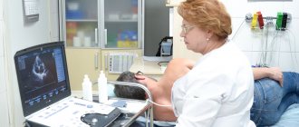
Chest pain, discomfort and shortness of breath are a serious reason to see a cardiologist
Auscultation reveals signs of tachycardia, the presence of vascular murmurs and dullness of percussion sound, which is described in more detail in the proposed video in this article. Laboratory examination methods are given in the table below.
Table. Diagnosis of coelomic pericardial cyst:
| Method | A comment |
| Registers changes in a graphical pattern during the formation of cysts that are fused to the pericardium. |
| First, photographs of the chest are studied, then, if necessary, an x-ray of the heart with contrast of the esophagus may be prescribed, which provides better visualization. As a rule, the negative shows a round shadow in the area of the cardiophrenic sinus. |
| Ultrasound of the heart helps to see neoplasms measuring a centimeter in diameter, and the relationship of the cyst with the heart is visible. The technique is positively characterized by the fact that it is fast, non-invasive, and the price is quite low. |
| Computed tomography of the chest may be useful in complex cases or in preparation for surgery. |
On X-ray photographs, a coelomic pericardial cyst is visible as a round formation. Its inner contour is usually fused with the shadow of the heart, and the lower one with the diaphragm. The external outlines are always clear and clearly visible.
The note. If diagnosis is difficult, thoracoscopy may be performed to clarify the disease.
In some cases, puncturing is required to make an accurate diagnosis. The resulting fluid samples are subjected to thorough laboratory analysis, with an emphasis on biochemical and electrolyte composition.
Differential diagnosis is carried out with heart tumors, hydatid cysts, diaphragmatic hernia and lipoma (abdominomediastinal).
Mediastinal cysts
Radiation diagnostic methods still play a leading role in the diagnosis of tumors and mediastinal cysts. We include multiplanar fluoroscopy and three-plane chest radiography into the radiology examination; when confirming a suspicion of a tumor, we always use computed tomography or magnetic resonance imaging. In some cases, invasive angiography is required to clarify the diagnosis. Fluoroscopy and chest radiography make it possible to identify a formation in the mediastinum, localize it in one or another section and thereby suggest the most likely nosological diagnosis. For more accurate localization of the formation, it is advisable to perform these studies under conditions of contrasting the esophagus in order to identify or exclude its deviation, which is not uncommon with formations of the posterior mediastinum. In addition, we gain insight into the condition of the lungs (hypoventilation due to airway compression), pleural cavities (reactive pleurisy, hydrothorax) and diaphragm function (compression of the phrenic nerves). However, it is most often impossible to differentiate a tumor from a cyst, as well as to judge the tumor’s invasion of surrounding structures on the basis of routine x-ray examination. In addition, it must be taken into account that routine X-ray examination does not always allow one to differentiate mediastinal formations from intrapulmonary and intrapleural ones; in such cases, tomography in any version is necessary.
Standard mediastinal tomography , pneumomediastinography, pneumomediastinotomography, which until recently were the methods of choice, are now almost completely replaced from the diagnosis of mediastinal formations by modern, more accurate methods of radiation diagnostics. Computed tomography and magnetic resonance imaging, confirming and clarifying the localization of the formation, provide answers to these questions: analysis of tomograms with high accuracy demonstrates the connection of mediastinal formations with certain organs and structures, and densitometry makes it possible to distinguish denser tumor tissue from the less dense fluid contents of the cyst . Contrasting vessels at the time of examination and the use of magnetic resonance imaging increase the accuracy of visualization of great vessels, which is important for excluding or identifying their invasion or compression. Magnetic resonance imaging seems to be the method of choice also in cases of suspected neurogenic tumor of the posterior mediastinum, since in such situations it is always necessary to exclude an hourglass tumor, and in relation to nervous tissue, magnetic resonance imaging is more sensitive than computed tomography. However, nosological diagnosis based on these high-precision research methods can also be only speculative. Invasive angiography is indicated in those relatively rare cases when it is necessary to exclude or identify invasion or compression of great vessels, or to differentiate the formation from a vascular aneurysm, and computed tomography or magnetic resonance imaging does not provide a definite answer to these questions.
Endoscopic and morphological diagnostics Endoscopic methods in the diagnosis of mediastinal formations are important not only, and perhaps not so much as a means of visual assessment, but as a way to obtain pathological material for morphological studies. Therefore, we will consider them in combination with other methods of morphological diagnosis, in order of increasing invasiveness of the method.
Bronchoscopy is the least invasive, but also the least informative of the instrumental methods for diagnosing mediastinal formations. This study makes it possible to identify and visually quantify the degree of compression of the airways, and also determines the invasion of the trachea and large bronchi by invasive mediastinal tumors. Endoscopically visible deviation of the trachea, bulging of one or another of its walls, expansion of its bifurcation serve as indirect signs of mediastinal formation and indicate its localization. However, a histological diagnosis can be made using bronchoscopy in no more than 25% of cases (Adler et al., 1983). We consider bronchoscopy indicated in all cases of mediastinal formations, which, according to radiological diagnostics, appear to be associated with the respiratory tract.
Fine-needle aspiration biopsy is a more invasive method, but significantly more informative than bronchoscopy. Cytological examination of the punctate obtained by this method provides morphological diagnosis of mediastinal formations, according to the literature, in 80 - 90% of cases. The diagnosis of a mediastinal cyst is confirmed immediately by puncture. Fine-needle biopsy, performed under the control of computed tomography, can reach almost all anatomical parts of the mediastinum. If necessary, puncture with a thin needle can be performed not only by percutaneous, but also by transesophageal, transtracheal or transbronchial access. An important advantage of the method is the ability to perform the procedure on an outpatient basis using local anesthesia, which is especially important for patients with signs of mediastinal compression. Based on the above advantages, many authors consider it necessary in all cases of mediastinal formations to begin morphological diagnosis with fine-needle aspiration biopsy, using more invasive methods only in cases where puncture of the formation does not provide a definite diagnosis.
The disadvantages of puncture biopsy include, first of all, a certain number of complications accompanying it: for example, according to Adler et al (1983), certain complications are observed in 21.4% of puncture biopsies of the mediastinum, including pneumothorax - in 3.6% patients. In addition, the amount of pathological material aspirated through a fine needle is usually very small, which limits its ability to be studied by cytological examination. In addition, if biopsy is a local research method, the results of which largely depend on which point of the visible substrate the biopsy instrument reaches, then puncture biopsy is even more local. Obviously, the last two circumstances are responsible for the not so rare discrepancies between puncture biopsy and postoperative histological diagnoses. Thus, puncture biopsy is justified for all mediastinal formations and is especially indicated in cases where preoperative morphological diagnosis is important for treatment tactics, but the results of this study, unfortunately, cannot be absolute. A biopsy of peripheral lymph nodes (if there are pathological changes in them: enlargement, hardening or pain) is of particular importance for the diagnosis of lymphomas.
Parasternal mediastinotomy, an abdominal surgery requiring hospitalization and general anesthesia, is a variant of open excisional biopsy. The amount of pathological material obtained in this case is usually sufficient for a full-fledged morphological study. However, the possibilities of visualizing a pathological formation in the mediastinum from this approach are very limited, and the range of application of diagnostic mediastinotomy is reduced to a biopsy of formations of the anterior mediastinum adjacent parasternally to the anterior chest wall. The method is used infrequently, and the introduction of endovideosurgery into clinical practice will apparently completely replace mediastinotomy.
Mediastinoscopy in the diagnosis of tumors and mediastinal cysts also has rather limited use. Visualization and excisional biopsy by classical transcervical mediastinoscopy are available only in the middle and upper part of the anterior mediastinum. Due to the limitations of the review and instrumental manipulations, it is possible to make a nosological diagnosis in slightly more than half of the cases. At the same time, many consider mediastinoscopy to be contraindicated for tumors of the anterior mediastinum due to the real possibility of damage to the thymic capsule; in this case, if the tumor is a thymoma, contamination of the mediastinal tissue is inevitable. Thoracoscopy seems to be the most informative diagnostic method, combining the ability to directly visualize mediastinal formations from various angles with excisional biopsy under visual control. The application of this endoscopic technique to the diagnosis and then to the treatment of tumors and mediastinal cysts has expanded significantly with the advent of videothoracoscopy in surgical clinics.
This technique provides the surgeon with a significantly wider view and, as a result, a more accurate selection of the site for biopsy compared to mediastinoscopy and mediastinotomy. Operative videothoracoscopy makes it possible not only to determine the relationship of the mediastinal formation with surrounding structures, but also to identify, in some cases, invasion of the latter. According to various authors, videothoracoscopy with biopsy provides a reliable diagnosis of tumors and mediastinal cysts in 86 - 100% of cases. At the same time, it must be remembered that thoracoscopy is an intracavitary surgical intervention that requires general anesthesia, one-lung artificial ventilation and is accompanied by a certain percentage of complications.
Other diagnostic methods . Other diagnostic techniques have very limited use in the case of tumors and mediastinal cysts. Of historical interest is the Mondor test, aimed at studying disorders of the autonomic innervation of the chest in neurogenic tumors of the posterior mediastinum. There are reports in the literature about certain diagnostic capabilities of radioisotope methods (Ferguson et al., 1987), tumor markers (Kohman et al., 1993), and the diagnosis of parathyroid cysts based on determining the level of parathyroid hormone in their contents (Ramos-Gabatin et al. , 1985). But all these reports refer to casuistically rare cases and therefore are not included in the usual algorithm for examining patients with mediastinal masses.
So, diagnosis of a mediastinal tumor or cyst begins with a standard x-ray examination, the main purpose of which is to answer the question: are we really dealing with a mediastinal formation?
The next stage is modern methods of non-invasive radiation diagnostics: computed tomography and magnetic resonance imaging, which should clarify the localization of the formation, its structure, consistency, identify its relationships with surrounding organs and anatomical structures, as well as the nature of these relationships. Endoscopic and biopsy methods, confirming the diagnosis, should answer the question about the nosology of the formation and thereby provide grounds for choosing treatment tactics.
However, as can be seen from the above, none of the preoperative diagnostic methods, including even such informative ones as videothoracoscopy with biopsy, guarantees against errors and discrepancies with the final diagnosis when it comes to mediastinal tumors and cysts. In connection with this circumstance, the last means of surgical diagnosis—early surgical exploration, which is usually immediately followed by removal of the formation—is of particular importance. Regarding staging studies, as we mentioned above, most malignant and invasive mediastinal tumors usually do not produce the hematogenous metastases so typical of other malignancies and rarely produce distant implants. Since there are simply no typical localizations of the latter, the development of screening methods for searching for implants is very difficult. Therefore, staging studies are usually planned after a morphological diagnosis has been established, based on the type of tumor and its tendency to spread.
The required minimum examination for suspected mediastinal formation includes: • multiplanar fluoroscopy and three-plane radiography of the chest organs under contrast conditions of the esophagus; • computed tomography of the mediastinum, if invasion of large vessels is suspected - under conditions of bolus contrast of the latter; for tumors of the posterior mediastinum that are suspected to be neurogenic, magnetic resonance imaging is advisable; • bronchoscopy - in case of formation of the mediastinum, demonstrating intimate connections with the trachea or large bronchi. • If the above diagnostic measures do not allow us to establish the nosology of the formation, modern requirements dictate the need to move to more invasive methods of morphological diagnosis: • transthoracic fine-needle aspiration biopsy under the control of a computer or magnetic resonance imaging scanner; • diagnostic video thoracoscopy; • wide thoraco- or sternotomy, combining diagnostic and therapeutic purposes.
The main role in the diagnosis of pericardial cysts and diverticula, as well as other mediastinal neoplasms, is played by X-ray examination. Multiaxial fluoroscopy and radiography of the chest cavity allows one to determine the localization of the formation, its relationship to neighboring organs, its shape and changes in its position during respiratory movements.
An informative research method is CT. This modern research method allows not only to identify the formation, determine its size, but also to assess the thickness of the walls of the cyst, the nature of the contents, and the relationship with neighboring anatomical formations.
Another modern research method is ultrasound echocardiography (EchoCG). Using this widespread, informative, non-burdensome research method for the patient, it is possible to identify thin-walled, cavity, fluid-filled formations, evaluate their relationship with the heart and other mediastinal organs, and identify communication with the pericardial cavity.
Differential diagnosis of pericardial cysts is quite difficult. They should be distinguished from dermoid cysts of the mediastinum, lung cysts, and aortic aneurysms. The integrated use of the listed research methods makes it possible to exclude most of these diseases and establish the true nature of the changes.
Treatment
The only effective treatment is surgical excision, because puncturing or sclerosing coelomic pericardial cysts is impractical. In the first case, relapses occur, and in the second, there are real risks of the formation of constrictive pericarditis, which is formed when the sclerotizing substance penetrates the pericardial cavities.
The operation is performed using anterolateral or lateral thoracotomy. Radical enucleation of the cyst makes it possible to get rid of it once and for all. When the size of the neoplasm is large, it is first necessary to remove the fluid using a puncture.
Heart surgery
4.Treatment
The method of choice in the treatment of lung cysts is surgery; Medications are only important as supportive and additional therapy. Usually, breast-conserving cystectomy is sought, but in approximately 30% of cases the situation forces removal of part of the lung. In case of severe complications (eg, pyopneumothorax, i.e. rupture of the cyst with leakage of purulent contents), the lesion is drained, accompanied by a powerful antibiotic response.
The prognosis is mostly favorable, but the most severe cases result in death (about 5%), sometimes even before intervention, or result in disability (up to 25%). Therefore, when symptoms of respiratory failure appear and especially when they rapidly increase, assistance should be provided as a matter of urgency.
Forecast
Although the anomaly is small in size and stable, it does not interfere with the patient’s life. In this case, the patient is constantly monitored and must undergo preventive examinations by a cardiologist.
If there are complaints, an immediate examination and surgical removal of the coelomic pericardial cyst are performed. During the rehabilitation period, the condition rapidly improved after surgery, and the recovery process itself proceeded smoothly, negative symptoms regressed.
The prognosis is difficult in case of complications in the form of suppuration, rupture of the capsule and penetration of exudate into the pleura or respiratory organs.
1.General information
According to generally accepted medical concepts, a cyst is an abnormal, unnatural cavity in any tissue or organ, filled with gas or liquid. Unlike an abscess (a sealed purulent-inflammatory melting in the tissue), a cyst is not necessarily accompanied by inflammation and clinical symptoms; the presence of such a “quiet” cyst may remain unknown to the carrier for a long time.
A cyst in the lung tissue, surrounded by a fibrous and alveolar-epithelial membrane, in general, corresponds to all these characteristics, however, taking into account the peculiarities of the structure and functioning of the respiratory organs, it also has its own specific etiopathogenesis, clinical picture, course and outcome. Cysts in the lungs, as a rule, are initially detected during a preventive or diagnostic X-ray examination in childhood or young age, less often in adults and the elderly. And although a lung cyst is not one of the most common pulmonary diseases, this does not make it any less dangerous.
A must read! Help with hospitalization and treatment!