The meaning of the ancient Greek roots from which the term “chorioretinitis” is derived is as follows. The choroid is a network of blood vessels in the posterior wall of the eyeball that nourishes the retinal (retinal) tissue; The ending “-itis” in medical terminology always indicates inflammation. Thus, chorioretinitis is an inflammatory process that extends to both the retina and its vascular system. According to the type of course, chorioretinitis is divided into acute and chronic; by genesis (origin) - congenital and acquired.
Diagnostics
During ophthalmoscopy, you can detect retinal opacities in the macular area, which have a round or oval shape. The size of the opacification is 0.5-2 times the diameter of the optic nerve head. Usually the lesion protrudes slightly forward, and the vessels along its edge bend. The border of the lesion is emphasized using a light reflex.
The cloudiness can be expressed in varying degrees (from slight to intense gray). The defect is not always located in the foveal zone; it can be located paramacularly or eccentrically.
After a few months or days, the disease enters the second stage. This is called the precipitate stage. At this time, visual acuity can gradually recover, but the relative scotoma remains. There are no more retinal opacities in the fundus area, only gray-white small-pointed spots called precipitates remain.
At the third stage of chorioretinitis, metamorphopsia and central scotoma disappear, and visual acuity is restored. Both retinal edema and precipitates sometimes resolve without a trace, but dyspigmentation usually persists. The macula becomes mottled due to small clumps of pigment, areas of discoloration, and yellowish flat defects.
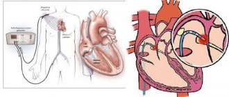
The process may involve one or both eyes, sometimes it has a recurrent course, and therefore the prognosis may be unfavorable. During an outpatient examination, it is quite difficult to identify signs of central serous chorioretinitis, since it has scanty symptoms. In this case, special examination techniques (ophthalmoscopy, biomicroscopy, fluorescein angiography) come to the rescue.
Differential diagnosis should be carried out with retrobulbar neuritis, juvenile macular degeneration, as well as other diseases. Characteristic features of central serous chorioretinitis include: widening of the optical section of the retina, the presence of transudate between the retina and the pigment epithelium, and small defects in the basal lamina. Fluorescein angiography reveals pinpoint dye leakage (so-called fluorescent flags).
Symptoms
The clinical picture of chorioretinitis significantly depends on the location of the inflammatory focus. Thus, central chorioretinitis is characterized by predominant damage to the macula (“macula”, the most light-sensitive and specialized area of the retina, responsible for the clarity of the central field of vision), equatorial and peripheral. There are also several options based on the nature of the focality: focal, multifocal (disseminated, divided into several separate foci) and diffuse, involving the entire retinal tissue without clear focal boundaries. Acute chorioretinitis can last up to three months, chronic chorioretinitis has a tendency to frequent relapses.
The content of subjective complaints is also determined by the localization of the inflammatory process. Thus, peripheral chorioretinitis often does not manifest itself at the level of subjective sensations at all - and in this case it is diagnosed by chance, during a consultation for another reason or during a medical examination. Macular chorioretinitis, on the contrary, manifests itself with multiple and varied visual disturbances: “fog before the eyes”, scotomas (blind or dark areas in the visual field), photopsia (illusory sparks or flashes of light), a noticeable decrease in the acuity and quality of vision. “Night blindness” is quite typical, as well as a distorted perception of the size and shape of objects observed by the patient.
Symptoms of this kind, no matter what the final diagnosis turns out to be, require immediate attention to an ophthalmologist.
Treatment
Treatment of the disease is usually complex; it must take into account the etiology of the pathology. Treatment that involves blocking defects in the basal lamina using laser coagulation is considered pathogenetic. After this manipulation, retinal edema disappears after 4-10 days. Additionally, medications are used that normalize the permeability of blood vessels and capillaries (Aevit, Ascorutin), dehydrating drugs (hypertonic solution of glucose, sodium chloride, glycerin, fonurite), vasodilating agents (nicotinamide, no-spa). To stimulate trophic processes, vitamins, ATP, cocarboxylase, and heparin are used. For retinal edema, corticosteroids are used in the form of a retrobulbar injection.
By contacting the Moscow Eye Clinic, each patient can be sure that some of the best Russian specialists will be responsible for the results of treatment. The high reputation of the clinic and thousands of grateful patients will certainly add to your confidence in the right choice. The most modern equipment for the diagnosis and treatment of eye diseases and an individual approach to the problems of each patient are a guarantee of high treatment results at the Moscow Eye Clinic. We provide diagnostics and treatment for children over 4 years of age and adults.
Possible complications of chorioretinitis
Complications of chorioretinitis can occur both with improper treatment and during the rehabilitation period after surgery on the eye, these include:
- retinal detachment;
- retinal vein blockage;
- neovascular membrane;
- repeated hemorrhages in the eyeball;
- complete loss of vision;
- inability to navigate in space and terrain;
- confusion of colors and shapes.
Some unpleasant symptoms may gradually decrease over time, and after a couple of months a person’s ability to navigate the area and distinguish colors and shapes is normalized.
If, after treatment, the initial symptoms reappear, then you will urgently need to visit an ophthalmologist to exclude a recurrence of the inflammatory process in the eyeball.
Our doctors who will solve your vision problems:
Fomenko Natalia Ivanovna
Chief physician of the clinic, ophthalmologist of the highest category, ophthalmic surgeon. Surgical treatment of cataracts, glaucoma and other eye diseases.
Yakovleva Yulia Valerievna Refractive surgeon, specialist in laser vision correction (LASIK, Femto-LASIK) for myopia, farsightedness and astigmatism.
You can find out the cost of a particular procedure or make an appointment at the Moscow Eye Clinic by calling in Moscow 8 (499) 322-36-36 (daily from 9:00 to 21:00) or using the ONLINE REGISTRATION FORM.
Causes
The main factors under the influence of which the retinal-vascular complex can become inflamed include:
- local and systemic infections caused by bacterial or viral pathogens (toxoplasmosis, herpes, and many others); the most common local primary sources of chorioretinitis are untreated infectious foci in adjacent organs - the nasopharynx and oral cavity;
- radiation (radiation) injury;
- intoxication, incl. products of decay of one’s own tissues (in particular, long-term non-resorbable blood after intraocular hemorrhage);
- allergic reactions;
- autoimmune disorders and diseases;
- immune deficiency (AIDS, exhaustion of various origins, recovery period after a major operation, etc.);
- ophthalmotrauma.
Prevention of chorioretinitis
To reduce the risk of developing chorioretinitis, each person must adhere to several rules of prevention, which are as follows:
- treat infectious diseases in a timely manner (especially acute respiratory viral infections);
- in case of a sharp decrease in vision, immediately consult an ophthalmologist;
- do not stay in the cold for a long time;
- compliance with personal hygiene rules (especially hand washing);
- undergo routine medical examinations annually.
In particular, you need to visit the dentist regularly. Advanced caries can provoke inflammatory and purulent processes in the oral cavity, which can subsequently spread to the visual structures.
The prognosis of chorioretinitis with timely diagnosis and adequate treatment is favorable in most cases.
Folk remedies
The use of tinctures and decoctions can speed up the healing process. It is recommended to use vasodilators and vitamins. This will complement the conservative course of treatment, but will not replace it.
An infusion of hazel bark is a vasodilator. The crushed bark is steamed with boiling water, wrapped and allowed to rest for about an hour. 10 g of crushed bark is consumed per glass of water. The product is taken orally in a teaspoon several times a day.
To strengthen the immune status, take vitamins. Riboflavin, folic acid, and retinol have a good effect. You need to eat fresh vegetables and fruits: squeeze juice out of them, stew them. If the pharmacy does not have riboflavin, it can be found in fermented milk products.
Prevention is regular examination by an ophthalmologist, timely treatment of ENT diseases and elimination of infection.
Conservative therapy
Treatment of chorioretinitis is carried out after a detailed diagnosis, based on its data. The therapeutic regimen depends on the patient’s condition, his clinical picture, and the presence/absence of serious internal diseases of a systemic or autoimmune nature.
Conservative treatment involves:
- the use of ophthalmic drops that eliminate the inflammatory process;
- individually selected set of antibiotics;
- the use of mydriotic drugs to control intraocular pressure;
- biogenic stimulants, reparants for activation of regenerative processes in the choroid and retina;
- hormonal medications for acute pathology.
For the syphilitic form, Doxycycline, Erythromycin, and Penicillin are prescribed. To cure toxoplasmosis chorioretinitis, Pyrimethamine or Sulfadimezine is prescribed. For tuberculosis, hormones and chemical medications are prescribed. The viral form is treated with antiviral medications and immunomodulators are prescribed.
To accelerate cell regeneration, enzymatic agents are prescribed: Fibrinolysin, Lidaza.
For acute chorioretinitis, medications with hydrocortisone are prescribed. To speed up tissue healing, ultrasound therapy and physiotherapy are prescribed.
If the eye has been injured by external mechanical force, surgical correction is performed. Perhaps the patient needs bone grafting, which involves joining the fragments of the orbit.
In case of severe clouding of the ocular media, the vitreous body is removed. Laser coagulation of the retina is also prescribed to prevent the formation of complications. At the stage of active healing of the tissues of the visual apparatus, ultrasound treatment is prescribed.
Forecast
How optimistic is the prognosis for healing? The earlier treatment is undertaken, the more optimistic the prognosis. In the old form, scars form that cannot be reabsorbed. This disease can pass without a trace, or can lead to complete blindness.
The prognosis is determined not only by timely seeking medical help and taking medications - much depends on the person’s immune status, age, lifestyle, and the presence/absence of bad habits.
Changes occurring in the organ of vision, or Pathogenesis
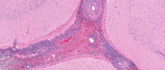
Gray-yellow foci of inflammation with blurred boundaries appear on the retina, gradually growing towards the vitreous body and infiltrating into it. At this moment, microhemorrhages are possible in the areas surrounding the lesion. Gradually, as the degree of activity of the process decreases, the boundaries of the lesion become more defined, pigmentation appears around it, and after some time the choroidal tissue becomes thinner, this area of the retina atrophies and ceases to function, and a scar is formed.
The appearance of soft exudate as a result of blockage of small arteries indicates the secondary development of the inflammatory process and the development of retinal ischemia.
Photo: https://pixabay.com/photos/eye-blue-eye-iris-pupil-face-1173863/
Gradually, structural deformation of parts of the vitreous body occurs with the formation of opacities of varying degrees of severity. The rougher the vitreous body becomes, the greater the chance of developing complications associated with the formation of adhesions between it and the retina.
Clinical picture
How does the disease manifest itself, what symptoms are characteristic of the pathology of the retina and choroid?
The symptoms are as follows:
- progressive decrease in visualization quality;
- the appearance of luminous figures and spots before the eyes;
- the appearance of “blind” areas in the field of view;
- distortion and change in the shape of familiar objects;
- increased intraocular pressure;
- retinal disinsertion;
- purulent inflammation of the inner membranes of the eyes;
- night blindness in the evening;
- vitreous hemorrhages;
- complete blindness.
Floating spots may appear before the eyes, and vision becomes clouded. During the daytime, patients do not complain of decreased visual acuity if the foci of inflammation are located in the periphery. But in the evening, the clarity of visualization noticeably decreases. The most common symptom of chorioretinitis is the appearance of spots before the eyes.
Most patients complain of the appearance of chorioretinitis after infectious and autoimmune diseases.
However, ophthalmologists are not always able to make the correct diagnosis, since a decrease in visual acuity with chorioretinitis does not occur immediately. The patient sounds the alarm only when “blind” spots appear in the field of vision.
Bottom line
Chorioretinitis is a dangerous inflammatory ophthalmological disease of the posterior part of the eyeball, which can lead to complete loss of visual ability. The pathology is characterized by the formation of foci of inflammation of the choroid (choroid) and retina (retina).
Symptoms of the pathology are “blind” spots in the field of vision, flickering of dark or luminous dots before the eyes, flashes of light, distortion of the shape of familiar objects. However, more often it is asymptomatic and discovered by chance at a preventive appointment with an ophthalmologist.
Chorioretinitis can appear for various reasons: as an allergy to substances, as a result of trauma to the visual apparatus with tissue rupture, or as an infection.
Hormones, antibiotics, and anti-inflammatory drugs are used in treatment: the therapeutic regimen depends on the characteristics of the disease. An integrated approach to treatment consists of using antibiotics, placing ointment in the conjunctival sac, administering medications behind the eyeball, and administering antiviral injections intramuscularly.
Sources used:
- Morphological foundations of ophthalmoscopic diagnostics / V.N. Arkhangelsk. - M.
- Eye diseases. For general practitioners. Reference manual / Yu.S. Astakhov, G.V. Angelopulo, O.A. Jaliashvili. - M.: SpetsLit, 2004.
- Physiology and pathology of the retina: Primary mechanisms of vision / G.G. Demirchoglian. - M.
- Shaikh S, Trese MT (2004). "West Nile virus chorioretinitis." Br J Ophthalmol.
What manifestations are characteristic of chorioretinitis?
Complaints made by patients depend on the localization of the process and etiology. The main complaint is a sharp decrease or blurred vision, a feeling of a “film” before the eyes, especially in the dark and twilight.
Patients are concerned about the appearance of spots before the eyes, bright sudden flashes in the eyes, intensifying with the movement of the eyeballs, distortion of objects, lines, shapes - macropsia, micropsia, metamorphsia.
Features of the course of the disease depending on the etiology
Toxoplasmosis chorioretinitis
This is the most severe chorioretinitis of the eye, the onset of which occurs in utero, so it is almost always congenital. The toxoplasmosis microorganism affects not only the eye, but the structures of the central nervous system.
Photo: https://pixabay.com/photos/woman-headscarf-exotic-beautiful-2558823/
When examining the fundus of patients with toxoplasmosis chorioretinitis, rough lesions are revealed in the central zone and on the periphery; they have clear boundaries with pronounced pigmentation at the edges. In severe cases, new chorioretinal lesions appear on top of the old ones, accompanied by the formation of hemorrhages in the retina and vitreous body.
Tuberculous chorioretinitis
This chorioretinitis develops secondary to the background of an already existing underlying tuberculous process in the lungs. It manifests itself by the appearance of cotton wool-shaped lesions in the fundus of the eye, which become more distinct over time. It does not have any specific differences from other chorioretinitis.
Syphilitic chorioretinitis
With this type of chorioretinitis, the fundus picture is quite characteristic - small black and white spots alternate on the retina. This picture is formed due to white foci of retinal and choroidal atrophy and black foci of pigment deposition and is called “salt and pepper.”
Chorioretinitis in AIDS
The cause of this chorioretinitis is cytomegalovirus, which is activated in the body against the background of acquired immunodeficiency. Necrotic foci form on the retina, alternating with traces of constant hemorrhages in the retina.
Myopic chorioretinitis
Myopic chorioretinitis does not develop against the background of any infectious agent, but due to structural changes in the posterior part of the eyeball due to its growth and stretching. The greater the rate of myopia progression and its degree, the higher the risk of developing myopic chorioretinitis. More often, this type of inflammation develops in adults, after 40-50 years.
Chorioretinitis in children
Chorioretinitis is most often congenital. Unfortunately, it is rarely diagnosed in the early stages of development, since the child may not complain of decreased or blurred vision.
An experienced ophthalmologist may suspect chorioretinitis, but it is not possible to conduct additional examination due to the young age of such patients. Parents and doctors begin to sound the alarm when the child’s eye begins to squint.