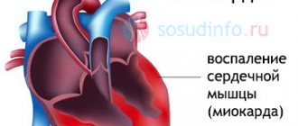
Treatment of heart rhythm disorders
TREATMENT OF AFIBLIAR ARRHYTHMIA
Conservative (drug) therapy. High-tech cardiac surgical treatment - radiofrequency ablation (RFA), which radically solves the problem. The operation is performed using minimally invasive transvascular access, using the modern non-fluoroscopic mapping system "CARTO", which helps to accurately find the area of the required impact.
TREATMENT OF PAROXYSMAL TACHYCARDIA
Training in emergency care for paroxysmal tachycardia. Drug treatment. Cardiac surgery - depending on the clinical situation and the nature of the attacks: radiofrequency ablation of arrhythmogenic zones or implantation of a cardioverter-defibrillator for ventricular tachycardia.
TREATMENT OF SVC SYNDROME
Drug therapy. Minimally invasive cardiac surgery - catheter RFA with precise identification and coagulation of the pathological focus - in almost 100% of cases permanently eliminates SVC syndrome.
TREATMENT OF EXTRASYSTOLIA
The main treatment for extrasystole is pharmacotherapy and treatment of primary diseases that provoke extrasystole. For very frequent ventricular extrasystole – radiofrequency ablation of arrhythmogenic zones.
TREATMENT OF HEART BLOCKS
Pharmacotherapy, treatment of the underlying disease - the causes of conduction disorders. Surgical treatment is the installation of dual-chamber pacemakers (PACs) in the heart, which restore the erratic heart rhythm at the right time.
TREATMENT OF WEAK SINUS NODE SYNDROME, CARDIOGENIC syncope
Conservative treatment of the disease that caused the rhythm disturbance, drug therapy. Cardiac surgery – implantation of two-chamber pacemakers into the heart, ensuring a stable heart rhythm.
PREVENTION OF SUDDEN DEATH FROM CARDIAC ARREST
Installation of cardioverter-defibrillators in the heart, maintaining the correct rhythm and reducing the risk of cardiac accidents.
We do
Radiofrequency ablation of cardiac arrhythmias
Radiofrequency ablation (RFA) is a minimally invasive, highly effective surgical intervention in the heart, performed under the control of X-ray equipment. The procedure is carried out to treat extrasystole, supraventricular and ventricular heart rhythm disturbances. The therapeutic effect is achieved through a targeted effect on arrhythmogenic zones of the heart, high-frequency current (radiofrequency energy) with a temperature of 40-55 degrees or cooling (cryo-procedure) to -80 degrees. Catheter ablation is a very effective method of treating tachyarrhythmias and, being a fairly safe procedure, can permanently eliminate the cause of heart rhythm disturbances. In the vast majority of cases, this allows you to avoid constant medication use and still lead an active lifestyle.
Indications for RFA
Supraventricular rhythm disturbances:
- atrioventricular nodal reentrant tachycardia (AVNRT),
- atrioventricular reentrant tachycardia (AVRT) associated with the functioning of the accessory pathway (AP)
- atrial tachycardia (AT)
- atrial fibrillation (AF)
- atrial flutter (AF)
- atrial extrasystole
Ventricular arrhythmias:
- ventricular extrasystole
- idiopathic ventricular tachycardia
- ventricular tachycardia due to structural heart diseases.
Preparation for surgery : − it is necessary to refrain from eating and drinking water for 6-8 hours before the procedure. If necessary, in agreement with your doctor, you can take a few small sips of water if you need to wash down the medicine (for arterial hypertension, diabetes, etc.); − 2-3 days before the operation you will need to stop taking antiarrhythmic drugs (in consultation with the doctor); − be sure to tell your doctor or nurse if you are allergic to medications.
Catheters are inserted under local and intravenous anesthesia, usually in the groin (femoral vein access), neck (jugular vein access) or subclavian vein. The procedure uses special long flexible electrodes that can both record the electrical activity of the heart and stimulate the heart during electrophysiological testing (EPS). They are performed in the heart under fluoroscopic control.
EPI is part of a procedure performed to accurately diagnose cardiac arrhythmias.
By stimulating the heart, the doctor induces an arrhythmia and studies it. EPI helps to identify the localization of pathological pathways or ectopic foci. This is called "mapping". The EF laboratory has all the necessary equipment and medications for studying and stopping arrhythmias.
After EPI and identification of the source of arrhythmia, targeted radiofrequency or cryoablation is performed. This leads to the formation of a scar (3-4 mm in diameter), which does not conduct the electrical impulse and eliminates the arrhythmia.
To monitor the effectiveness of the procedure, repeated EPI is performed. If the arrhythmogenic focus is successfully eliminated, the procedure is completed.
Treatment of arrhythmia using ablation is a fairly lengthy procedure, usually requiring from 30 minutes to several hours.
After the procedure is completed and the electrodes are removed, pressure bandages are applied in the puncture area to prevent bleeding. On the first day, bed rest should be observed, which allows to stabilize the patient’s condition.
Atrioventricular nodal reentrant tachycardia (AVNRT)
The normal propagation of the impulse from the sinus node to the ventricles passes through the region of the atrioventricular node. The heart valve rings separate the atrial myocardium from the ventricles and prevent direct passage of the impulse.
In patients with nodal tachycardia in the atrioventricular node, there are two pathways - slow and fast. And under certain conditions, a situation may arise when the impulse begins to quickly spin inside the node itself, and then almost simultaneously spread to the atria and ventricles.
The essence of the operation is to eliminate the “slow” path of impulse conduction in the AV node to prevent the tachycardia from spiraling. The effectiveness of the operation is 95-98%.
WPW syndrome (Wolff-Parkinson-White syndrome)
WPW syndrome is the second most common form of supraventricular tachycardia (after atrioventricular nodal tachycardia). In 1930, L. Wolff, J. Parkinson and P. White described the ECG syndrome of “functional bundle branch block” and short PQ interval, which is observed in young, physically healthy individuals suffering from attacks of tachycardia.
The anatomical substrate of WPW syndrome is the accessory atrioventricular (atrioventricular) junctions (ADJ), “bundles of Kent” or so-called “muscle bridges”. Additional atrioventricular connections are pathways between the myocardium of the atria and ventricles, existing in addition to the structures of the atrioventricular node.
The essence of the operation is to eliminate the “additional, abnormal” path of impulse conduction in the AV sulcus. The effectiveness of the operation is more than 98%.
Complications of the procedure. Like any surgical procedure, radiofrequency ablation is associated with certain risks. However, the risk is small (less than 1%) and the procedure is considered relatively safe: some patients experience bleeding at the site of insertion of the electrodes - this can cause the formation of a hematoma, in rare cases the procedure can lead to more serious complications associated with damage to the heart wall and blood vessels . Depending on the type and location of the pathological pathway, there is a risk of damage to the conduction system of the heart. Therefore, in some cases it may be necessary to implant a pacemaker. These complications are quite rare and the expected benefit from the procedure most often prevails over them. Doctors in our department do everything possible to reduce the risk of complications. Catheter ablation is a very effective method of treating tachyarrhythmias and, being a fairly safe procedure, can permanently eliminate the cause of heart rhythm disturbances. In most cases, this allows you to avoid constant medication use and still maintain an active lifestyle.
Ventricular heart rhythm disturbances
Ventricular heart rhythm disturbances can be in the form of extraordinary ventricular extrasystoles (VCs) or in the form of ventricular tachycardias (VT). RFA is performed in symptomatic patients and, most often, does not depend on the number of PVCs and when the patient with VT is hemodynamically stable. Preparation and surgery are almost the same as for supraventricular cardiac arrhythmias.
However, the effectiveness of the RFA procedure directly depends on the localization of the arrhythmogenic focus and on the patient’s concomitant structural diseases (coronary heart disease, valvular defects, cardiopathy, etc.)
Atrial fibrillation
More than 3 million people in Russia suffer from the most common form of tachyarrhythmia - atrial fibrillation or atrial fibrillation. Along with the need for frequent hospitalization, emergency calls, a sharp decline in the quality of life and the development of ischemic strokes in at least 40,000 patients a year, the state spends huge amounts of money without returning these patients to a socially active lifestyle.
After 60 years of life, up to 6% of the population suffers from this disease and despite the constant use of antiarrhythmic drugs and blood-thinning anticoagulants - 50-60% of patients remain symptomatic and the “sword of Dodocles” hangs over them - either sudden death or disability, progressively increasing from the moment onset of the disease.
Today, cardiologists and cardiac surgeons, or rather specialists in the field of interventional arrhythmology, have found a new approach to diagnosis (also called electrophysiological) and elimination of the so-called paroxysmal and early persistent form of atrial fibrillation, when arrhythmogenic foci that trigger the arrhythmia are localized in the vessels flowing into the left atrium . Using various systems of electrophysiological diagnostics, using catheter technology (without a scalpel), specialists from the City Clinical Hospital named after. I.V. Davydovsky perform these procedures in previously incurable patients who have suffered from this arrhythmia for decades.
Currently, we are only at the beginning of the use of modern technologies for diagnosing and treating heart diseases and arrhythmias using so-called “bloodless surgery”, which does not require incisions.
Preparing for surgery:
includes a full diagnostic examination of the patient - ECG, echocardiography, Holter monitoring, blood tests. It is very important that the patient receives anticoagulant therapy (warfarin, Eliquis, Pradaxa or Xarelto) at least 3-4 weeks before surgery, since the operation will be performed in the “arterial chamber” of the heart. All medications the patient receives are discontinued in the clinic by the attending physician. At the clinic, the patient undergoes computed tomography and/or transesophageal echocardiography to exclude a “thrombus in the left atrial appendage. The presence of a blood clot is an “absolute” contraindication to surgical interventions and requires a change in anticoagulant therapy.
The procedure is performed through venous access (femoral, subclavian, jugular veins). Diagnostic electrodes are inserted into the region of the coronary sinus and a long introducer into the cavity of the right atrium. Under the control of X-ray, transesophageal or intracardiac echocardiography, a puncture of the interatrial septum is performed and instruments are passed into the cavity of the left atrium.
Depending on the anatomy of the left atrium, either Radiofrequency or Cryo-isolation of the pulmonary veins (PV) is performed. At the moment, the methods show equal effectiveness. The choice of PV isolation technique occurs before or during the procedure. If the patient underwent computed tomography before the operation, the anatomy of the left atrium and pulmonary veins allows you to select the desired technique (if TEE is performed before surgery, angiography of the left atrium is performed during the operation).
- if the left atrium is not enlarged in size and the pulmonary veins flow into the left atrium with separate orifices, the choice falls on cryo-isolation;
— if the atrium is enlarged or the PVs have a “collector” or “vestibular” entrance, or the size of the pulmonary veins is more than 25 mm (cryoballoon size 28 mm), we choose the radiofrequency isolation technique.
The operation is performed under intravenous anesthesia (propafol, fentanyl), the patient is completely awake, but does not experience pain. All pulmonary veins are sequentially isolated (cryo- or radiofrequency destruction), the effectiveness of electrical isolation is checked, and a repeat electrophysiological study is performed.
After the procedure is completed, the administration of anesthetic drugs ends, and the patient is transferred for 5-6 hours (sometimes longer) to the intensive care unit, where he is monitored. Then a control echocardiogram is performed and the patient is transferred to the ward under the supervision of the attending doctor. Another day of observation is carried out, antiarrhythmic and anticoagulant therapy is selected - then the patient is discharged. The patient is in the hospital for 3-4 days from the start of hospitalization. Changes in the length of hospitalization depend on the orders of the treating cardiologist. In the postoperative period, episodes of chest pain, dizziness, interruptions in heart function and attacks of arrhythmia associated with the operation (edema) may occur, which are not signs of a “relapse” of arrhythmia.
This is followed by a 3-month “blind period”, when the formation and normalization of the “isolation line” occurs. This is a period of rehabilitation and, during this time, additional interventions in the heart are not performed. Efficacy is assessed after this period and after discontinuation or reduction of dosages of antiarrhythmic drugs.
The overall effectiveness of primary procedures for isolating the pulmonary veins in paroxysmal forms of atrial fibrillation is 65-70%. This is due to restoration of conduction in isolated structures, other arrhythmias, progression of the disease, etc. Therefore, some patients (up to 50%) require a repeat procedure to identify and eliminate conduction breakthroughs in the pulmonary veins or concomitant “extrapulmonary” arrhythmias. The effectiveness of secondary surgery and improvement in quality of life is achieved in 90% of patients. It is advisable to carry out cardiac monitoring 3, 6 months and 1 year after the last operation. Cancellation of medications is carried out only with the consent of the treating doctor. Often, anticoagulant (thinning) therapy remains for long-term use and does not depend on the results of the operation (calculated using a specific CHADS2-VASc scale).
The number of complications in the City Clinical Hospital named after. I.V. Davydovsky is less than 1% and includes hemopericardium (accumulation of fluid in the heart sac), hematomas in the puncture area, phrenic nerve paresis (only during cryo-procedures). All complications are effectively managed during the hospital period and may lead to a delay in discharge. There were no deaths observed in our clinic.
Atrial flutter
The goals of management of patients with atrial flutter (AFL) are similar to the management of AF. Based on available data, the risk of stroke in patients with atrial flutter is not significantly different from that in AF. In addition, many patients with atrial flutter have concomitant AF (with this combination, the procedure is performed together). Thus, in patients with atrial flutter, anticoagulant therapy should be used in the same way as in patients with AF. Frequency control in AFL is achieved with the same drugs as in AF, but higher doses of drugs are usually required. Typical forms of AFL have a similar mechanism and “spin” around the tricuspid valve clockwise and counterclockwise.
In order to effectively eliminate this arrhythmia, it is necessary to draw a “demarcation” ablation line in the “narrowest” and “slowest” place of the tachycardia circle - the cava-tricuspid isthmus (from the tricuspid valve to the inferior vena cava).
Ablation of the cava-tricuspid isthmus in isthmus-dependent AFL effectively preserves sinus rhythm with an efficiency of 90-95%. The procedure, as with AF, is performed under intravenous anesthesia, transvenous access (femoral, subclavian, jugular veins).
This procedure is effective in reducing recurrences of AF in selected patients and will help avoid unnecessary hospitalizations. Isthmus ablation is a relatively safe and more effective method than antiarrhythmic drug therapy and is recommended for recurrent atrial flutter. Catheter ablation of left atrial macro-reentry tachycardia is a more complex procedure, with a lower success rate and a high rate of postoperative relapse.
Features of treatment of arrhythmia at the Yauza Clinical Hospital
The patient can undergo a full diagnosis, high-quality treatment for arrhythmia (including surgery), and also be under the supervision of specialists (cardiologists, cardiac surgeons) for a year after the operation.
During the period of outpatient observation after surgical treatment, the patient can constantly (if necessary, almost around the clock) be in touch with the doctor.
About cardiac surgical treatment of arrhythmia
Minimally invasive operations with access through a vessel. Duration: 2-3 hours.- High-tech equipment - the non-fluoroscopic mapping system "CARTO" allows you to conduct an intracardiac electrophysiological study directly during surgery to accurately determine the location of the pathological impulse causing the arrhythmia, coagulate it, restoring normal sinus rhythm.
- Hospital stay – 1-1.5 days. Postoperative rehabilitation – 2-7 days.
- Operations can be performed on elderly people (over 65 years old).
- Minimally invasive cardiac surgical treatment is possible even against the background of somatic diseases.
- The department is headed by one of the leading operating arrhythmologists in Russia, Doctor of Medical Sciences, Professor A.V. Ardashev.
Performed for the first time in Russia
- laser angioplasty for stenotic and occlusive lesions of the coronary arteries of the heart in ischemic heart disease - February 1989
- removal of a foreign body from the lumen of a coronary artery during a complicated angioplasty operation - April 1991
- stenting of the coronary artery for atherosclerotic lesions of the coronary artery – January 1992
- implantation of an original device to close a cardiac septal defect – March 1995
- retrograde recanalization through the collateral canal of a blocked coronary artery – September 2006
- hydrodynamic recanalization for coronary artery occlusion – March 2007
Experience in endovascular operations for coronary artery disease and structural pathologies of the heart - more than 6,000 successful operations!
How it all began . . .
1988 He performed his first operation on the coronary arteries of the heart as a clinical resident at the All-Union Scientific Center of Surgery of the USSR Academy of Medical Sciences, under the guidance of prof. Rabkina I.Kh. After a successful start, further intensive development of new minimally invasive methods of treating coronary heart disease and structural pathologies. The first operation using a laser beam and the thinnest light guide to eliminate atherosclerotic lesions, first during coronary bypass surgery, then using a puncture (puncture) in the femoral artery.
Then there was an unexpected twist.
It’s not for nothing that they say that sometimes incidents can decide the rest of your life. 1993 Bronstein A.S. was present during the operation of the young patient. and after a successful operation and discharge of the patient, a week later a phone call came with an invitation to establish a minimally invasive cardiac surgery service at the Center headed by Alexander Semenovich. It must be said that the surprise of the proposal set in complete stupor, because there was also a good prospect on the basis of the All-Russian Research Center for Surgery. However, I preferred more “creative freedom” and would like to test my strength in establishing a full-fledged minimally invasive cardiology service. Further events developed quite intensively, the defense of a doctoral thesis, two monographs, organization and participation in 6 Russian symposia, the first operations “live” with transmission to the meeting room, where there were colleagues taking part in scientific conferences. The first operations performed abroad (in particular, at the onze lieve vrouwe Gathius hospital in Amsterdam with Prof. Kiemeneij F.), participation and presentation of reports on our achievements in the field of minimally invasive cardiology at numerous international prestigious symposia and conferences (such as EUROpcr in Paris and Barcelona, TCT in the USA and South Korea, CCT in Japan, etc.). Participation in more than 10 international clinical studies. First stent.
. . Nowadays, implantation of prostheses (stents) into the coronary arteries of the heart has become commonplace, but in the early 90s, implantation of an artificial prosthesis into the artery of the heart seemed as fantastic as flying to the moon. . . Some said that this was very dangerous, since it increased the risk of a blood clot in the artery, with clear results, others doubted the effectiveness of these prostheses themselves. But, nevertheless, progress cannot be stopped.
One day at the beginning of 1992, almost immediately after the New Year holidays, Professor Yu.V. Tarichko came up to me, then we worked together at the Russian Surgery Center of the Academy of Medical Sciences, and said that his good friend, a researcher at one of the universities in Moscow, was suffering angina pectoris and he should be examined. After performing a diagnostic coronary angiography, he found himself with a rather dangerous lesion - an almost critical narrowing of the largest artery of the heart, the closure of which could lead to catastrophic consequences. We discussed the situation and treatment tactics together with surgeons and cardiologists and decided to perform balloon angioplasty (expansion and elimination of narrowing with an inflated balloon). At that time we had the largest experience in balloon coronary angioplasty in Russia (more than 700 operations). So, on January 24 we took it to the table and... . . During balloon angioplasty, a detachment of the inner surface of the vessel occurred (the so-called dissection of the vessel wall) with almost complete blocking of blood flow in the artery. You can imagine the situation in the operating room; there was no panic, but the situation seemed insoluble. The patient could not have been kept until the open bypass operation, since it took about 40-50 minutes to prepare the operating room for the open operation and during this time the heart could stop; there were no other ways to save the situation with the help of surgery at that time; it remained to be treated with medications unknown and absolutely unguaranteed outcome, and nevertheless, resuscitation measures were started, stress reigned in the operating room, dangerous rhythm disturbances had already begun, defibrillation twice.
Resuscitators were preparing for intubation and external cardiac massage. Prof. stood next to me. Tarichko, watching this picture, apparently regretted in his heart that they had made such a decision regarding the treatment of his friend. And suddenly it dawned on me: we have an intravascular coronary prosthesis, then this sample was given to me from Johnson & Johnson for testing. It was a Palmaz-Shatz prosthesis, which by that time was installed in only 2 or 3 clinics in the world. But in Russia, no one has ever implanted a stent! And we took a risk, because the situation was practically critical. . Preparation for implantation, as I was later told, took only 5 minutes. . I was stressed and just lost track of time. But I clearly remember the moment of control angiography after implantation, blood flow was restored, the artery was completely filled with blood, the heart began to contract at full strength, the rhythm had improved. The first feeling was that a person had been saved from an inevitable outcome. The first stent in Russia turned out to be so lucky.
By the way, this patient is still alive and well, 18 years have passed!!! and there is reason for pride - the patient was not only saved, but also given 18 years of comfortable life. What could be even better for a surgeon?