Diseases of the cardiovascular system
With pain in the chest on the right side, almost every doctor will first suspect the presence of cardiac pathologies, especially if the patient is no longer young, and the patient himself leads a sedentary, unhealthy lifestyle and has a history of cardiovascular and endocrine diseases.
Aortic aneurysm
An aneurysm is a weak section of the wall of the main artery in our body. In this place the wall stretches, bulges, and at some point it can either burst or tear. This condition is life-threatening, therefore, at the first suspicion of an aneurysm, the patient will be prescribed an X-ray or MRI, where the pathology will be identified and a decision will be made on the need for surgical treatment.
Angina pectoris
If there is pain in the upper right side of the chest, this may indicate the presence of angina pectoris: it occurs due to impaired blood supply to the myocardium and is often a consequence of atherosclerosis (the formation of plaques on the inner walls of blood vessels).
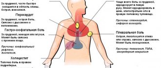
Pericarditis
The pathology is caused by the accumulation of fluid in the cavity of the heart muscle (pericardium), as a result of which its function is impaired. This condition is most often observed in men, and at a young age - from 20 to 50 years.
Myocardial infarction
Sudden acute pain in the chest on the right, more often in women and less often in men, which does not go away even after taking strong painkillers, indicates myocardial infarction. This is a deadly condition caused by impaired blood supply to the heart muscle.
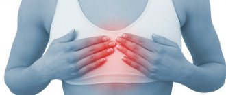
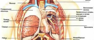
Chest pain is any pain or discomfort in the chest area. It can be caused by various diseases, including pathology of the heart, blood vessels, pericardium, lungs, pleura, trachea, esophagus, muscles, ribs, and nerves. In some cases, chest pain is a sign of damage to organs outside the chest, such as the stomach, gallbladder, or pancreas.
Chest pain is very diverse: sharp, dull, aching, cutting, stabbing, pulling, bursting, burning or pressure. Painful sensations vary among different diseases, but pain is not a specific symptom of a particular disease. The characteristics of pain may vary depending on the age, gender of the patient, concomitant diseases, and psychological characteristics. Identifying the immediate cause of chest pain is often difficult and requires a number of diagnostic procedures.
It is one of the most alarming symptoms, as it can be a manifestation of severe, life-threatening conditions that require emergency medical care, in particular myocardial infarction.
Synonyms Russian
Thoracalgia, chest pain, chest pain
English synonyms
Chest pain, pain in the chest, thoracalgia.
Symptoms
Chest pain can be of different types. Sometimes it radiates to the arm, shoulder, shoulder blade, back, neck. The patient may complain not only of pain, but also of tightness, burning, and discomfort in the chest area.
Unpleasant sensations may intensify when coughing, deep breathing, swallowing, pressing on the chest, changing body position (constant or periodic). Chest pain and discomfort may be accompanied by a number of additional symptoms, depending on the underlying disease: belching or bitterness in the mouth, nausea, vomiting, difficulty swallowing.
General information about the disease
Chest pain can be a manifestation of various diseases, each of which requires a specific medical approach.
- Acute myocardial infarction (heart attack). Acute chest pain in people over 40 years of age is most often associated with this disease. Myocardial infarction occurs when a section of the myocardium is damaged and killed as a result of impaired circulation in the coronary vessels. Most often it manifests itself as acute pain behind the sternum or to the left of the sternum, which radiates to the back, neck, shoulder, arm and does not decrease when taking nitroglycerin or at rest. Symptoms vary from patient to patient. Elderly women are characterized by atypical symptoms: severe weakness, nausea and vomiting, rapid breathing, abdominal pain.
- Angina pectoris. A condition in which, as a result of atherosclerosis and narrowing of the coronary vessels, the blood supply to the heart muscle is disrupted. Pain during angina pectoris resembles that during myocardial infarction, but occurs during physical activity, decreases with rest and is relieved by nitroglycerin.
- Dissecting aortic aneurysm. The aorta is a large vessel that carries blood from the left ventricle of the heart to organs and tissues. With a dissecting aneurysm, a rupture of the intima (inner lining) of the aorta occurs with penetration of blood into other layers of the aortic wall and subsequent dissection of the wall, which most often leads to complete rupture of the aorta and massive internal bleeding. The disease in most cases ends in death within a few hours or days, even with timely diagnosis and timely treatment.
Dissecting aortic aneurysm is most often a consequence of long-term arterial hypertension, and can also occur with Marfan syndrome, as a result of chest trauma, during pregnancy, or as a later complication of heart surgery.
Pain with dissecting aortic aneurysm is similar to pain with myocardial infarction and angina pectoris, can last for several hours or days, and does not decrease with rest or with nitroglycerin.
- Pulmonary embolism. Blockage of the pulmonary artery or its branches by a thrombus, through which venous blood flows from the right ventricle to the lungs for oxygen saturation. As a result, gas exchange is disrupted, hypoxia occurs, and pressure in the pulmonary arteries increases. Chest pain occurs suddenly, intensifies with deep inspiration, is accompanied by rapid breathing and, in some cases, hemoptysis. The risk of thromboembolism increases after surgery, prolonged forced immobility, pregnancy, taking oral contraceptives, especially in combination with smoking, and cancer.
- Pneumothorax. The accumulation of air or other gas in the pleural cavity, a slit-like space between the membranes lining the surface of the lungs and the inner surface of the chest. Accompanied by acute chest pain, rapid breathing, anxiety, loss of consciousness.
- Pericarditis. Inflammation of the heart sac (pericardium), that is, the serous membrane of the heart. Pain occurs due to friction of the inflamed pericardial layers. Pericarditis can be a consequence of a viral infection, rheumatoid arthritis, systemic lupus erythematosus, or renal failure. Idiopathic pericarditis, that is, pericarditis of unknown etiology, is common. The pain is acute, occurs only in the initial stages of the disease, and may be accompanied by rapid breathing, fever, and malaise.
- Mitral valve prolapse. Pathology of the valve, which is located between the left atrium and the left ventricle of the heart. In some people, when the left ventricle contracts, the mitral valve bends into the atrium and some of the blood from the left ventricle flows back into the left atrium. For most patients, this does not cause discomfort, but some experience increased heart rate and chest pain that does not depend on physical activity and does not radiate, unlike angina.
- Pneumonia. Inflammation of the lung tissue. Chest pain with pneumonia is usually one-sided, worsens with coughing, and is accompanied by fever, malaise, and cough.
- Esophagitis. Inflammation of the esophagus. Accompanied by chest pain and difficulty swallowing. Symptoms do not improve with antacids.
- Gastroesophageal reflux disease. A chronic disease in which acidic stomach contents reflux into the esophagus, leading to damage to the lower esophagus. In this case, acute, cutting pain in the chest along the esophagus, heaviness, discomfort in the chest, belching, bitterness in the mouth, difficulty swallowing, and dry cough may occur.
- Pleurisy. Inflammation of the pleura. Friction of the inflamed pleura causes pain. Pleurisy can be the result of a viral or bacterial infection, cancer, chemotherapy or radiation therapy, or rheumatoid arthritis.
- Fractured ribs. In this case, the pain intensifies with deep breathing and movement.
- Other causes: pancreatitis, cholelithiasis, depression.
Who is at risk?
- People over 40 years old.
- Obese people.
- Patients with arterial hypertension.
- People with high levels of cholesterol in the blood.
- Have recently undergone surgery.
- Those suffering from alcoholism.
- Smokers.
- Pregnant women.
- Suffering from cardiac arrhythmia.
- People with cancer.
- Taking certain medications.
- People with chronic lung diseases.
Diagnostics
Chest pain is not a specific symptom and can clearly indicate a particular disease. However, when this sign appears, the doctor must first of all exclude a number of life-threatening conditions that require immediate help. Sometimes only additional laboratory and instrumental studies can accurately determine the cause of chest pain.
Laboratory research
- General blood analysis. Leukocytosis (with pleurisy, pneumonia), anemia (with dissecting aortic aneurysm), thrombocytosis and erythremia (with pulmonary embolism) can be detected.
- Erythrocyte sedimentation rate (ESR). Nonspecific indicator of inflammation. ESR can be increased with pleurisy, pericarditis, pneumonia and other diseases.
- C-reactive protein. Increased in inflammatory diseases, as well as in myocardial infarction. With angina, the level of C-reactive protein does not change.
- NT-proBNP (brain sodium uretic propeptide). A protein, the main part of which is found in myocardial cells. It is a precursor of natriuretic peptide, responsible for the excretion of sodium in the urine. This indicator is used to assess the risk of heart failure, identify the initial stages of heart failure, and evaluate therapy. Is highly specific. May be elevated during myocardial infarction.
- Troponin I. Troponin is a protein involved in muscle contraction. The cardiac form of troponin is found in the heart muscle and is released when the myocardium is damaged. It may be increased in myocardial infarction and other diseases accompanied by the destruction of cardiomyocytes.
- Myoglobin. A protein similar in structure to hemoglobin and responsible for the deposition of oxygen in muscle tissue, including the heart muscle. It increases when muscle tissue is damaged, in the first hours after myocardial infarction.
- Alanine aminotransferase (ALT). An enzyme that is found primarily in the liver, as well as in skeletal muscles, kidneys and myocardium. An increase in ALT indicates liver damage, but may also indicate myocardial infarction and serves as an indicator of the extent of damage to the heart muscle.
- Aspartate aminotransferase (AST). This enzyme is found mainly in the myocardium, skeletal muscles, and liver. An increase in AST levels is a sign of myocardial infarction. The AST value corresponds to the degree of damage to the heart muscle.
- Generic creatine kinase. An enzyme involved in energy metabolism reactions. Its different isoforms are found in different tissues of the human body. An increase in the level of total creatine kinase is observed in myocardial infarction and myopathies.
- Creatine kinase MB. An isoform of creatine kinase, which is found mainly in the myocardium and tissues of the nervous system. Its level corresponds to the extent of myocardial damage.
- Lactate dehydrogenase (LDH) total. An enzyme that is involved in energy metabolism and is found in almost all tissues of the body. Different types of LDH are present in different organs. Total lactate dehydrogenase may be elevated in myocardial infarction and liver disease.
- Lactate dehydrogenase 1, 2 (LDH 1, 2 fractions). These are types of lactate dehydrogenase, the increase of which is a more specific indicator of myocardial and kidney damage.
- Lipase. Pancreatic enzyme. Increased lipase levels are specific to pancreatic diseases.
- Total cholesterol. This is the main indicator of fat metabolism in the body. Used to diagnose atherosclerosis and liver diseases.
- D-dimer. Fibrin breakdown product. It is an indicator of fibrinolytic activity of the blood. The level of D-dimer may change with pulmonary embolism, dissecting aortic aneurysm.
- The main blood electrolytes are potassium, sodium, chlorine, calcium. Changes in the level of blood electrolytes may indicate pathology of the kidneys, adrenal glands, endocrine diseases, and malignant neoplasms.
- Urea, serum creatinine. These are the end products of nitrogen metabolism, which are excreted from the body by the kidneys. Their increase may indicate kidney pathology.
Instrumental research methods
- Electrocardiography (ECG). Changes in the ECG are detected during myocardial infarction, angina pectoris, and pericarditis. Helps determine the location and extent of myocardial damage.
- X-ray, computed tomography (CT), magnetic resonance imaging (MRI), ultrasound examination of the chest organs. These are imaging methods that allow you to assess the condition of the chest organs, identify injuries, neoplasms, signs of internal bleeding and other pathological changes.
- Transesophageal echocardiography. An ultrasound examination in which a probe is inserted into the esophagus. With its help, the condition of the heart, its valves, and large vessels is assessed. It has great diagnostic value for pulmonary embolism and aortic aneurysm.
- Angiography. X-ray examination of blood vessels using a non-toxic contrast agent, clearly visible on the images. Allows you to assess the condition and patency of blood vessels, including coronary ones.
Treatment
Treatment depends on the underlying disease, the symptom of which is chest pain. Therapy can consist of both the use of appropriate medications and surgical procedures.
Prevention
There is no specific prevention for most diseases accompanied by chest pain. However, to reduce the risk of their development, quitting smoking and alcohol, sufficient physical activity, a healthy diet, and timely preventive medical examinations are useful.
Recommended tests
- General blood analysis
- Erythrocyte sedimentation rate (ESR)
- C-reactive protein (quantitative)
- NT-proBNP (quantitative)
- Troponin I
- Myoglobin
- Alanine aminotransferase (ALT)
- Aspartate aminotransferase (AST)
- Creatine kinase total
- Creatine kinase MB
- Lactate dehydrogenase (LDH) total
- Lactate dehydrogenase 1, 2 (LDH 1, 2 fractions)
- Lipase
- Total cholesterol
- D-dimer
- Serum potassium
- Serum sodium
- Chlorine in serum
- Serum calcium
- Urea in serum
- Serum creatinine
- Laboratory diagnosis of acute coronary syndrome and myocardial infarction (optimal)
Digestive diseases
Quite often, a patient’s complaints that he has pain on the right side of the chest can signal problems with the gastrointestinal tract. As a rule, such diseases are accompanied by a number of very characteristic additional symptoms - nausea and vomiting, increased body temperature, and upset stool.
Hepatitis
A dangerous disease that affects the liver develops as a result of transfusion of infected blood into the human body (forms B and C), and is also transmitted through dirty hands (form A). A characteristic symptom of hepatitis is not only pain, but also yellowing of the whites of the eyes, associated with an increase in the amount of bilirubin in the blood.
Stomach ulcer
It may appear as the body’s response to the abuse of spicy, fatty and fried foods, as well as alcohol. Often, peptic ulcer disease is preceded by other gastrointestinal lesions. The pathology is characterized by the formation of a perforation in the wall of the stomach, through which its contents enter the abdominal cavity. As a rule, with an ulcer, the patient complains that the pain is most intense on the right side (including in the chest) during or after eating.
Pancreatitis
Pancreatitis (inflammation of the pancreas) in our country is not treated very responsibly, believing that it is enough to relieve pain symptoms. In fact, such a careless attitude towards the disease can lead to a number of more serious problems that will require urgent surgical intervention.
Methods for diagnosing intercostal neuralgia
If you suspect intercostal neuralgia, you should consult a neurologist.
In most cases, the diagnosis is established based on the patient’s complaints, medical history and examination of the patient directly at the appointment. The doctor pays attention to the patient’s posture: in an effort to reduce pain by reducing pressure on the affected nerve, the patient tilts the body in the opposite direction. The intercostal spaces are palpated in the area where the pain is located. Loss of sensitivity and blanching of the skin is established.
Sometimes instrumental and laboratory diagnostics may be required:
ECG
An ECG is performed in case of pain on the left side. The goal is to rule out heart problems. In combination with an ECG, echocardiography (ultrasound of the heart) may be prescribed. More information about the diagnostic method
Chest X-ray
A chest x-ray is performed to exclude diseases of the lungs and pleura.
More information about the diagnostic method
Ultrasound of the abdominal organs
A survey ultrasound examination can exclude diseases of the abdominal organs.
More information about the diagnostic method
Neuromyography
Electroneurography is prescribed, as a rule, if the traumatic nature of intercostal neuralgia is suspected.
X-ray of the spine
X-ray of the thoracic spine allows us to determine the causes of radicular syndrome. A CT scan or MRI may also be ordered to evaluate the condition of the spine.
More information about the diagnostic method
Blood test for antibodies to the herpes virus
It is carried out if there is a suspicion of a viral origin of intercostal neuralgia.
Sign up for diagnostics To accurately diagnose the disease, make an appointment with specialists from the Family Doctor network.
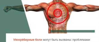
Spinal problems
The back is also often able to “give” under the ribs or into the chest. This occurs if there is an injury, sprain after intense physical activity, and as the main concomitant symptom of some pathological conditions.
Intercostal neuralgia
This term refers to compression of the roots of the intercostal nerves. As a result, the latter become irritated and respond with intense pain in the chest on the right or left, often radiating under the shoulder blade.
Intervertebral hernia
This is a musculoskeletal disease in which there is a gradual thinning of the wall of the fibrous ring and, as a result, its rupture.
Scoliosis
The “sickness” that our parents and school teachers used to scare us as children is not as harmless as it might seem. Deforming over time, the spine often produces more or less intense pain and various pathologies.
Ankylosing spondylitis
A situation in which pain in the back and/or chest on the right is a frequent companion to this pathology, manifested by inflammation of the intervertebral joints. The disease is chronic, limits joint mobility, stunts growth and ultimately leads to disability.
Causes of intercostal neuralgia
All causes of intercostal neuralgia can be reduced either to a pinched nerve root (the nerve is compressed at the point of exit from the spinal canal) or to irritation or compression of a nerve already in the intercostal space.
Intercostal nerve radicular syndrome is usually caused by one of the following:
- osteochondrosis of the thoracic spine;
- intervertebral hernia;
- tumor processes;
- other diseases of the spine.
Much more often, intercostal neuralgia occurs as a result of factors affecting the nerve in the intercostal space. The main reasons here are:
- chest injuries;
- herpes infection (herpes zoster);
- muscular-tonic syndrome due to excessive physical activity or working in an awkward position;
- hypothermia;
- for women - wearing tight underwear (bra). First of all, those who have a poorly developed subcutaneous fat layer should be careful;
- compression of the nerve by a growing tumor (pleura, chest wall) or an aneurysm of the descending thoracic aorta;
- factors affecting the entire nervous system (poisoning, hypovitaminosis).
Factors that increase the risk of intercostal neuralgia are:
- diabetes;
- problems with immunity;
- age-related changes in blood vessels;
- alcoholism;
- hormonal disorders;
- stress, depressed mental state;
- long-term use of potent medications.
Treatment
It can be conservative or surgical, depending on the diagnosis. Some types of pathologies - for example, hepatitis - require long-term use of antibiotics and strong antiviral drugs, others are based on normalizing the diet and maintaining a healthy lifestyle. The specialists of the Energo clinic, who are true professionals in their field, will definitely identify the causes of back and chest pain, select a method that is suitable for you to solve the problem, and give useful recommendations regarding the period of recovery or remission in the case of a chronic illness.
What to do?
The first rule when detecting pain is not to make a diagnosis yourself. If there is pressure, what to do:
- In case of prolonged or acute sensations, call an ambulance.
- While waiting for doctors, lie down with a bolster or pillow under your head.
- Loosen clothing and provide oxygen access.
- If you suspect that the sensations are due to heart disease, take a nitroglycerin tablet.
It is strictly forbidden to leave the patient unattended. If the situation is not critical, then you need to visit a doctor yourself as soon as possible.