© Author: A. Olesya Valerievna, candidate of medical sciences, practicing physician, teacher at a medical university, especially for SosudInfo.ru (about the authors)
Adrenergic agonists constitute a large group of pharmacological drugs that have a stimulating effect on adrenergic receptors located in internal organs and vascular walls. The effect of their influence is determined by the excitation of the corresponding protein molecules, which causes changes in the metabolism and functioning of organs and systems.
Adrenergic receptors are found in all tissues of the body; they are specific protein molecules on the surface of cell membranes. The effect on adrenergic receptors of adrenaline and norepinephrine (natural catecholamines of the body) causes a variety of therapeutic and even toxic effects.
With adrenergic stimulation, both spasm and vasodilation, relaxation of smooth muscles or, conversely , contraction of striated muscles can occur. Adrenergic agonists change the secretion of mucus by glandular cells, increase the conductivity and excitability of muscle fibers, etc.
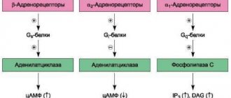
The effects mediated by adrenergic agonists are very diverse and depend on the type of receptor that is stimulated in a particular case. The body contains α-1, α-2, β-1, β-2, β-3 receptors. The influence and interaction of adrenaline and norepinephrine with each of these molecules are complex biochemical mechanisms, which we will not dwell on, specifying only the most important effects from stimulation of specific adrenergic receptors.
α1 receptors are located predominantly on small arterial-type vessels (arterioles), and their stimulation leads to vascular spasm and a decrease in the permeability of capillary walls. The result of the action of drugs that stimulate these proteins is an increase in blood pressure, a decrease in swelling and the intensity of the inflammatory reaction.
α2 receptors have a slightly different meaning. They are sensitive to both adrenaline and norepinephrine, but combining them with a mediator causes the opposite effect, that is, by binding to the receptor, adrenaline causes a decrease in its own secretion. The effect on α2 molecules leads to a decrease in blood pressure, dilation of blood vessels, and increased permeability.
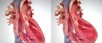
The predominant localization of β1-adrenergic receptors is the heart, therefore the effect of their stimulation will be to change its work - increased contractions, increased heart rate, accelerated conduction along the nerve fibers of the myocardium. β1 stimulation will also result in an increase in blood pressure. In addition to the heart, β1 receptors are located in the kidneys.
β2-adrenergic receptors are present in the bronchi, and their activation causes expansion of the bronchial tree and relief of spasm. β3 receptors are present in adipose tissue and promote the breakdown of fat with the release of energy and heat.
There are different groups of adrenomimetics: alpha- and beta-adrenergic agonists, mixed-action drugs, selective and non-selective.
Adrenomimetics are able to bind to receptors themselves, completely reproducing the effect of endogenous mediators (adrenaline, norepinephrine) - direct-acting drugs. In other cases, the medicine acts indirectly: it enhances the production of natural mediators, prevents their destruction and reuptake, which helps to increase the concentration of the mediator at the nerve endings and enhance its effects (indirect action).
Indications for the use of adrenergic agonists may include:
- Acute heart failure, shock, sudden drop in blood pressure, cardiac arrest;
- Bronchial asthma and other diseases of the respiratory system accompanied by bronchospasm; acute inflammatory processes of the nasal and eye mucosa, glaucoma;
- Hypoglycemic coma;
- Conducting local anesthesia.
Non-selective adrenergic agonists
Non-selective adrenergic agonists are capable of stimulating both alpha and beta receptors, causing a wide range of changes in many organs and tissues. These include adrenaline and norepinephrine.
Epinephrine activates all types of adrenergic receptors, but is considered primarily a beta agonist. Its main effects:
- Narrowing of blood vessels in the skin, mucous membranes, abdominal organs and enlargement of the lumens of blood vessels in the brain, heart and muscles;
- Increased myocardial contractility and heart rate;
- Expansion of the lumens of the bronchi, reduction of mucus production by the bronchial glands, reduction of edema.
Adrenaline is used mainly for the purpose of providing first aid and emergency care for acute allergic reactions, including anaphylactic shock, cardiac arrest (intracardiac), hypoglycemic coma. Adrenaline is added to anesthetic drugs to increase their duration of action.
The effects of norepinephrine are in many ways similar to adrenaline, but less pronounced. Both drugs have the same effect on the smooth muscles of internal organs and metabolism. Norepinephrine increases myocardial contractility, constricts blood vessels and increases blood pressure, but the heart rate may even decrease, due to the activation of other heart cell receptors.
The main use of norepinephrine is limited to the need to raise blood pressure in cases of shock, injury, or poisoning. However, caution should be exercised due to the risk of hypotension, renal failure if dosing is inadequate, and skin necrosis at the injection site due to narrowing of small microvasculature vessels.
Alpha adrenergic agonists
Alpha adrenergic agonists are represented by drugs that act mainly on alpha adrenergic receptors, and they can be selective (only on one type) and non-selective (acting on both α1 and α2 molecules). Norepinephrine, which also stimulates beta receptors, is considered a non-selective drug.
Selective alpha1-adrenergic agonists include mezatone, etilephrine, and midodrine. Drugs in this group have a good anti-shock effect by increasing vascular tone and spasm of small arteries, therefore they are prescribed for severe hypotension and shock. Their local use is accompanied by vasoconstriction; they can be effective in the treatment of allergic rhinitis and glaucoma.
Drugs that excite alpha2 receptors are more common due to the possibility of predominantly local use. The most famous representatives of this class of adrenergic agonists are naphthyzine, galazolin, xylometazoline, and visine. These drugs are widely used to treat acute inflammatory processes of the nose and eyes. Indications for their use include allergic and infectious rhinitis, sinusitis, and conjunctivitis.
Due to the rapid onset of the effect and the availability of these drugs, they are very popular as medicines that can quickly relieve such an unpleasant symptom as nasal congestion. However, you should be careful when using them, because with excessive and prolonged use of such drops, not only drug resistance develops, but also atrophic changes in the mucosa, which can be irreversible.
The possibility of local reactions in the form of irritation and atrophy of the mucous membrane, as well as systemic effects (increased pressure, changes in heart rhythm) does not allow their long-term use, and they are also contraindicated for infants, people with hypertension, glaucoma, and diabetes. It is clear that both hypertensive and diabetic patients still use the same nasal drops as everyone else, but they should be very careful. Special products are produced for children that contain a safe dose of adrenergic agonists, and mothers must ensure that the child does not get an excess amount of them.
Selective centrally acting alpha2-adrenergic agonists not only have a systemic effect on the body, they can pass through the blood-brain barrier and activate adrenergic receptors directly in the brain. Their main effects are:
- Reduce blood pressure and heart rate;
- Normalizes heart rate;
- They have a sedative and pronounced analgesic effect;
- Reduce the secretion of saliva and tear fluid;
- Reduce the secretion of water in the small intestine.
Methyldopa, clonidine, guanfacine, catapresan, and dopegit are widely used and are used in the treatment of arterial hypertension. Their ability to reduce salivary secretion, provide an anesthetic effect and soothe allows them to be used as additional drugs during anesthesia and as anesthetics during spinal anesthesia.
Structural and functional damage in glaucoma
During the development of glaucoma, damage to visual functions is associated with damage to the RGC complex [4]; damage to photoreceptors in glaucoma has not been confirmed [5]. The main mechanism of death of GCS is excitotoxicity (English to excite - excite, activate) - a pathological process leading to damage and death of nerve cells under the influence of neurotransmitters that can hyperactivate NMDA and AMPA receptors (N-methyl-D-aspartate; α- amino-3-hydroxy-5-methyl-4-isoxazolepropionic acid). Excitotoxicity was first described by Lucas in an experiment on mice in 1957 [6], and in 1996, Dreyer showed an increase in glutamate levels in the vitreous body of patients with glaucoma [7]. The excessive intake of calcium ions into the cell that occurs during excitotoxicity activates a number of enzymes (phospholipases, endonucleases, proteases) that destroy cytosolic structures and initiate apoptosis. The most prominent example of an endogenous excitotoxin is glutamate (a salt of glutamic acid), the most common excitatory neurotransmitter in the vertebrate nervous system. Increasing evidence indicates that retinal neurodegeneration due to glutamate excitotoxicity and/or glutamate-induced oxidative stress is associated with mitochondrial DNA (mtDNA) dysfunction [8–10]. However, the molecular mechanisms underlying these effects are not well understood. In the first trimester of pregnancy, RGC axons find their way to the thalamus (in primates, to the midbrain) due to the expression of certain genes [11], electrical impulses [12], and the influence of glia [13]. Axons that do not reach their target undergo apoptosis [14], while those that reach it, under the influence of neurotrophins, form a projection of their topographic relative position in the thalamus. Due to this close interaction, the death of a particular axon in glaucomatous neuropathy is accompanied by the death of neighboring RGCs [15]. Thus, the initial number of GCS is determined by the cells that survived the process of embryogenesis. A relatively larger number of GCS could delay the onset of clinical signs of glaucoma, but this advantage is offset by a more pronounced response to an increase in IOP in the large optic nerve head, associated with a larger volume of GCS [16]. The 2014 European Glaucoma Guidelines describe the dynamics of changes in the number of GCS as the main criterion for determining the principles of treatment of glaucoma: in healthy eyes without glaucoma, with a natural decrease in GCS with age, their number never decreases to a level at which significant visual deterioration occurs. With the accelerated death of GCS, characteristic of glaucoma, their critically low level is reached throughout life, leading first to visual field defects and then to blindness [17]. Changes in visual fields in patients with glaucoma are a classic example of the association of structural (sectoral lesions of the nerve fiber layer) and functional damage to the eye. Modern research methods make it possible to rethink the classical Graefe triad, more fully studying the relationship between the increase in IOP, changes in visual fields and damage to the RGC [18, 19]. A decrease in the photosensitivity of the retina and the appearance of loss of the visual field, observed on static perimetry, correlates with the thinning of the RGC and the layer of retinal nerve fibers (the coefficient of determination in regression analysis R2 is 0.303 and 0.406, respectively), verified by optical coherence tomography [20]. The relative risk (the ratio of the risk of an event in one group relative to the risk in another group) of developing visual field defects with progressive thinning of the retinal nerve fiber layer using various assessment methods is 3.95–8.44 (using Early Manifest Glaucoma Trial criteria) [21 ]. The rate of progression increases by 0.02% per year for every decibel decrease in photosensitivity [22]. Features of retinal damage in glaucoma - characteristic arcuate defects in photosensitivity in the central region and defects in the inferior nasal region (the most detrimental to the quality of life [23]) - are used in the development of test program algorithms for perimeters and programs for determining progression [19, 24]. The presence of pseudoexfoliations, older age, and higher mean IOP are associated with more rapid disease progression. However, in a sample of 362 patients with IOP dynamics over 7.8 years from 20.15 to 18.10 mm Hg. Art. a negative trend was observed in 89% of cases. At the same time, the dynamics of the decrease in photosensitivity varied from -30.4 to +1.6 decibels per year, with peaks at -0.3 and -0.7 decibels per year [25]. Changes in visual functions significantly reduce the quality of life of patients: in a survey of 3,700 patients with ophthalmological diseases, patients with glaucoma rated their condition at 62.6 points out of 100, which is noticeably lower than for other common eye diseases (78.1 points for refractive errors, 74 .4 - for cataracts, 72.7 - for retinal pathology) [26]. When tested using the SF36 questionnaire with differentiated scales, patients with glaucoma showed the lowest category of vitality, social activity and emotional state (social functioning, role-emotional functioning) [27]. Damage to binocular vision significantly affects the quality of life: a loss of binocular light sensitivity by 0.1 decibels per year increases the risk of a significant decrease in quality of life by almost a third [28]; a decrease in binocular light sensitivity by 1 decibel leads to a decrease in quality of life by approximately 2.8 points out of 100 (according to the results of testing using the NEI VFQ-25 questionnaire) [29]. When summarizing a number of studies on the quality of life in patients with glaucoma, it becomes clear that even the fact of diagnosing an incurable disease, potentially leading to blindness, can reduce the quality of life, and the ability of patients to self-care and maintain habitual daily activities directly depends on photosensitivity [30 ]. Based on the above, modern guidelines on glaucoma postulate the goal of its treatment to preserve visual functions by preventing the death of RGCs [17, 31]. The tasks that need to be addressed on the way to this goal are neuroprotective treatment (both indirect by achieving the target IOP and direct) and visual field monitoring. It is necessary to use programs for threshold study of the visual field of automatic static perimeters (30–2 or 24–2 for Humphrey, G1 or G2 for Octopus); in suprathreshold screening modes or with Goldmann kinetic perimetry, small defects often remain undetected, getting lost among the isopter boundaries [32, 33]. To establish the rate of GC loss in patients with newly diagnosed glaucoma, the European Glaucoma Guidelines recommend that static perimetry be performed at least three times in the first two years; automatic methods for assessing progression (event analysis, trend analysis) require at least five studies for calculation. Having determined the stage of the disease, rate of progression and target IOP, with further observation it is possible to perform static perimetry 2 times a year or less often.
Beta-agonists
Beta-adrenergic receptors are located mainly in the heart (β1) and smooth muscles of the bronchi, uterus, bladder, and vascular walls (β2). β-adrenergic agonists can be selective, acting only on one type of receptor, and non-selective.
The mechanism of action of beta-agonists is associated with the activation of beta receptors of vascular walls and internal organs. The main effects of these drugs are to increase the frequency and strength of heart contractions, increase blood pressure, and improve cardiac conduction. Beta-agonists effectively relax the smooth muscles of the bronchi and uterus, therefore they are successfully used in the treatment of bronchial asthma, threat of miscarriage and increased uterine tone during pregnancy.
Non-selective beta-agonists include isadrin and orciprenaline, which stimulate β1 and β2 receptors. Isadrin is used in emergency cardiology to increase heart rate in cases of severe bradycardia or atrioventricular block. Previously, it was also prescribed for bronchial asthma, but now, due to the likelihood of adverse reactions from the heart, preference is given to selective beta2-agonists. Isadrin is contraindicated in coronary heart disease, and this disease often accompanies bronchial asthma in elderly patients.
Orciprenaline (alupent) is prescribed for the treatment of bronchial obstruction in asthma, in cases of emergency cardiac conditions - bradycardia, cardiac arrest, atrioventricular block.
Dobutamine is a selective beta1-adrenergic agonist used in emergency situations in cardiology. It is indicated in cases of acute and chronic decompensated heart failure.
Selective beta2-agonists are widely used . Drugs with this action relax predominantly the smooth muscles of the bronchi, which is why they are also called bronchodilators.
Bronchodilators can have a quick effect, then they are used to relieve attacks of bronchial asthma and allow you to quickly relieve the symptoms of suffocation. The most common are salbutamol and terbutaline, manufactured in inhalation forms. These drugs cannot be used constantly and in high doses, since side effects such as tachycardia and nausea are possible.
Long-acting bronchodilators (salmeterol, volmax) have a significant advantage over the above-mentioned medications: they can be prescribed for a long time as the basic treatment of bronchial asthma, provide a long-lasting effect and prevent the occurrence of attacks of shortness of breath and suffocation.
Salmeterol has the longest effect, reaching 12 hours or more. The drug binds to the receptor and is able to stimulate it many times, so a high dose of salmeterol is not required.
To reduce the tone of the uterus at the risk of premature birth, disruption of its contractions during contractions with the likelihood of acute fetal hypoxia, ginipral is prescribed, which stimulates beta-adrenergic receptors of the myometrium. Side effects of ginipral may include dizziness, tremors, cardiac arrhythmias, renal dysfunction, and hypotension.
Adrenergic agonists of indirect action
In addition to agents that directly bind to adrenergic receptors, there are others that indirectly have their effect by blocking the breakdown of natural mediators (adrenaline, norepinephrine), increasing their release, and reducing the reuptake of “excess” amounts of adrenergic stimulants.
Among the indirect adrenergic agonists, ephedrine, imipramine, and drugs from the group of monoamine oxidase inhibitors are used. The latter are prescribed as antidepressants.
Ephedrine is very similar in action to adrenaline, and its advantages are the possibility of oral administration and a longer lasting pharmacological effect. The difference lies in the stimulating effect on the brain, which is manifested by excitement and an increase in the tone of the breathing center. Ephedrine is prescribed to relieve attacks of bronchial asthma, hypotension, shock, and local treatment for rhinitis is possible.
The ability of some adrenergic agonists to penetrate the blood-brain barrier and have a direct effect there allows them to be used in psychotherapeutic practice as antidepressants. Widely prescribed monoamine oxidase inhibitors prevent the destruction of serotonin, norepinephrine and other endogenous amines, thereby increasing their concentration at the receptors.
Nialamid, tetrindole, and moclobemide are used to treat depression. Imipramine, which belongs to the group of tricyclic antidepressants, reduces the reuptake of neurotransmitters, increasing the concentration of serotonin, norepinephrine, and dopamine at the site of transmission of nerve impulses.
Adrenergic agonists not only have a good therapeutic effect in many pathological conditions, but are also very dangerous due to some side effects, including arrhythmias, hypotension or hypertensive crisis, psychomotor agitation, etc., therefore drugs of these groups should be used only as prescribed by a doctor. They should be used with extreme caution by persons suffering from diabetes mellitus, severe cerebral atherosclerosis, arterial hypertension, and thyroid pathology.
Introduction
Glaucoma is the most common cause of irreversible vision loss [1]. As ophthalmology has advanced, the understanding of the pathogenesis of glaucoma has evolved from decreased vision associated with increased intraocular pressure (IOP) to the development of optic neuropathy. According to the modern concept, apoptosis of retinal ganglion cells (RGCs) is a key factor in the pathogenesis of glaucoma. To date, the only modifiable risk factor for the development of glaucoma remains a decrease in IOP (indirect neuroprotection) [2]. However, according to a number of studies, in particular CNTGS (Collaborative Normal Tension Glaucoma Study; 160 eyes randomized into treatment and control groups), some patients with glaucoma, despite effective IOP control, continue to lose vision [3]. As a result of this fact, it is increasingly suggested that risk factors independent of IOP play an important role in the development of glaucoma. The direct drug effect on the mechanism of GCS apoptosis is direct neuroprotection.