Saturation (English saturation - “saturation”) of blood with oxygen shows how much of this vital gas for organs is currently contained and circulating in the blood. The indicator is expressed as a percentage. Low saturation, including during coronavirus, indicates hypoxemia, which requires urgent action. It is necessary to increase the level of oxygen in the lungs and blood to prevent complications and sometimes to save the patient's life.
Blood oxygen saturation is measured with a special device - a pulse oximeter. With the help of this express analysis, emergency doctors can obtain objective information about the state of the patient’s respiratory and circulatory system, as well as quickly make a decision on hospitalization and oxygen support. It is not always possible to quickly perform a CT scan of the lungs. If a patient’s saturation is clearly reduced during COVID-19, this most likely indicates a viral infection of the lungs—pneumonia.
Measuring blood oxygen saturation with a pulse oximeter device allows emergency teams to identify seriously ill patients with likely extensive lung damage caused by COVID-19, and also quickly make decisions about hospitalization and the necessary additional oxygen support.
If a patient with coronavirus has such a device at home, then he can carry out monitoring on his own, however, it is important to understand that pulse oximetry (measurement of saturation) does not replace a visual assessment of the condition of the lungs (CT of the lungs), and an incorrect interpretation of this indicator can only frighten the patient and mislead ambulance or cost the patient his life.
A patient with a new coronavirus infection and doctors should not focus solely on the saturation indicator - the measurement result highly depends on a number of third-party factors: the sensitivity of the device, lighting, and the color of the patient’s skin. Meanwhile, if you rely on this indicator as the main one (without spirometry and CT), there is a high probability of an inadequate assessment of a person’s health - in severe pneumonia, the saturation may remain normal for some time, and then drop sharply. At night, saturation decreases even in a healthy person.
In this material we will examine the main issues related to saturation during coronavirus.
What is saturation?
Saturation is an indicator of the saturation of blood with oxygen, which comes from the pulmonary alveoli. Together with blood, oxygen is transported to organs and tissues. A decrease in saturation during COVID-19 indicates hypoxemia, the likely cause of which is viral infection of the lungs. The hypothesis can be confirmed or refuted by computed tomography - during a visual assessment of the lungs.
Treatment of coronavirus with oxygen
Oxygen therapy is the main method of treating inpatients in serious condition, as it alleviates the patient’s general condition, reduces shortness of breath and normalizes blood gas composition.
If the blood oxygen deficiency is 80-94%, an oxygen mask or gas supply through nasal cannulas is sufficient, and if the decrease is less than 80%, mechanical ventilation is used.
Oxygen and helium - relief for lungs during COVID-19
With the use of oxygen, everything is clear - air mixtures with a high content of this gas (O2 in air is only 21%) make it easier for severe patients to breathe and increase blood saturation with oxygen. Even with severely damaged lungs, concentrated oxygen mixtures provide improvement.
Pulmonologist, Academician of the Russian Academy of Sciences Alexander Chuchalin spoke about the benefits of helium during Covid. He explains that helium is a small molecule that easily penetrates the airways and facilitates the delivery of O2 from the alveoli to the blood. Helium improves the condition of patients with respiratory failure, increases saturation and speeds up recovery.
WHO experts recommend starting oxygen support for COVID-19 if saturation is 90% or less . Russian protocols for the treatment of coronavirus (version 9 of October 26) indicate that a value of less than 94% is a reason for hospitalization and oxygen therapy.
Transfusion of oxygenated blood
Transfusions of blood products with good oxygen saturation help quickly eliminate the shock state of the organs and compensate for the resulting disorders. This treatment is indicated if oxygen support does not produce the expected results due to too large an area of damage to the lung tissue. In addition to blood, modified hemoglobin preparations that increase saturation are rarely used to treat patients.
Transfusion of blood enriched with atomic oxygen is indicated when saturation is less than 50%.
What is the normal saturation level for a healthy person?
The norm for a healthy person is SpO2 = 95-99 (or 100)%. The rate of blood oxygen saturation depends on the individual characteristics of the human body, for example, on the presence or absence of anemia, apnea, chronic diseases of the respiratory and cardiovascular systems, bad habits, and age. At night, each person's saturation decreases, and the differences can be significant. For example, in people with chronic diseases of the respiratory system (COPD, apnea), who have adapted to a constant lack of oxygen, the rate can drop to 90% (in the deep sleep phase).*
According to the observations of doctors working in hospitals with seriously ill patients who are on oxygen, the most dangerous time is from 3 to 7 am. At this time, the largest number of deaths are recorded due to decreased saturation, or more precisely due to oxygen hypoxemia.
RE Gries, LJ Brooks, Normal oxyhemoglobin saturation during sleep. How low does it go? K. Szabó, F. Ihász, The effect of reduced oxygen saturation during sleep on depression, 2020
Reviews of coronavirus patients with lack of oxygen
I have asthma, so I bought a pulse oximeter 2 years ago.
When I fell ill with coronavirus, on the second day I began to experience shortness of breath and chest pain. The device showed only 89%, so we immediately called an ambulance and they took me to the hospital. I felt bad for about 2 weeks, I was constantly out of breath, only with an oxygen mask it became easier. After discharge, I suffered from shortness of breath for another month, and my asthma worsened due to Covid. Only 2 months after the onset of the disease, the condition returned to normal. Elena, 59 years old
I was sick with coronavirus without pneumonia, but periodically there was a feeling that there was not enough air.
I was very scared, bought a pulse oximeter and measured my oxygen every 3-4 hours, but the readings were normal. As the doctor told me, this happens against the background of fever and weakness. Fortunately, everything went without complications, I was cured in 2 weeks. Yuri, 47 years old
At first, Covid was mild for me.
I had a slight cough and a sore throat. After 5 days, I suddenly woke up at night because there was not enough air. Immediately called an ambulance. When the doctors arrived, my oxygen was only 88%. It turned out that bilateral pneumonia had begun, although there were no special symptoms. I was treated for a long time, almost 3 weeks in the hospital, and another 2 weeks at home. I still feel short of breath when I walk fast and exercise, although I do breathing exercises and go to physiotherapy. Maxim, 36 years old
I was sick with coronavirus pneumonia, but I had no risk factors, so I was treated at home at first.
The doctor advised me to buy a pulse oximeter and measure oxygen 2-3 times every day. For the first 3 days everything was fine, and then shortness of breath began, the indicators began to drop to 91-92%. I had to go to the hospital, they put me on an oxygen mask for several days, and it was normal to breathe with it. Natalya, 53 years old
Share your opinion or experience - write comments. Did you like the article? – Share it with your friends on social networks.
Lack of oxygen in the blood is a common problem with coronavirus infection, which occurs due to damage to the lungs. The pathology is manifested by shortness of breath, bluish skin, tachycardia, dizziness and fainting. Portable pulse oximeters and fitness trackers are used to monitor saturation levels at home. If the indicators decrease to less than 94%, different types of oxygen support are indicated, and if oxygenation drops to 80%, the patient is transferred to mechanical ventilation.
Why does saturation decrease during coronavirus?
Not all patients with COVID-19 have a decrease in saturation, but only with the development of a complication - viral pneumonia. A decrease in saturation indicates probable respiratory failure. If a coronavirus infection has penetrated the lung tissue, and a person’s immune system cannot cope with it, a destructive process begins in the lungs - the alveolar septa (and interstitium) are damaged and inflamed, and the alveoli themselves are filled with liquid exudate - normally they are filled with air and are the starting point of transportation oxygen to organs, including the heart and brain. Since there is no damage to the bronchial tree with coronavirus, a decrease in saturation in a patient may indicate a reduction in the functional areas of the lung tissue.
If coronavirus saturation is below 95%, the patient may be hospitalized.
Doctor of Medical Sciences, Professor of the Department of Phthisiology and Pulmonology of the Medical Faculty of Moscow State Medical University, Sergey Lvovich Babak
Degree of oxygen deficiency relative to saturation (SpO2) - pulse oximeter readings
| Degree | SpO2,% (pulse oximetry readings) |
| Norm | more than or equal to 95% |
| 1st degree | 90-94% |
| 2nd degree | 75-89% |
| 3rd degree | less than 75% |
| Hypoxemic coma | less than 60% |
*Recommendations, the required oxygen flow, the regimen and duration of oxygen therapy for COPD are prescribed by the attending physician! Oxygen therapy at home is carried out using oxygen concentrators under the control of pulse oximeter readings.
— My name is Babak Sergey Lvovich. I am a professor at the Department of Phthisiology and Pulmonology of the Medical Faculty of Moscow State University of Medicine A.I. Evdokimov. I have several questions that I would like to devote the remaining time to. The role of oxygen in human daily life. The fact is that those mechanisms that we usually evaluate as oxidative are impossible without oxygen. Life is built around oxygen.
It exists in different forms. There are concepts of atomic oxygen, and there are concepts of molecular oxygen. The most curious thing is that molecular oxygen in the air in the lungs turns into atomic oxygen, which penetrates the blood and delivers it to the muscles. And already inside the muscles, it actively participates in the craps chain, allowing the body to receive the necessary proteins, fats, carbohydrates and nutrients by oxidizing products entering the body with food, water, liquids, and so on. Therefore, this delivery of oxygen by the lungs to the blood performs the function of gas exchange.
This is the most important function, and in short, why we breathe. We breathe only to maintain the constancy of atomic oxygen inside our body. The human lungs are adapted to inhale air at a pressure of one atmosphere containing 21% oxygen, almost 80% nitrogen and not containing any additional other impurities in the form of smoke, in the form of solid particles, and so on. But having a humidity of no higher than 60% at a temperature of about 22 degrees.
There are so many conditions necessary for the lungs in order to transform molecular oxygen into atomic oxygen and create a constant saturation of arterial blood with oxygen. If a person, for example, smokes or inhales some dust particles, or some other impurity components in the air, then the lungs react very harshly to this and do not allow such people to have an adequate level of arterial blood oxygen saturation. That is, it seems to be fighting to ensure that we still breathe the freshest air without pathogenic impurities or foreign particles. The second very important component that should be discussed when we talk about the role of oxygen in daily human life concerns environmental humidity and temperature.
The fact is that humans are adapted to live and survive in different climatic conditions. In conditions of very high humidity, in conditions of low humidity, in conditions of cold temperatures, in conditions of very hot temperatures. In fact, this is a unique creature with a high adaptive reserve. Almost all pulmonary diseases can be accompanied by the development of respiratory failure.
The essence of respiratory failure comes down to the fact that there is a discrepancy between the need for oxygen and the ability to deliver oxygen to the arterial blood. The partial tension of arterial blood with oxygen is less than 55 ml of mercury or an increase in the partial tension of carbon dioxide in the arterial blood is above 45 ml of mercury. These two parameters indicate that a person has experienced some degree of respiratory failure.
To our joy, there is an indirect method, but it is quite accurate, by which we can also find out what the degree of respiratory failure is. This method is called pulse oximetry. Pulse oximetry reflects the saturation of arterial blood with oxygen in the degree of saturation. This degree can also suggest the degree of respiratory failure, for example, arterial blood oxygen saturation in the range from 90 to 93% corresponds to a partial blood oxygen tension of 60 to 80 ml of mercury. Which corresponds to zero degree of respiratory failure.
The parameter of reducing blood saturation to 85% will correspond to the first degree of respiratory failure or a reduction to the level of 50 ml of mercury. A parameter of up to 80% blood saturation usually corresponds to the second degree of respiratory failure and 75% below blood oxygen saturation corresponds to the third degree of respiratory failure. It is believed that no matter how the patient feels, the degree of saturation of arterial blood with oxygen
should not be below 90% arterial blood oxygen saturation. The disease will proceed differently in a person if his arterial blood oxygen saturation is below 90%, that is, a certain degree of respiratory failure will occur.
What diseases are usually accompanied by respiratory failure? First of all, obstructive pulmonary diseases. These include bronchial asthma, they include obstructive bronchitis, they include chronic obstructive pulmonary disease, they include bronchiectasis, they include cystic fibrosis. How common is respiratory failure in the population?
It is impossible to give a direct answer here. Because we are talking about the prevalence of the disease, not the prevalence of the syndrome. Respiratory failure is a syndrome and it is quite difficult to separately calculate the prevalence of the syndrome. If we are talking about the comparison of diseases in which respiratory failure can occur, then this is almost 80% of all pulmonary diseases we encounter among the human population.
Therefore, we can say that the data is extrapolated from the given blood. To say that respiratory failure is a common phenomenon in obstructive pulmonary diseases. What underlies the development of respiratory failure? Primarily there are two main mechanisms. The mechanism of narrowing of the bronchi and the impossibility of bleeding out air containing 21% oxygen and the second mechanism is very important, this is the impossibility of oxygen penetrating through the alveolar membranes.
Here are the two main components influencing the development of respiratory failure. Therefore, we divide it into two different types that occur with obstructive pulmonary diseases, which occur with interstitial lesions of the lung tissue. Let's try to decipher the obstructive component of the development of respiratory failure. What is this connected with? First of all, it is due to the fact that in a number of diseases there is a narrowing of the lumen of the bronchial tree, a narrowing of the lumen of the bronchial tubes.
It's caused by bronchospasm, it's caused by swelling, mucus buildup. These three mechanisms lead to a narrowing of the lumen and the impossibility of air entering the respiratory tract. Therefore, even under normal conditions, when there is enough oxygen in the air to ensure the gas exchange function, it physically cannot penetrate the lower part of the respiratory system and saturate the blood with oxygen. Due to the fact that the development of certain respiratory volumes necessary to maintain the gas exchange function is not achieved.
The second situation is completely different; it is associated with intersocial lesions of the lung tissue. When tidal volume is reduced due to compression of the lung. The lung seems to be compressed a little on one side, and on the other side the membranes thicken and oxygen, at a pressure of one atmosphere, cannot penetrate through the membranes and penetrates worse than it should penetrate, cannot adequately saturate the arterial blood with oxygen. In both cases, increasing the concentration of the oxygen mixture supplied to the lungs leads to a very interesting effect.
Oxygen penetrates into the blood at a higher rate and practically the person loses respiratory failure. Therefore, we are talking specifically about devices in this case that are capable of creating an increased concentration of oxygen in the exhaled mixture; they are called an oxygen concentrator. A separate category is respiratory failure caused not by the oxygen component, but by the accumulation of carbon dioxide, it is called hypercapnic respiratory failure.
The first type of respiratory failure that we talked about before is called hypoxemic or hypoxic respiratory failure, where oxygen does not penetrate into the blood, low concentrations. And the second type of respiratory failure is called hypercapnic, associated with the accumulation of carbon dioxide. The culprit of the protogynesis of the development of this type of respiratory failure is precisely the respiratory muscle. A person cannot physically create an excursion adequate to the need for air oxygen to penetrate the respiratory tract.
This is usually associated with neuromuscular diseases, very often associated with obesity or with damage to the skeletal structure of the chest. Also plays an important role in the expansion of the lungs. How does respiratory failure manifest itself clinically? First of all, a person feels a feeling of lack of air, which has an organic name - shortness of breath. Shortness of breath occurs at rest, shortness of breath occurs during physical activity, so we grade this shortness of breath on a certain scale. We assign a point score, the higher the score, the more severe the person’s shortness of breath
In total, the scale provides four points, starting from two points, shortness of breath is chronic and is a reason to seriously think about the causes of such shortness of breath. Clinical shortness of breath manifests itself, if you look at such a patient with shortness of breath, you will see that there is usually bluish skin, blue lips, and often puffs.
True, in some diseases , chronic obstructive pulmonary diseases, in which shortness of breath is very characteristic, we even distinguish two different phenotypes of such a disease. One phenotype is called pink puffing patients, and the other is called blue panting patients. The pink ones that puff are called Pinkpuffers, and the blue ones that pant are called Blue Blowers.
So, Blue Blowers usually have a hypoxemic type of respiratory failure, they are cyanotic, the air supply is very useful for them. Pink-puffing patients more often have a hypercapnic type of respiratory failure with CO2 accumulation and oxygen in this case is not very useful. But on the contrary, it is necessary to have ways to strengthen the disabling part. That is, by changing the ventilation of the lungs in order to flush out CO2 in such patients, since the accumulation of oxygen in the blood causes an increase in the level of CO2 in the blood.
Frequency and seasonality of diseases causing respiratory failure. If we talk about the frequency and seasonality of these diseases, then, in my opinion, these diseases should still be divided into two main categories: obstructive diseases and restrictive diseases with damage to the lungs. If we are talking about the obstructiveness of the disease, then of course, first of all, they are associated with changes in humidity and ambient temperature.
Since this leads to the fact that sputum can swell in the lumen of the bronchus and clog small bronchi, this causes disturbances in the flow of air through the bronchial tree. Therefore, patients usually have chronic obstructive bronchitis twice a year. COPD has this type of exacerbation associated with climate change. A very important component influencing the frequency of exacerbations is continued smoking; such patients have obstructive diseases.
Regular inhalations of toxic gases and fumes support very pronounced inflammation in the respiratory tract and it overlaps with the course of treatment of the disease itself, causing an increase in the frequency of exacerbations. In this case of exacerbation of the disease, there is a sharp increase in shortness of breath, an increase in the secretion of sputum mucus more than usual, this causes the patient to begin to choke and experience varying degrees of respiratory failure.
With which he usually comes to our hospital or is subject to treatment at home. Seasonality in this case is not as important as maintaining those factors that can maintain inflammation of the respiratory tract. The situation is completely different with such an obstructive disease as bronchial asthma. This is a separate category of patients who are usually allergic and have hay fever, and at the moment of flowering of herbs, plants and flora, to which they react very sharply, they experience an exacerbation of bronchial asthma.
Exacerbations are associated specifically with the allergic component, and much attention is paid to the concept of a hypoallergenic regime in patients with asthma, maintaining this and combating hay fever or a reaction to flowering plants, all kinds of herbs, trees, and so on. If we are talking about restrictive diseases, such as pulmonary fibrosis, then they have neither frequency nor seasonality of exacerbation, the process is associated with something else.
The process is often associated with an additional infection, which the patient can get due to a cold or a viral infection. We are essentially talking about pneumonia, pneumonia. Pneumonia in such patients is very severe and very often patients are tormented by destructive diseases; when they get pneumonia, they get a very pronounced degree of respiratory failure. And they literally die from lack of oxygen in the arterial blood.
It must be said that oxygen is a medicine. Like every medicine, it must be considered as a kind of poison, which is given little by little under certain conditions. Since the principle of doing no harm should work in this case too. You can’t just breathe in a certain volume or flow of oxygen. In this way, you can seriously disrupt the humidity of the respiratory tract and disrupt the structure of the respiratory tract, causing yourself serious harm. Oxygen is a powerful oxidizing agent. I would really like our listeners and viewers to remember that the ozone you are talking about: “It’s very good to breathe ozone.”
- This is a big deal! Tragic mistake! There are many people who specially ozonate the room, creating so-called three-molecular oxygen. They damage the pulmonary apparatus so severely that they can eventually die from severe damage to the lung tissue from ozone breathing. Therefore, any oxygen therapy requires clear, specific intervention from a doctor.
Flow intensity. What flow intensity should be set in order to achieve success in oxygen therapy?
The oxygen flow should be such that arterial oxygen saturation figures range from 90% to 95% arterial oxygen saturation. If you can achieve this flow of one and a half liters per minute, that is enough. There is no need to increase the flow to 2 liters, 3 liters, 4 liters. If 3 liters are needed for this, conditions must be created so that the patient receives 3 liters. Therefore, in each specific case, there is a titration or selection of the oxygen flow that creates normal blood oxygen saturation figures. It is believed that flows in excess of one and a half liters per minute are unsafe. That is, they require a special air humidification system, since they can dry out the respiratory tract. And it requires warming, because it will lead to cooling of the respiratory tract.
Let me give you a simple example. For example, cooling the respiratory tract by one degree, that is, 37.4 there becomes 36.4. This leads to a decrease in air humidity by 12%. A decrease of 12% actually dries out the mucus, it becomes in the form of crusts, these crusts will never leave the lower respiratory tract, and respiratory plugs form. Or we call it a mucus plug.
Therefore, it is very important that we properly deliver oxygen to the airways. Properly humidify and, if necessary, properly warm the delivered air so as not to cause hypothermia of the respiratory tract. You need to consult a specialist, a doctor first of all, who knows this technology. And set the parameters necessary for this type of treatment.
How to prescribe oxygen therapy, which patients to prescribe and how to choose the right level? There is a concept of diphomysioma test, if the diffusion of oxygen decreases, we see a significant decrease. That is, the percentage of blood becomes below 55 ml. mercury, then long-term oxygen therapy is indicated for such patients. What is the best way to titrate the level of such therapy? During titration, the oxyinter course is used, which allows one to accurately determine the oxygen flow that maintains normal levels of oxygen saturation in arterial blood.
The need for long-term therapy arises in all patients with respiratory failure starting from the second stage. Since at this stage the arterial blood oxygen tension decreases, usually below 55 ml. mercury column. In fact, these are all patients admitted to the hospital with exacerbation of chronic obstructive pulmonary disease, exacerbation of obstructive bronchitis or with severe attacks of bronchial asthma. They will need oxygen therapy.
If we are talking about the duration of such a maneuver, the duration of this technique, it is important to look at the life-sustaining technique and the technique carried out for some time. Naturally, if we expect that the patient’s respiratory function will be restored and gas exchange will be restored, then we will cancel such therapy.
Usually when therapy takes about two, three weeks of oxygen therapy. We carry out this therapy in the hospital and upon discharge the patients do not receive further oxygen. But a number of patients, especially with interstitial lung lesions with severe obstructive disorders, when it is impossible to replenish gas exchange, require lifelong use of this type of therapy.
And then they are forced to use oxygen concentrators at home. This is an important factor in prolonging the life of such patients. It has been studied and shown that the use of an oxygen concentrator at home prolongs the patient’s life by 15-20 years. This is significant for such patients, while the degree and risk of exacerbations are reduced by up to four times.
That is, if a patient has a minor exacerbation per year, using long-term oxygen therapy for virtually the entire year, he does not experience any serious exacerbations of disease requiring hospitalization or a change in the amount of drug therapy.
This is a significant contribution of the duration of oxygen therapy or oxygen therapy to the doctrine of treatment of patients with chronic respiratory failure. There are oxygen concentrators operating in the range from one liter to five liters per minute with a high output concentration. Creating conditions for good saturation of arterial blood with oxygen. They are expensive and the patient does not have the money to purchase such a device; he is limited to simple concentrators that either work unstably, with a low oxygen concentration at the outlet, or do not provide a flow of, say, five to three and a half, four liters per minute.
What does this lead to? It leads to the fact that the real oxygen concentration in the inhaled mixture drops to a very low value and is virtually no different from room air. And we know very well that the patient’s room air is not enough to relieve gas exchange disorders in such a patient. And respiratory failure progresses in such patients, despite the fact that they supposedly use oxygen concentrators in their lives and are treated with the help of concentrators. In this case, we suggest renting an oxygen concentrator; the cost of renting an oxygen concentrator starts from 6,000 rubles per month.
Therefore, it is the reliability, the percentage reliable benefit of oxygen, the wide variation of flows of oxygen devices that allows you to have some maneuver. In order to select for each patient, in each specific case, adequate, reliable oxygen therapy for a very long period of use. One of the companies that has legalized such a line is the Agmung company. Which adopted the doctrine of various oxygen concentrators for various treatment methods.
For example, there is a model line of concentrators for hospitals and home use, for example, where fairly high flows are combined with a very high concentration of the inhaled oxygen mixture.
| Atmung 3L-I (LFY-I-3A) | Atmung 03-C (LFY-I-3A-11) | Atmung 5L-H (LFY-I-5F-11) | Atmung 5L-F (LFY-I-5A-01) |
And there are oxygen concentrators for home use, small, portable, low noise, when the flow ranges from one to three liters per minute.
| Atmung Oxybar | Atmung Oxybar Auto | Armed 8F-1 | Armed 7F-1L |
I note that usually for home use, flows of more than one and a half liters per minute are not used. Therefore, oxygen supplied in a flow of even three liters per minute is twice the patient’s needs, which provides a guarantee of reliability and stability for such patients, even in emergency situations happened at home. It is important to understand that sometimes patients themselves must know how to behave correctly in the current situation. For example, with a feverish patient, he puts a thermometer or thermometer under the armpit or in the mouth and determines the temperature for himself, understands that with a temperature of 37. he behaves according to one, with a temperature of 38 for another, 39 for a third.
Question: — How should a patient with respiratory failure who is receiving long-term oxygen therapy behave correctly?
For this, there are the concepts of pulse oximeters, a small portable device located on the phalanx of the finger, and allowing to measure the saturation of arterial blood with oxygen. So, if the patient feels increasing shortness of breath without receiving oxygen, puts a pulse oximeter on the phalanx of the finger and sees that the pulse and oximetry indicators begin to decrease below 90%. This is a reason to reconsider the scope of such therapy, but in the presence or after consultation with your doctor who prescribed this type of long-term oxygen therapy.
If he feels some kind of ailment, some kind of weakness, fatigue, but pulse oximetry is maintained above 90% of arterial blood oxygen saturation, then there is no need to change the volume of such therapy. These symptoms are associated with another manifestation of the disease, for example, with not receiving a bronchodilator, receiving hormonal therapy, or impaired mucus drainage in the respiratory system, but are in no way associated with long-term oxygen therapy.
Such a simple method of monitoring well-being and blood oxygen saturation makes the patient confident in the regularity and reliability of this type of treatment.
How long does it take to supply oxygen to a person's respiratory tract?
Professor Ludo in the early 80s in France conducted a huge clinical study on a huge sample of patients and it was established. That with long-term oxygen therapy, it is necessary to supply oxygen to the respiratory tract twenty hours a day, at least twenty hours a day, so that respiratory failure undergoes correction.
At the same time, if we reduce the number of hours of oxygen therapy to 15 or less, then this is equivalent to as if we did not conduct such sessions of long-term oxygen therapy at all.
That is, the boundaries of behavior range from 15 to 24 hours a day. And the desired duration is twenty hours during which the patient breathes a certain concentration of oxygen to relieve any degree of respiratory failure.
Why measure saturation during coronavirus?
Coronavirus saturation is measured to quickly detect life-threatening hypoxemia. In this way, the severity of the disease is determined and a decision is made on further actions: hospitalization, oxygen support, computed tomography.
In foreign literature there is the term “silent hypoxemia”, which appeared only recently, during the COVID-19 pandemic, when it became clear that a fairly large percentage of patients were admitted to the hospital with an acute lack of oxygen, disproportionate to the symptoms. It turns out that patients can breathe, do not choke, there is no strong cough and no fever, while the lungs are severely damaged, saturation is critically low, and additional oxygen is needed.
Can a patient with symptoms of coronavirus somehow suspect that he has a lack of oxygen due to pneumonia? Yes.
What is saturation during COVID-19 and why is oxygen measured?
A new strain of coronavirus causes interstitial lung damage - inflammation of the septa of the alveoli and the small blood vessels located there, which disrupts normal gas exchange. In this case, the person does not receive enough oxygen, he develops hypoxemia (lack of O2 in the blood) and hypoxia (lack of O2 in the tissues). These are dangerous conditions that cause painful subjective symptoms and provoke severe complications.
Oxygen saturation during coronavirus is the main indicator that doctors use to determine the severity of the disease, timely diagnosis of pneumonia and respiratory failure, and determine indications for oxygen support or mechanical ventilation.
The danger of the virus is that it can cause asymptomatic hypoxia. As Galina Goltsman, a doctor at the Israeli hospital Shamir Asaf A, explains: “There is a phenomenon that in medicine is called “happy hypoxia” - when the patient may not notice low saturation, while initially feeling good.” Emergency physician Richard Levitan said the same thing in an interview with The New York Times. According to his clinical observations, in some patients, bilateral pneumonia initially proceeded without symptoms, although there were already saturation disturbances.
Symptoms of decreased saturation
- Try holding your breath for a few seconds - if you can't help but breathe as long as before, and this action is causing difficulty, it makes sense to measure your saturation.
- Your breathing and heart rate have increased.
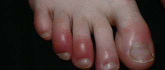
- The skin turned pale and acquired a bluish tint (face, lips, fingertips).
- You feel very tired and sleepy.
- My head hurts and feels dizzy, I have problems with memory and concentration.
- You are experiencing some symptoms of a respiratory system disease: shortness of breath, discomfort and unusual sensations in the chest, coughing.
What to do if saturation drops?
Don't panic about low saturation - normal vital signs can be quickly restored, and even a reading of 70% within a few days is compatible with life, and the chances may be even higher if the patient has, for example, chronic obstructive pulmonary disease, and to a low level oxygen, his body has already adapted. Saturation may drop for several days.
However, if during coronavirus the saturation drops to 95%, 93, 90...%, and all measurements are made correctly (it is important to check that the pulse oximeter has an adequate battery level, and that the device itself is registered as a medical device and not purchased from dubious manufacturer) - you need to call an ambulance.
What can affect readings when measured with a pulse oximeter
The accuracy of measurement readings is affected by:
- Choosing a location. Excessively bright sunlight or the rays of operating lamps may prevent reliable readings from being obtained. To perform the procedure, it is better to choose a room with moderate lighting.
- Human movement, shaking. Human movement, including trembling, will lead to incorrect interpretation of data. In these cases, the sensor's contact with the body shifts. It is optimal to measure indicators in a supine position. If for any reason the patient is trembling, the procedure should be performed after the trembling has stopped.
- Low battery. Errors cannot be avoided if the device’s battery is discharged; it is not advisable to measure with a discharged device. Before use, make sure there is enough charge. The charge level is shown on the screen by a special icon.
- Mounting the device. Errors in measurements can be caused by insufficient fixation on the body or excessive compression, for example, when the finger is pinched too tightly. The use of force to fix the device is unacceptable. It is also important that the distance between the LED and the photodetector is equal throughout the entire area.
- Reduced tissue perfusion. This situation entails a decrease or disappearance of the pulse wave.
- Carbon monoxide poisoning. In people who inhale smoke, the amount of hemoglobin bound to carbon monoxide increases significantly. Most instruments cannot detect the difference, so the saturation level will be exceeded.
- Availability of dyes. Gel, varnish, and shellac on nails provoke underestimated values, since the coloring substance absorbs light. Errors occur when nails are painted, as well as in cases of skin pigmentation with henna or other mixtures.
- Profound anemia. At values below 50 g/l, 100% saturation indicators are observed even with a lack of oxygen in the blood.
Using a pulse oximeter, we can promptly consult a doctor for help with coronavirus. Today, a medical device should be present in every family. Detailed information such as temperature, pulse and blood oxygen saturation will help the specialist prescribe appropriate treatment even during a remote consultation.
Why can pulse oximetry data be misleading?
It is important to understand that the result of pulse oximetry can be influenced by: the sensitivity of the device (including battery charge), lighting, and the color of the patient’s skin (the darker, the higher the indicator, which does not reflect the real state of affairs).
The concept of “saturation rate” is very arbitrary. It happens that patients with signs of coronavirus infection and a mobile non-invasive device for measuring saturation begin to panic and call an ambulance if the oxygen in the blood drops, for example, to 93%. To do this, let’s look at the data from the conditional norm table:
Details for instructions for use
Each pulse oximeter package contains the necessary documentation, including instructions for use. It is not difficult to understand, since the device is compact and simple. Most home pulse oximeters run on AA batteries or rechargeable batteries. To use the device, you must insert the batteries and wait a few minutes. Then you should pay attention to the device's charge level - if it is low, the readings may not be accurate enough. Next, you just need to follow a few simple steps yourself or with an assistant.
- The first step is to take a stationary position, sitting or lying down, with your arm supported. It is recommended to remain in this position for several minutes before measuring to obtain more accurate readings.
- The second step is to place your finger in the special hole of the pulse oximeter. It must be clean and free of nail polish and other coatings that could affect the measurement result. The sensor will operate for 20–30 seconds, depending on the model of the device, and during this time the finger must remain motionless.
- When the results are displayed on the monitor, the measurements are completed. It is recommended to record the data in order to keep a constant record of heart rate and saturation levels. After this, the pulse oximeter should be turned off and placed in a box to keep it away from sunlight and dust.
The device can be used independently or with an assistant. Even simple portable pulse oximeters can measure pulse and saturation in bedridden patients, as well as during sleep. For longer measurements, it is recommended to use instruments that continuously read data and display it on the screen. The monitor is attached to the wrist, and the data is accurate even if you do not hold your finger in a stationary position. These pulse oximeters are effective for diagnosing sleep apnea and for monitoring heart rate and saturation levels around the clock.
How to understand pulse oximeter readings
As a result of the measurement, all data is displayed on the monitor screen. The first indicator that a pulse oximeter reads is the pulse. It is displayed in the number of heartbeats per minute. In an adult, the normal pulse ranges from 60 to 90 beats per minute. It can increase after physical exertion, with chronic diseases of the heart and blood vessels, and also as a result of a sharp decrease in oxygen levels in the blood. The pulse is not a constant value and can change frequently, so to obtain more accurate data it is recommended to measure it several times at intervals of 3-4 minutes.
The second indicator for which portable pulse oximeters are used is saturation (SpO2). It is displayed as a percentage and indicates the level of oxygenated hemoglobin. In a healthy person at rest, it will correspond to 95% or more. However, if the monitor displays a result of 98–100%, it is worth repeating the procedure - the device may be faulty. The saturation level from 90 to 94% corresponds to 1 degree of hypoxia, from 75 to 89% - 2 degrees, from 60 to 75% - 3 degrees. When saturation decreases to 60%, hypoxic coma is observed, which poses a danger to the patient’s life. It is recommended to consult a doctor if the monitor displays a saturation level of 94% or lower - even minor oxygen deprivation can significantly affect the functioning of internal organs and tissues.
When is hospitalization and oxygen support needed?
- If the indicator has dropped to 93% and the patient feels unwell (hypoxemia, respiratory failure, respiratory symptoms are severe).
- With a significant percentage of lung damage on CT (> 50%, with CT-3, CT-4).
- Old age of the patient.
- Concomitant chronic diseases of the respiratory system.
- Concomitant chronic diseases of the cardiovascular system.
- Initially low saturation due to anemia.
- Pregnant women.
- Patients with immunodeficiency.
- Obese patients.
- For diabetes mellitus.
A decrease in saturation is dangerous primarily for these groups of patients.
If a hospitalized patient's saturation is low and does not rise even with oxygen, then, according to the current recommendations of resuscitators, the patient undergoes tracheal intubation.
Indications for use of the device
Pulse oximetry is a diagnostic area that is informative for various diseases of the respiratory system. To assess the patient's condition and the degree of hypoxia (oxygen starvation) of tissues, the saturation level is measured. Pulse oximeters are also necessary for diagnosis and during treatment of coronavirus, which occurs with pneumonia. In addition, these measurements can be informative in the following areas:
- during oxygen therapy, to assess its effectiveness;
- during anesthesia and operations, as well as during rehabilitation;
- in neonatology, especially for examining premature babies;
- for chronic heart failure;
- with proven apnea (stopping breathing) during sleep;
- with congenital and acquired heart defects.
Patients with chronic diseases that can cause hypoxia should purchase a pocket pulse oximeter and keep it in a portable first aid kit. The measurements will be informative and will allow you to quickly determine the deterioration of your health, since the device shows data at the time of use. If you carry out the procedure daily, you can monitor the dynamics of indicators and choose more effective treatment.
Application for Covid-19
Coronavirus
– a dangerous disease that occurs primarily in the lungs.
The inflammatory process leads to a deterioration in gas exchange, so the blood is not saturated with sufficient oxygen. In addition, the damaged areas undergo fibrosis over time and are gradually replaced by low-functional connective tissue. A decrease in saturation to 91% or lower is one of the characteristic signs of a coronavirus infection, which occurs in moderate or severe form. It is recommended to measure this indicator when the following symptoms appear:
- pallor of the skin and mucous membranes;
- weakness, dizziness, especially with sudden movements;
- wheezing in the lungs, cough, fever;
- deterioration of sense of smell;
- rapid heartbeat, uneven pulse, possible fainting.
A pulse oximeter must be in the home medicine cabinet for people at risk: the elderly, patients with chronic diseases of the cardiovascular and respiratory systems, as well as other internal organs. With its help, you can promptly identify dangerous oxygen metabolism disorders and seek medical help.
In addition, the device should definitely be used in the post-coronavirus period, during rehabilitation. Inflammatory processes, high temperature, general weakness and decreased immunity lead to the fact that tissues are restored slowly. Foci of fibrosis form in the lungs - islands of connective tissue that cannot participate in the breathing process. Despite the fact that the virus can be removed from the body, the patient continues to experience symptoms of oxygen starvation if the healthy surface of the lungs does not have time to saturate hemoglobin with oxygen to the required extent.
Is it possible to quickly increase the saturation on your own?
Before the ambulance and oxygen therapy arrive, the patient can take the following measures:
1. Do breathing exercises
Sit up straight, lower your shoulders, straighten up and try to relax.
Exercise 1
- Raise one hand up and grab the floor lamp/beam/door handle to secure the position of your hand.
- Place your other hand on the diaphragm. Breathe deeply, lifting your diaphragm as you inhale.
Exercise 2
- Continue to hold one arm up while keeping the other extended forward.
- As you inhale, turn your body towards your outstretched arm (if your left hand is raised, turn your body to the left and vice versa).
Exercise 3
- Breathe deeply with one arm raised up. The hand is fixed on any object (floor lamp) or wall.
Open the windows and ventilate the room.
Lie on your stomach for up to 30 minutes.
Important! Due to individual anatomical characteristics, elderly patients should not lie on their stomach - compression of the respiratory organ is possible.
There is no evidence that any medications can effectively increase saturation in a coronavirus patient. However, in the absence of individual intolerance and the presence of aspirin in the home medicine cabinet, the use of this drug is acceptable.
The anticoagulant aspirin is associated with a reduction in the need for mechanical ventilation and admission to the intensive care unit, as well as a reduction in mortality in patients hospitalized with COVID-19.
In general, doctors who worked with seriously ill patients have repeatedly noted that oxygen therapy, or rather the constant stay of the patient “on oxygen,” is less effective than breathing exercises. The patient must breathe independently, under the supervision of medical personnel, “kneading” and stimulating the lungs.
Important! Performing the popular exercise with “inflating” balloons is not permissible in cases of lung damage KT-2, KT-3, KT-4 and especially in elderly patients, since the damaged and overstretched pulmonary matrix may simply not withstand the load.
How to increase saturation after viral pneumonia?
If even after suffering from coronavirus the saturation is slightly reduced, then this is normal - the lung tissue needs time to restore the previous vital capacity of the respiratory organ. Breathing exercises (see Strelnikova’s set of breathing exercises) and walks in the fresh air with moderate physical activity are extremely useful.
To prevent aggressive adhesions in the lungs in patients with fibrous changes evident on CT; usually with CT-4, CT-3, less often with CT-2 and very rarely with CT-1, antioxidant therapy for pulmonary fibrosis is prescribed, which includes a diet enriched with antioxidants, acetylcysteine, and vitamins E (if there are no allergies).
To clarify the diagnosis and causes of reduced saturation after coronavirus, CT control is important.
What to do with low saturation
At critical levels, life-sustaining drugs and equipment should be used. This is done by the medical staff.
If it is low, it is recommended to do the following:
- breathe fresh and clean air, do breathing exercises;
- drink water in small sips and take blood thinning medications (as prescribed by your doctor);
- consume foods high in iron (buckwheat, meat, apples).
Regular breathing training, combined with physical exercise, will strengthen the body and make it easier to endure any disease due to the improved function of oxygen supply to all tissues in need.
When else does a person have low saturation?
- For heart defects;
- With hypoventilation of the lungs (with a slowdown in the frequency of exhalations and inhalations);
- For anemia;
- For chronic diseases of the lungs and bronchi (COPD, emphysema);
- If the oxygen concentration in the air is reduced;
- For diffuse disorders;
- Under excessive loads;
- When smoking;
- If you are overweight;
- When there are changes in atmospheric pressure;
- At night (from 3 to 7 o’clock) and in the deep sleep phase;
- When he is under general anesthesia.
How is saturation measured?
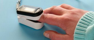
Saturation is measured with a pulse oximeter. Ambulance teams are equipped with mobile devices. You can also purchase it for home monitoring. The device resembles a clothespin that is attached to your finger.
Within a minute, a sensor with LEDs reads data, namely the color of the blood (hemoglobin), which changes depending on the saturation, as well as a specific pulsating light signal that changes depending on changes in blood pressure.
The pulse oximeter display shows two numbers - the top one shows the percentage of oxygen in the blood, the bottom one shows the pulse.
Saturation is measured in a sitting or lying position, the patient's hand should lie on the surface and not hang in the air.
Hospitals also use invasive devices to help laboratory technicians measure blood gases. To do this, it is taken from an artery or vein.
Description of pulse oximeters
Portable pulse oximeters resemble a small clothespin that fits onto your finger. They simultaneously measure two vital signs: pulse and saturation. The measurement methods are non-invasive, that is, they do not require skin puncture, blood sampling or other painful procedures. The device runs on batteries and can be stored in a home medicine cabinet. There are several types of similar devices, and it is worth familiarizing yourself with each of them before purchasing.
A clip-on pulse oximeter is the most common option and is suitable for use in hospitals and at home. You can purchase such a device in our store and use it according to the instructions. For professional use, you can also find pulse oximeters for round-the-clock monitoring of saturation levels. They are also attached to the finger, but a cord extends from them to a bracelet located on the wrist. The indicators are constantly displayed on a small monitor. There is also professional-sized equipment that is used during the treatment of patients with lung diseases in a hospital.