Home > Encyclopedia of Oncology > Alphafetoprotein: tumor marker
Alphafetoprotein tumor marker is one of those that is among the twenty most informative among the total number (and there are more than 200 of them).
Despite the fact that it is produced in some quantity by the body, its specificity for pathologies is quite high. Therefore, it is used to diagnose cancer of the liver, pancreas, testicles (ovaries in women). Also, its study is used during pregnancy for the purpose of early detection of abnormalities in the fetus. Consult an Israeli specialist
Reference values
For children, men and non-pregnant women, normal values are presented in the table.
| Normal blood values for alpha-fetoprotein, IU/ml | ||
| Age | Men | Women |
| up to 4 weeks | < 13600 | < 15740 |
| from 4 weeks to a year | < 23,5 | < 64,3 |
| from 1 year and older | < 7,29 | < 7,29 |
For pregnant women, depending on the stage of pregnancy, normal values are presented in the table.
| Normal Alpha Fetoprotein Values in Pregnant Women | |
| gestational age | alpha-fetoprotein, IU/ml |
| up to 12 weeks | less than 15 |
| 13 to 15 weeks | 15 – 60 |
| from 15 to 19 weeks | 15 – 95 |
| from 20 to 24 weeks | 27 – 125 |
| from 25 to 27 weeks | 52 – 140 |
| from 28 to 30 weeks | 67 – 150 |
| from 31 to 32 weeks | 100 – 250 |
Alpha-fetoprotein refers to the so-called embryonic proteins, which are synthesized by embryonic cells.
AFP performs many important functions, ensuring the full development of the fetus. Alpha-fetoprotein enters the blood of a pregnant woman through the placenta (the “baby place” that forms during pregnancy and connects the mother and fetus, providing the baby with essential nutrients) and from the amniotic fluid (amniotic fluid, which plays an important role in the metabolism of nutrients and fetal protection). Accordingly, with an increase in the concentration of fetal alpha-fetoprotein, its values in the mother’s blood increase, increasing from the 10th week of pregnancy and reaching maximum values by the 32-34th week. Alpha-fetoprotein testing in pregnant women is carried out as part of a “triple test” together with hCG (human chorionic gonadotropin) and E3 (free estriol) in the second trimester of pregnancy (between 15 and 20 weeks) to identify the risk of abnormal development of the fetus.
What does alphafetoprotein tumor marker show?
Checking the alpha-fetoprotein tumor marker is prescribed in the following cases:
- Comprehensive diagnostics of the pelvic and abdominal organs in case of frequent malaise due to the absence of adequate causes.
- Monitoring the course of pregnancy in case of suspected pathologies in the fetus based on the results of other tests.
- When there is a family history of cancer.
- Monitoring the effectiveness of treatment and recovery after a course of procedures.
- Monitoring the process of metastasis or confirming its absence.
- Visiting cancer clinics is a thing of the past.
- Chronic alcoholism.
- Chronic hepatitis.
Everyone does not need to be tested for this tumor marker. Only a doctor can determine who needs it.
To pass the test, you need to properly prepare for this process. There are no special requirements. The main condition is to donate blood on an empty stomach in the morning from 8 to 11-12 hours. It is advisable to follow a diet, and 12 hours before the test, exclude all food and drink, except clean water without gas. Water is also acceptable on the day of blood collection.
3-4 days before, you need to stop taking medications in consultation with your doctor, stop drinking alcohol and smoking. Significant physical activity and stress are undesirable.
Before going for analysis, you should not undergo massage or any medical examinations.
Compliance with the rules and requirements for preparing for analysis increases the accuracy of the research results.
Reasons for the decrease in alpha-fetoprotein in the blood of pregnant women
- Down syndrome in the fetus,
- delayed fetal development,
- fetal death
- false pregnancy.
AFP is used as a tumor marker in adult men and non-pregnant women.
As the tumor process develops, cancer cells begin to synthesize alpha-fetoprotein, so an increase in its concentration in the blood indicates the presence of a malignant neoplasm. Alpha-fetoprotein was first described as a human tumor-associated protein in 1964. It was then discovered that elevations of alpha-fetoprotein above the values typically found in healthy individuals occur in several malignant diseases such as testicular cancer, liver cancer (primary hepatocellular carcinoma), with metastases of other tumors to the liver.
Patients with liver cirrhosis (a disorder of the liver structure, manifested by functional failure), chronic hepatitis B and C are at risk of developing liver cancer. The determination of alpha-fetoprotein in them is of great practical importance.
The level of alpha-fetoprotein in liver cancer and testicular cancer well reflects the dynamics of therapy. A significant decrease in alpha-fetoprotein after chemotherapy indicates the effectiveness of treatment.
Sources:
- Rodriguez-Diaz JL, Rosas-Camargo V, Vega-Vega O, Morales-Espinosa D, Mendez-Reguera A, Martínez-Tlahuel JL, et al. Clinical and pathological factors associated with the development of hepatocellular carcinoma in patients with hepatitis virus-related cirrhosis: a long-term follow-up study. Clin Oncol (R Coll Radiol) 2007; 19:197-203.
- Chereshnev V.A., Rodionov S.Yu., Cherkasov V.A., Malyutina N.N., Orlov O.A. Alpha fetoprotein. Ekaterinburg: Ural Branch of the Russian Academy of Sciences, 2004. 376 p.
- N. Salesi, B. Di Cocco, F. Alghisi, F. Calabretta, G. Bossone. Testis cancer markers. Clinical use. Minerva Med. 2002 Oct; 93(5):365-9.
What is an AFP marker?
Alpha-fetoprotein tumor marker (Alpha-fetoprotein) is a specific protein compound produced by the cells of the yellow sac in the body of the embryo. Its maximum level is reached at 34 weeks of pregnancy, after which it gradually decreases. By the first year of a baby’s life, the amount of alpha protein stabilizes and is the same as that of a healthy adult.
In the body of adults, the tumor marker is produced mainly by liver cells, which is why it is called a liver marker and an analysis is prescribed to detect it if liver cancer, hepatitis and other complex diseases are suspected. Also, the tumor marker alphafetoprotein is also informative in diagnosing possible oncology of the testicles in men, ovaries in women, and pancreas. Accordingly, it refers to the markers of the listed organs.
Testing for only one tumor marker is not sufficient to make a diagnosis. Therefore, if the reaction is positive, do not panic. It could just be inflammation!
Please note that this protein is sensitive not only to neoplasms in adults and pathologies in the embryo, but also to inflammation and various dysfunctions of these organs.
Indications
The tumor marker AFP in the blood is studied for the following reasons:
- Diagnosis of primary hepatocellular carcinoma (liver cancer);
- high risk of developing liver tumors (with liver cirrhosis, chronic hepatitis);
- suspicion of cancer (confirmation of the diagnosis of testicular teratoblastoma, low-grade tumors);
- predicting the likelihood of cancer metastasis to the liver;
- definition of antitumor therapy;
- screening for the quality of removal of malignant tissues after radical surgery;
- predicting the clinical picture of the disease, its progression, the likelihood of relapse or remission;
- diagnosis of diseases of the lungs, liver, uterus, mammary glands, etc.;
- monitoring the condition and development of the fetus in the second trimester of pregnancy.
An obstetrician-gynecologist, oncologist and other specialized specialists, as necessary, refer you for the study and interpret the results.
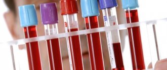
How to donate blood for tumor markers
The attending physician should give specific recommendations on restrictions before blood collection. They depend on which specific protein will be studied.
The general rules are as follows:
- donate blood at least 8 hours after your last meal;
- try not to be nervous during the procedure;
- interrupt serious training for a week;
- stop drinking alcohol three days before;
- It is advisable not to smoke immediately before collecting biomaterial;
- If in the next week the patient underwent x-rays or other examinations, the doctor must be informed about this.
Other requirements may need to be met. Thus, if you suspect a prostate tumor, it is advisable to abstain from sexual intercourse for 7 days; it is better not to test blood for tumor markers of the female reproductive system during menstruation.
High AFP in pregnant women
An increased level of AFP by more than 2-3 times is observed in the following diseases:
- anencephaly (severe pathology of the formation of cranial bones and cerebral hemispheres);
- hydrocephalus;
- spinal malformation (spina bifida);
- kidney and liver defects in the embryo;
- atresia of the esophagus or intestines;
- umbilical hernia, gastroschisis (defect of the anterior abdominal wall),
- teratocarcinoma (cancer) of the yolk sac;
- pathology of the placenta;
- encephalocele (cranial hernia);
- threat of miscarriage or premature delivery;
- large fruit;
- multiple pregnancy, etc.
Important: to diagnose pathology of fetal development, it is necessary to know the exact duration of pregnancy. The AFP level alone cannot serve as a diagnostic criterion.
Why do blood tests for tumor markers?
Analysis for tumor markers is often used as an evaluative test to determine positive (or negative) dynamics in cancer therapy. Based on its results, relapse can be prevented.
Markers allow you to:
- confirm or refute the assumptions of preliminary diagnostics;
- notice the formation of metastases in the absence of other symptoms;
- evaluate the effectiveness of the prescribed therapeutic course.
The study is not recommended as an independent diagnosis due to the large number of false positive (false negative) results. But, in combination with other methods, it helps to identify cancer at an early stage.
Degree of reliability of markers in oncology
Analysis of the content of these substances is very informative for assessing the prescribed therapeutic course for cancer. In primary diagnosis, none of them is used independently. This is due to the lack of specificity of the markers.
The reliability of the readings of specific proteins can be considered using the example of the most common of them. These include the PSA test. This tumor marker should be tested in men for timely detection of prostate cancer. In case of malignant tumors of the gland, this indicator actually exceeds the norm. But it also increases if a benign neoplasm forms in the prostate. As medical practice shows, prostate cancer was not confirmed in most men with elevated PSA levels.
Attention: there are cases when unscrupulous doctors in dubious clinics prescribe unnecessary therapeutic procedures. They not only require serious financial investments, but can also provoke serious complications!
Another indicative marker is CA-125. It grows in the blood of women who have ovarian cancer, but can increase in a large number of other pathologies not related to oncology.
Medicine knows of cases when, in the late stages of the formation of malignant neoplasms in the ovaries, a blood test did not show deviations in the results of any of the 28 protein compounds specific to this disease.
CA-125 is not recommended for testing in the initial stages of the disease in the absence of characteristic symptoms. To analyze the course of the diagnosed pathology, it is considered together with other proteins, as well as ultrasound results.
As part of a comprehensive examination, women with poor heredity or gene mutations need to take tumor markers every six months.
In general, the question of how much one can trust tumor markers remains open today. When interpreting research results, you should pay attention to a number of factors that can distort the data:
- the level of protein content is affected by inflammatory diseases in acute or chronic form;
- CA-125 increases during menstruation;
- smokers are characterized by a twofold increase in the CEA index;
- A normal test result does not guarantee the absence of a tumor.
These are the most common nuances, but there may be other “subtleties”. Only a qualified doctor who can correctly decipher the results can understand them.
General information
Tumor markers are specific elements that appear in human biological fluids against the background of the development of benign and malignant processes. These may include hormones and enzymes, but the most common are proteins, such as alpha-fetoprotein (AFP).
AFP is produced by the cells of the fertilized egg in the body of a pregnant woman, but can also be found in a child or man. It indicates the likelihood of developing a malignant process and allows you to diagnose cancer at an early stage. Also, a blood test for AFP helps assess the effectiveness of antitumor treatment, identifies early metastases and indicates the condition of the fetus during pregnancy. etc.
At the moment, medicine knows two hundred tumor markers. One of them, AFP, is a protein macromolecule to which a carbohydrate or fat component is attached. AFP is produced by malignant cells and then enters the blood, where its level can be determined using an enzyme-linked immunosorbent assay (ELISA).
Regular testing of a pregnant woman's blood for AFP allows us to monitor some of the immune reactions of the mother's body. Since alpha-fetoprotein is produced by the embryo during pregnancy, the expectant mother's immune system often identifies the fetus with a foreign agent and tries to attack it. That is why increased AFP in pregnant women should be considered normal, and its low values, on the contrary, may indicate fetal malformations.
The tumor marker AFP is also detected in the body of adults and children, since it begins to be produced in the liver before birth (during embryonic development) and throughout life. Therefore, this indicator is one of the main criteria in the diagnosis of oncological pathologies of the liver and gastrointestinal tract. The significance of AFP also lies in the fact that it has independent antitumor activity - it can bind and remove malignant cells of the liver, uterus, respiratory system, mammary glands, etc.
The half-life of AFP is about 5 days. Therefore, the study of tumor markers for several weeks after chemotherapy, radiation therapy or surgical procedures allows us to monitor the effectiveness of treatment. If alpha-fetoprotein levels continue to rise, the prognosis for the patient is poor. If the intensity of the decrease in AFP is low, then tumor particles may remain in the patient’s body or the process of metastasis has begun.
The biomaterial for AFP is blood serum. But other biological media can be used periodically: the secretion of the pleural cavity of the lungs, bile, urine, ascitic or amniotic fluid.
Low AFP during pregnancy
An indicator that is too low indicates the following deviations:
- malnutrition (malnutrition);
- hypoxia (oxygen starvation);
- Down syndrome;
- Edwards syndrome (trisomy 18 chromosome),
- Patau syndrome (trisomy 13 chromosome),
- frozen pregnancy;
- intrauterine death;
- diabetes mellitus or gestational diabetes in a pregnant woman;
- obesity of various types;
- low placenta previa;
- endocrine disorders.