If coronary artery bypass surgery is recommended for a person with coronary artery disease, reviews of patients who have undergone therapy in Israel will provide an invaluable service. People's stories clearly demonstrate the state of medicine in Russia and abroad, describe the intricacies of treatment, the infrastructure of clinics, the professionalism of doctors, and the cost of procedures. Online reviews about coronary artery bypass surgery can be found on specialized forums where people with similar symptoms communicate, share stories of the development of the disease and facts of successful treatment, as well as their own fears and concerns. Among like-minded people it is easy to get support from understanding people. In this article about coronary artery bypass surgery, patient reviews are collected from various sources. We tried not to filter reviews; we took as a basis not advertising materials, but real stories from yesterday's patients.
Myth 2. After the operation you will have to “carry” yourself like a crystal vase
In fact . This is wrong. Usually, the very next day after surgery, the doctor warns: if you move little, complications are possible, such as pneumonia. The operated person immediately begins to learn to turn over in bed, sit up...
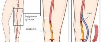
This is why shunts are placed so that the patient can walk without feeling pain. At first, of course, weakness and pain from the stitch interfere, but it is necessary to gradually increase physical activity. And then those movements that caused pain before the operation will be easy.
Myth 3. The pain may return
In fact . There is no need to wait for the pain to return, but it is better to imagine that it never happened at all. However, there is no need to perform “feats”. Everything should be in moderation. The patient must set realistic goals for himself: for example, today and tomorrow I will walk 50 meters, the next days - 75, then - 100...
Statistics show that not all patients, even after CABG, manage to get rid of angina. And this is not surprising: no matter how well the operation is performed, it is only one of the stages in the treatment of coronary heart disease. Doctors have not yet learned how to cleanse the heart vessels of atherosclerotic plaques, the main cause of the disease. Therefore, even after successful surgery, approximately half of patients may still have angina, manifested by chest pain during exercise. But the number of attacks and pills taken after CABG will still be less. So the patient’s quality of life will still improve. And most importantly, it will be possible to delay the onset of myocardial infarction, and therefore prolong life.
Consequences and complications
Complications can develop if the patient has concomitant pathology:
- diabetes;
- pathology of the renal system;
- diseases of the pulmonary system.
Most often, after surgery, bleeding occurs in the anastomotic area and rhythm disturbances are recorded. Possible complications:
- acute circulatory disorder in the brain and myocardium;
- thrombosis of the venous bed;
- renal failure;
- local complications in the form of wound infection and the formation of postoperative keloid scars;
- closure or narrowing of the shunt.
Myth 5. Smoking does not have much effect on the heart after surgery.
In fact . Quitting smoking prolongs the life of the shunts by several years. After all, the duration of operation of shunts is different for each patient. On average it is 5–7 years. This period largely depends on how much the person was able to change his life after the operation, and whether he follows the doctors’ recommendations.
Article on the topic
Prevent stroke: how to treat patients with atrial fibrillation
Proper nutrition (diet with limited animal fats), normalization of body weight, adequate physical activity, taking all necessary medications in total add several more years of active and fulfilling life.
There is no doubt that the effectiveness of coronary artery bypass grafting (CABG) depends on the presence of effectively functioning grafts, and the return of myocardial ischemia and mortality after surgery are associated with the timing of their patency [7]. If, after “open” myocardial revascularization, graft failure or obliteration of native coronary arteries (CA) occurs, and endovascular correction of these lesions is impossible, then the method of choice is repeat coronary artery bypass grafting (RCBG). However, repeated surgery, even if performed as planned, is accompanied by a higher incidence of complications compared to primary CABG and mortality from 1.5 to 5.2% [1, 4, 6, 17, 20]. This largely explains the small number of such interventions in Russia: according to L.A. Boqueria, they are still performed in isolated cases, although with some tendency to increase their quantity and quality. Thus, if in 2008, 96 re-CABG operations were performed in the country in 21 clinics (with an average mortality rate of 12.5%), then in 2010 - already 169 (with a mortality rate of 2.4%) and in 35 specialized institutions out of 92 [ 2, 3].
The purpose of the study was to study the results of re-CABG operations and analyze the structure of the resulting hospital complications.
Material and methods
In total, during the period from September 2002 (when the first re-CABG was performed at the research institute) to December 2011, 4451 CABG operations were performed, of which 48 (1.1%) were repeated (main study group). In table 1
indicators are presented that illustrate a significant deterioration in the clinical and functional state of patients before repeated myocardial revascularization in comparison with their initial status.
In 42 (87.5%) cases, reCABG was performed under conditions of artificial circulation, in 6 (12.5%) - on a beating heart.
The main indication for intervention was the return of angina pectoris during antianginal therapy due to the appearance of hemodynamically significant occlusive-stenotic lesions of coronary bypass grafts and coronary arteries when it was impossible to perform percutaneous coronary intervention (PCI). During the primary operation, in 31 cases, patients used mammarocoronary shunts (MCS), in 74 - venous shunts (VS), in 7 - grafts from the left radial artery (LA), in 1 - the right gastroepiploic artery. In 3 cases, a sequential shunt was formed (1 VS; 2 from LA), in 1 case a “Y”-shaped VS was formed. For bypass grafting, the internal mammary artery (IMA) was used in 21 cases, the autovenous vein in 69 cases, and the PA in 6 cases. During re-intervention, sequential (1 VS) and “Y”-shaped shunts (1 LA, 5 VS) were also used; in 5 cases, segmental reconstruction of a previously implanted conduit was performed. The myocardial revascularization index for primary and repeat CABG was the same - 2.4. The time between operations varied from 4 months to 18 years (average 94.3±62.6 months). In 2 (4.2%) patients, re-intervention was combined with resection of the left ventricular aneurysm and linear ventriculoplasty, in 1 (2.1%) with mitral valve replacement, in 1 (2.1%) with radiofrequency ablation, in 1 (2.1%) - with excision of the encysted hydropericardium, in another 1 (2.1%) - with sternum plastic surgery.
Considering that one of the objectives was to study the structure of hospital complications, and complete data on primary CABG in 18 (37.5%) patients were not available (in 6 the operations were performed in other clinics in Russia, in 12 archival information was not found due to the long-standing period), there was a need to form a control group. For this purpose, a control group of 107 patients operated on in the clinic of the Research Institute in 2010 (during the absence of deaths), who underwent only primary CABG, was formed from a continuous sample using the random number method. It should be noted that patients in the control group had no statistically significant differences in the main initial parameters (gender, average age, nature of interventions, etc.) with the group of patients who underwent re-CABG (Table 2)
.
Technical features of repeated operations on the coronary arteries.
There is no need to dwell in detail on the main technical aspects of reCS; they are described in sufficient detail in the manuals [1, 4]. However, summing up the little experience that our clinic has, we consider it necessary to highlight a number of points. In our opinion, the most difficult stage is resternotomy, especially if the pericardial cavity was not sutured during the initial operation (43.8% of such patients), or the postoperative period in patients was complicated by mediastinitis (4.2%). However, in all 48 cases we used a median resternotomy, although the surgical field was ready for retrograde cannulation of the femoral vessels. We believe that a thorough analysis of X-rays, tomographic images and angiograms, study of primary documentation (operation protocol, discharge), active “exhalation” and relative hypovolemia of the right heart at the stage of using a sternotomy saw can minimize this problem.
Recently, we have limited the cases of performing reCABG on a beating heart. We believe that under conditions of artificial circulation this operation is more convenient and simpler to perform. After sternotomy, mandatory partial exposure of the anterior surface of the heart and aorta, the edges of the wound (to reduce traction and tissue trauma when opening the wound), preparation of the IAV, we carry out local cardiolysis of the heart, necessary for cannulation with a double-lumen venous cannula, release the ascending aorta, coronary bypasses. Total cardiolysis is performed under conditions of parallel artificial circulation or after cardiac arrest. Inconvenience is caused by the release of the apex of the heart, so this stage is safer to carry out with a stopped heart. Preference is given to antegrade blood cardioplegia after the formation of each distal anastomosis. If difficulties arise in the formation of central anastomoses, we form them with a completely clamped aorta or use the unchanged “hoods” of the old VS anastomoses with the aorta. Currently, both during primary and repeat CABG, we strive to necessarily close the pericardial cavity. If necessary, we use the epoxy-treated KemPeriplas xenopericardial flap for this purpose.
results
Overall, the rate of repeat CABG was 1.1%. Patients who underwent this intervention had more risk factors, as evidenced by EuroSCORE scores, which were several times lower in primary CABG ( p
=0.001).
In addition, patients in the reCABG group showed progression of atherosclerosis in the coronary system (as evidenced by an increase in the number of significant lesions of the three main coronary arteries and stenosis of the trunk of the left coronary artery) and in non-cardiac areas (increase in the number of patients with lesions of the carotid arteries and arteries of the lower extremities) (see Table 1
)
There were no deaths or type I neurological disorders during the hospital period. Of the complications specific to this type of intervention, in 2 (4.2%) cases, during the development of the technique during resternotomy with a Gigli saw, the edge of the lung was damaged, in 2 (4.2%) functioning shunts were injured (1 MCS and 1 VS), in 2 (4.2%) patients, despite bypass surgery of the posterior wall of the left ventricle, showed electrocardiographic signs of myocardial infarction (MI) of the posterior wall of the left ventricle caused by ligation of stenotic but patent “old” VS; in another 2 (4.2%) cases, resternotomy was required due to postoperative bleeding. The remaining complications are rather typical for any CABG operations, as evidenced by a comparison of the structure of complications in the main and control groups (Table 3)
.
These include heart failure, paroxysms of atrial fibrillation, hydrothorax, hydropericardium, and local wound complications.
It should also be noted that 23 (47.9%) patients had no complications after reCABG. When comparing the results of operations in the groups as a whole, there were no differences in the incidence of perioperative complications, but their structure has qualitative differences. Thus, in the main group, a number of complications were caused precisely by repeated surgical trauma, while in the control group complications typical of open-heart surgery prevailed (Fig. 1)
.
Figure 1. Most likely causes of coronary artery bypass graft failure.
At the time of repeat revascularization, 32 (27.4%) coronary grafts remained patent, although some of them had degenerative changes. MCS most often remained viable - in 13 (41.9%) cases of their use, least often VS - in 17 (23%) cases. The cumulative patency of MCS 63 months after primary CABG was 72.2%, VS - 56.7%, LA - 53.6%. At 126 months after surgery, the actuarial patency curves of MCB and VS differed significantly, but statistically insignificantly - 23.2 and 27.9%, respectively; p
=0,548
(Fig. 2)
.
Figure 2. Actuarial curves of coronary artery bypass graft patency after primary coronary artery bypass graft surgery during follow-up up to 18 years (p=0.548).
When analyzing the causes of damage to coronary bypass grafts, we identified the following: progression of atherosclerosis; technical defects, primarily associated with rough “skeletonization” of grafts and their hydraulic “inflation”; atherosclerotic degeneration of shunts. In some cases, the reasons for shunt obstruction remained unknown.
Preoperative shuntography made it possible to visualize gross degenerative atherosclerotic changes (on average 147.3±61 months after primary implantation) in 9 VS and 1 LA graft (in 12.2 and 14.3% of cases of their use, respectively). In another 4 (8.3%) patients, 3 MCS and 1 VS had stenoses (≥50%) of anastomoses caused by intimal hyperplasia.
After repeated coronary artery surgeries, results were monitored in 44 (91.7%) patients over a period of 5 to 94 months (on average 33.1±22.7 months). During this period, 3 deaths were observed after 7, 21 and 88 months, respectively, from progressive chronic heart failure against the background of severe ischemic myocardial dysfunction, from ischemic stroke and from recurrent MI. The survival rate of patients up to 5 years after re-CABG was 93.3%, and up to 8 years - 46.7% (Fig. 3)
.
Figure 3. Cumulative survival of patients after repeat coronary artery bypass grafting (RCBG) up to 8 years.
Discussion
According to the Society of Thoracic Surgeons (STS) registry, the maximum number of reCABG operations was recorded in 1992 and 1993. - 10%, but already in 1997 and 2000 their number decreased to 8 and 7%, respectively [13]. The low rate of repeat CABG operations (1.1%) shown in this study reflects the general state of this problem in Russia. Thus, in 2010, the frequency of re-operations on coronary arteries in the structure of all CABGs performed in the country did not exceed 0.61% [2]. This is partly due to the aggressive use of percutaneous invasive interventions after previous CABG and better control of risk factors. However, PCI performed after CABG is characterized by less favorable immediate and long-term results compared to those without previous bypass surgery [6].
In the early years of coronary artery surgery (1967–1978), data from the Cleveland Clinic demonstrated graft failure at an average of 28 months after initial surgery. Subsequently, this period increased to 116 months [14]. Currently, the interval between primary and repeat CABG surgery is about 10 years [8]. In the presented study, the average time between primary CABG and reCABG was slightly shorter - 94.3 months, but apparently, in many ways we are only repeating the experience of our foreign colleagues.
After the primary operation, the likelihood of re-intervention on coronary artery disease depends on many reasons that are related to the patient, the technical features of the primary operation, adherence to medical control and correction of factors for the progression of atherosclerosis. Unfavorable factors associated with the risk of reCABG include indicators reflecting the high life expectancy of patients: young age, normal left ventricular ejection fraction, damage to one or two coronary arteries [10]. Similar factors are widely represented in the present study. It should be recognized that there is a high (29.2%) frequency of incorrect collection, evaluation and preparation of grafts used for primary CABG, especially LA conduits, which largely determined the low level of their primary patency. This was due to the lack of proper surgical experience at the stage of mastering the technique and insufficient technical support.
Atherosclerotic degeneration of the VS is a typical and common lesion in candidates for reCABG. Moreover, the risk of atherosclerotic dysfunction of the VS increases as the time from the primary operation increases [4]. Our data on the frequency and timing of manifestation of these complications do not differ from the literature data: after 10 years of observation, almost 50% of VS are exposed to occlusion or severe atherosclerotic degeneration [1].
There is a widespread opinion in the literature that re-CABG is accompanied by a greater number of complications compared to primary CABG, which is quite logical - by the time of repeated interventions, patients become older, their atherosclerosis progresses and concomitant pathologies are more often diagnosed; repeated surgery is technically more difficult to perform. It is not for nothing that in a multivariate analysis, “a history of CABG” is considered as a factor that increases mortality after CABG operations [9]. In our work, the analysis of complications after primary CABG and re-CABG did not reveal significant differences in their frequency. At the same time, a number of qualitative differences associated with re-intervention have been identified. If they are excluded, then the structure of hospital complications during primary and repeated interventions becomes homogeneous, reflecting complications typical of most heart surgeries [5].
In the last decade, relatively low mortality rates have been achieved during reCABG (2.5-3.9%), but they are noticeably higher than in patients of comparable severity during primary operations (1-1.6%) [6, 12, 16 , 17]. In the Australian Society of Cardiothoracic and Thoracic Surgeons registry, the rate of repeat CABG was 3.4%, and the perioperative mortality rate for reCABG was 4.8% compared with 1.8% for primary CABG ( p
<0,001) [20].
The high mortality rate in reCABG is primarily due to the high risk of perioperative myocardial infarction, in contrast to primary operations, in which the leading place is occupied by non-cardiac causes. Many causes of MI have been identified: incomplete revascularization of the myocardium due to distal lesions of the coronary arteries, thrombosis of the VS, hemodynamic insufficiency of arterial grafts, atheroembolism from the VS, damage to shunts, etc. [10]. We noted 2 (4.2%) cases of MI due to ligation of degeneratively changed but patent VS to the right coronary artery, despite bypassing its nearby branch. Now, in such a situation, we necessarily re-bypass the artery with a modified but passable primary bypass, forming an anastomosis slightly distal to the previous one.
Studies on reCABG have noted that female gender and urgency of surgery are associated with increased mortality [11, 19].
Mandatory use of MCB to the anterior descending artery reduces the risk of reoperation compared with the strategy of using only VBG.
Currently, it is believed that bimammary CABG reduces the likelihood of death and reoperation compared with the use of the left ITA alone, but long-term follow-up is still needed to answer this question [10]. In addition, there is no convincing evidence to support the comparative effectiveness of PCI or repeat bypass in patients who have previously undergone CABG [6].
Patients undergo reoperation at a later stage of progression of coronary atherosclerosis compared to the point at which primary CABG is performed. It is therefore not surprising that their long-term results are not as favorable as those of primary interventions. Survival after re-CABG is also worse than after primary CABG. W. Weintraub et al. [18] demonstrated the survival of patients after reCABG in 76% of observations after 5 years and in 55% after 10 years. The results of our work are close to these data. An Australian registry showed lower 6-year survival rates after re-CABG than after primary CABG ( p
=0.01), but after leveling for initial severity, the fact of reoperation did not affect the survival of patients during these periods [20].
It can also be noted that recently more and more studies have appeared (albeit on relatively small clinical material) in which there are no deaths during repeated CABG operations [15], which is quite consistent with the results of our study.
conclusions
The rate of repeat CABG operations was 1.1% of all CABG operations. With repeat CABG compared with primary CABG, there were no deaths and no statistically significant differences in the incidence of in-hospital complications. Qualitative differences in the structure of complications are primarily due to the fact of repeated surgical intervention. Despite the more complex category of patients, the results of repeat CABG indicate the relative safety of this operation.
Myth 6. After surgery you will be able to live without medication
In fact . People who have had CABG surgery should never stop taking their medications. Most of the drugs that are prescribed today after surgery are vital. To reduce the risk of shunts being blocked by blood clots, it is often necessary to take medications that reduce blood clotting.
Beta blockers are needed to reduce the heart's overworking. They lower blood pressure and slow the heart rate. However, any changes in treatment must be agreed with your doctor. It is too risky to solve such issues yourself.


