An objective assessment of the patient's condition, based on laboratory data, provides more accurate results than subjective data obtained from the patient. The results of laboratory tests allow not only to make a timely accurate diagnosis, but also to evaluate the quality of the therapy. That is why medical personnel need to ensure a high degree of reliability of the results.
Several factors can influence the degree of reliability:
- preliminary preparation of a person for blood collection;
- time of day at which the material was taken for laboratory research;
- the instruments used for sampling and the technique used to obtain the material;
- compliance with the sampling algorithm.
The main reason for the appearance of errors in the results of laboratory tests is non-compliance with the standards of the preanalytical stage of working with venous blood due to poor mastery of the technique for collecting biomaterial using vacuum systems.
Why is it important to use vacuum systems?
Laboratory diagnostics is carried out in three stages:
- Preanalytical.
- Analytical.
- Post-analytical.
The duration of the stages and the degree of their influence on the reliability of the data are different.
The longest is the first stage, occupying two-thirds of the duration of any study. Errors made at the preanalytical stage lead not only to an increase in time spent on making a diagnosis, but also to unnecessary waste of budget funds due to the appointment of a repeat procedure. They affect the entire process of correct diagnosis and evaluation of therapy.
The degree of reliability of the data obtained depends on a huge number of variables:
- personal characteristics of a person (gender, age, race, etc.);
- features of eating behavior before submitting laboratory material (fasting, abuse of a certain type of food, etc.);
- intensity of physical and emotional stress;
- natural changes in hormonal levels (phases of the menstrual cycle, pregnancy, menopause, etc.);
- weather and climatic conditions;
- medicines taken by humans;
- position of the patient at the time of material collection.
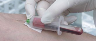
In addition to the above, the accuracy and correctness of the results depends on the technique of taking blood from a vein, the instruments used for this, the conditions of transportation and storage of the collected material.
When collecting blood from a vein using needles or syringes, it is impossible to standardize the technology for collecting the material. Using needles to collect venous blood can result in the collected material and pathogens of bloodborne infections getting into the hands of medical staff. This creates the danger of further transfer of pathogens to other patients. Taking biomaterial with a syringe practically eliminates this possibility, but when transferring it from a syringe to a test tube, hemolysis of red blood cells caused by mechanical action is possible.
Taking venous blood with a syringe does not exclude contact of medical staff with the patient’s blood and is therefore unsafe
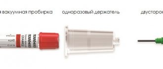
Thus, vacuum systems have become the optimal tool for collecting venous blood.
Price
The price of vacuum tubes depends on the material of manufacture, volume and filling. Their approximate cost is:
- with sodium citrate - from 7.5 to 11 rubles per piece, depending on the volume;
- with EDTA K-2 and EDTA K-3 – from 7.5 to 10.5;
- with a coagulation activator – from 7.5 to 8.5;
- with coagulation activator and gel – from 10.5 to 13.5.
A standard double-sided vacuum needle costs about 6 rubles, the same price for a needle with a transparent cannula and a butterfly needle with a luer adapter. The cost of a needle holder is about 4 rubles per piece.
Benefits of using negative pressure systems
All the benefits of negative pressure systems come from their design. Their use allows:
- completely eliminate contact of medical personnel with the patient’s blood during sampling;
- standardize the process of blood collection and sample preparation, create a simple algorithm of actions;
- reduce the number of operations spent on preparing a sample for testing in the laboratory;
- Primary tubes included in negative pressure systems can be used directly in many automated analyzers. This saves money on purchasing secondary plastic tubes and time on transferring samples into them;
- make the transportation and centrifugation of biomaterials safer, since the tubes are sealed and made of unbreakable materials;
- facilitate the identification and labeling of samples by type of examination, thanks to color coding of the caps of negative pressure systems;
- reduce laboratory material costs for the purchase and processing of additional secondary tubes;
- simplify staff training methods;
- reduce the occupational risk of infection;
- reduce the time spent collecting venous blood using the method discussed in the article.
Multi-colored test tubes made from high-strength modern materials ensure the safety of medical staff working with blood
Classification
Vacuum test tubes differ in the following characteristics:
- size (length and diameter);
- volume;
- purpose (hematological, biochemical analysis, blood clotting test, etc.);
- reagent filling the test tube;
- sample volume;
- color;
- cover type.
By filling, the test tubes are distinguished as follows:
- containing sodium citrate – intended for coagulological reactions;
- containing sodium citrate - to determine ESR;
- with EDTA K-2;
- with EDTA K-3;
- containing a blood clotting activator and gel;
- no filler.
Sequence of obtaining venous blood using vacuum systems
The process of collecting venous blood consists of three stages:
- preparation for the procedure;
- performing a fence;
- end of material collection.
At the preparation stage in the procedure for taking biomaterial from a vein, medical personnel need to:
- Treat your hands using the scheme provided by WHO.
- When working with blood, each person is considered a potential carrier of a blood-contact infection. Therefore, before starting the blood collection procedure, it is necessary to change into protective clothing.
- Fill out a referral for a blood test in the registration journal. This is necessary for labeling instruments and filling out documents related to one person. The referral contains the patient’s passport details, the date and time of the blood draw, the registration data of the analysis in the laboratory, and the details of the doctor who ordered the analysis.
- Compare the information in the referral with the data of a specific patient.
- Check whether the patient has given informed consent to the procedure, explain in detail the purpose and sequence of its implementation.
- Clarify the patient’s compliance with the rules of food restrictions adopted before taking the tests.
- Conveniently accommodate the patient.
- Prepare the workplace: arrange all the devices necessary for drawing blood, having first ensured their integrity and suitability for use (safety of sterility seals, expiration date, etc.). Select test tubes with the desired color coding and the required volume. Take a needle of the appropriate size.
- Wear a mask, safety glasses, and rubber gloves.
Correct positioning of the patient is one of the important principles of the correct blood sampling procedure.
After completing all the steps in the first stage, you can proceed to blood sampling.
POSSIBILITY OF INFECTION WHEN TAKING BLOOD
| Attention, I do not answer questions How to treat (write a prescription, prescribe treatment, etc.) Prescribing treatment in absentia can harm the patient and is legally illegal. What is this? Make a diagnosis from a photograph Only psychics, of which I am not one, can make a diagnosis from photographs. In some cases, a high-quality photo can only suggest a diagnosis, which must be confirmed (clarified) at an in-person appointment with a doctor. I do not answer questions asked in personal messages. In exceptional cases, correspondence on a paid basis is possible. Reception in Rostov-on-Don 344023, Rostov-on-Don, Lenin Ave., 251 Directions tel. +7, My sites Dermatologist-venereologist in Rostov-on-Don www.agapovmd.ru Information about sexually transmitted infections www.venuro.ru Forum sexually transmitted and skin diseases www.venderm.ru Information about ureaplasma and mycoplasma www.ureaplasma.info | There is a risk; the effectiveness of a condom in preventing the transmission of HIV infection is estimated at approximately 87% (according to various sources: from 60% to 90%.). The figures for the ineffectiveness of condoms: - for HIV infection up to 10% - 40%, - gonococcal, chlamydial infections and trichomoniasis up to 30%, - for genital herpes up to 70%, - for genital mycoplasmas up to 20%, were revealed in statistical studies. https://venuro.ru/preserv/preservativ.php The effectiveness of condoms against gonococcal and chlamydial infections with a known source of infection is provided by data that with constant use of a condom, infection with gonorrhea and chlamydia occurs in 30.3%, respectively, infection of women with human papillomavirus infection with constant use of condoms by their partners occurs in 37.8%, in 25% of cases use Male condoms prevent genital herpes in women, but not men. Pingmin W, Yuepu P, Jiwen Z. (2005) report the detection of genital mycoplasmas (Ureaplasma urealyticum, Mycoplasma hominis, Mycoplasma genitalium) in 16.67% of prostitutes who constantly use the male condom; 34.72% who use it regularly. A blood test should be performed using the ELISA method for HIV after 1, 3 and 6 months or using the PCR method. General smear - after 5 days, PCR scraping - after two weeks, blood for syphilis, hepatitis - after a month. The statistical risks of sexually transmitted HIV infection are as follows: With a single vaginal contact, the statistical risk for a woman is from 0.05% to 0.15% (from 5 to 15 cases per 10,000). The likelihood of a man getting infected from a woman is about three times lower. The average statistical risk of HIV transmission as a result of a single unprotected anal intercourse for the “receiving” partner ranges from 0.8% to 3.2% (from 8 to 32 cases per 1,000). For the “introducing” partner - 0.06%. When having unprotected oral sex with a man, the risk for the “receiving” partner is 0.04% (4 cases per 10,000). This risk is associated primarily with contact with the oral mucosa of sperm or pre-ejaculate. For the “introducing” partner, there is virtually no risk during oral sex, since he is only in contact with saliva (unless, of course, there is bleeding or open wounds in the “receiving” partner’s mouth). In the presence of sexually transmitted infections, the risk of HIV transmission through sexual contact increases by 2–5 times. Risks of HIV infection per 10,000 sexual acts without condoms. Oral sex - giving side -0.5 Oral sex - receiving side -1.0 Vaginal sex - giving side - 5.0 Vaginal sex - receiving side - 10.0 Anal sex - giving side - 6.5 Anal sex - receiving side - 50 Using condoms reduces infection by 20 times A log 10 decrease in viral load reduces the likelihood of infection by 2.5 times. Source: Pocket Guide Adult HIV/AIDS treatment 2008-2009. John G Barrett, MD editor, page 16/Videx K. CREATE NEW MESSAGE. But you are an unauthorized user. If you have registered previously, then “log in” (login form in the upper right part of the site). If this is your first time here, please register. If you register, you will be able to further track responses to your messages and continue the dialogue on interesting topics with other users and consultants. In addition, registration will allow you to conduct private correspondence with consultants and other users of the site. |
Algorithm for collecting biomaterial using a vacuum system
The second stage of the procedure is performed step by step:
Correct placement of the needle when collecting venous blood
- Examine the proposed venipuncture sites, select a point for the procedure, and palpate the vein. The ulnar veins are most often used, but if necessary, blood can be taken from the veins of the wrist, the back of the hand, above the thumb, etc.
- Secure the tourniquet 10 centimeters above the venipuncture site. When applying a tourniquet, women should not use the hand on the side of the mastectomy. Prolonged compression of tissues and blood vessels (more than two minutes) can lead to shifts in coagulogram parameters and the concentration of certain substances.
- Take the needle and remove the protective cap from it.
- Connect the needle to the holder.
- Ask the patient to clench his hand into a fist. You should not make sudden movements, as this can lead to changes in blood counts. If the vein is poorly visible, you can apply a warm napkin to your arm, or massage your arm from the hand to the elbow. If there are no vessels suitable for venipuncture on one arm, the other should be checked.
- Treat the puncture site with a disinfectant using circular movements from the center to the edge.
- Wait for the antiseptic to evaporate, or remove excess with a sterile dry cloth.
- Remove the protective colored cap from the vacuum system.
- Secure the vein by clasping your forearm. Place your thumb 3˗5 centimeters below the injection site. Stretch the skin.
- Insert the needle with the holder into the vein at an angle of 15°. When inserted correctly, blood will appear in the indicator chamber of the holder.
- Secure the test tube in the holder with the cap facing up. Under the influence of negative pressure, blood will begin to flow into the test tube.
- As soon as blood begins to accumulate in the test tube, loosen the tourniquet or remove it.
- Tell the patient to relax his arm and unclench his fist.
- When the flow of blood into the test tube stops, remove it from the holder.
- Mix the biomaterial with the preservative. Do not shake! The test tube can only be gently inverted.
- If several samples are taken from the patient, the holder with the needle is left in the vein and steps 11-15 are repeated sequentially.
The vacuum collection system allows you to collect several tubes of material without removing the needle
After completing all of the above steps, you can proceed to the final stage of blood sampling.
Stage of completion of the procedure At the final stage of taking biomaterial from a vein, medical personnel need to:
- Cover the venipuncture site with a dry sterile cloth.
- Remove the needle from the vein, cover it with a protective cap, and place it in a waste container.
- Apply a fixing bandage.
- Ask the patient how he is feeling. Provide assistance if necessary.
- Label the samples and sign each tube.
- Place samples in shipping containers and send to the laboratory.
How does a vacutainer work?
The operating principle of vacuum systems is similar to the Westergren method. After collecting clinical blood, the container is shaken so that the biological fluid is mixed with the chemical components to begin the reaction. This is important, especially for identifying the exact characteristics of red blood cells and their sedimentation rate.
Today, vacuum containers are used in pediatrics to take blood from a child, which makes the procedure painless, unlike a regular needle puncture. Children and even newborns behave calmly, without tears.
The vacuum closed-loop blood sampling system confidently replaces traditional finger or vein sampling because it is effective, convenient and safe. The sampling is carried out quickly, and this is also important for obtaining an accurate analysis result.